Received May 8, 2012, Revised June 21, 2012, Accepted for publication July 9, 2012
Corresponding author: Hyang-Joon Park, Department of Dermatology, VHS Medical Center, 53, Jinhwangdo-ro 61-gil, Gangdong-gu, Seoul 134-791, Korea. Tel: 82-2-2225-1388, Fax: 82-2-471-5514, E-mail:
parkhjmd@medimail.co.kr
This is an Open Access article distributed under the terms of the Creative Commons Attribution Non-Commercial License (http://
creativecommons.org/licenses/by-nc/3.0) which permits unrestricted non-commercial use, distribution, and reproduction in any medium, provided the original work is properly cited.
ORIGINAL ARTICLE
Extramammary Paget Disease: Minimal Surgical Therapy
Ji-Hyun Choi, Mihn-Sook Jue, Eun-Jung Kim, Ok-Ja Joh, Kye-Yong Song1, Hyang-Joon Park
Department of Dermatology, VHS Medical Center, 1Department of Pathology, College of Medicine, Chung-Ang University, Seoul, Korea
Background: Extramammary Paget disease (EMPD) is an uncommon malignant neoplasm affecting apocrine gland- bearing skin which usually occurs in the anogenital area of patients older than 50 years. Although Mohs micrographic surgery (MMS) is recommended for the treatment of EMPD, wide local excision has also been performed by many other surgeons including dermatosurgeons. However, the extent of an adequate resection margin is still under debate.
Objective: The efficacy of minimal surgical therapy con- sisting of a wide excision combined with preoperative multiple scouting biopsies and postoperative topical imiquimod was investigated for the treatment of EMPD in Korean patients. Methods: Between 2006 and 2012, 10 patients with primary EMPD were treated with wide surgical excision, with a surgical margin of less than 2.5 cm. Multiple preoperative scouting biopsies and postoperative topical imiquimod were also performed to delineate the lesional boundaries and to reduce the recurrence rate. Results:
During the 6-year follow-up period, complications and recurrences were not observed. Conclusion: Minimal surgical therapy may be an effective alternative when MMS is unavailable. (Ann Dermatol 25(2) 213∼217, 2013) -Keywords-
Extramammary Paget disease, Imiquimod, Minimal surgical therapy, Scouting biopsy
INTRODUCTION
Extramammary Paget disease (EMPD) typically appears clinically as erythematous, scaly or moist, eczematous patches with irregular borders1. Primary and secondary EMPD that develop into primary EMPD are thought to originate from intraepidermal apocrine glands without underlying malignancy, whereas those that develop into secondary EMPD are thought to be associated with underlying internal malignancy in the gastrointestinal or genitourinary tract. The occurrence of EMPD is quite rare in Asia2.
There is no consensus on the gold standard treatment for EMPD, but surgical removal is the usual treatment of choice. Indistinct borders, subclinical extensions and multiple foci associated with EMPD result in high recu- rrence rates after surgery3. Thus, Mohs micrographic surgery (MMS) or wide radical excision with surgical margins of 5 cm is recommended4. However, the latter may cause serious functional and aesthetic impairment due to the anatomical characteristics of the anogenital region.
In an effort to replace wide excision with 5 cm surgical margins, minimal surgical therapy was used in 10 patients with primary EMPD in which MMS was not possible. The results of a 6-year follow-up are reported.
MATERIALS AND METHODS
Patients
Between 2006 and 2012, 10 Korean patients were treated with our protocol. The characteristics of the 10 patients, 7 men and 3 women, are summarized in Table 1. The patients’ ages ranged from 60 to 81 years (mean age: 69.2 years). At the time of treatment, the lesions had been present between 4 months and 30 years (mean: 73.6 months). Clinical manifestations included erythema or eczema with exudation, and crusting and hyperpigmen- tation of the pubic and genital areas (Fig. 1).
Table 1. Clinical data of 10 patients
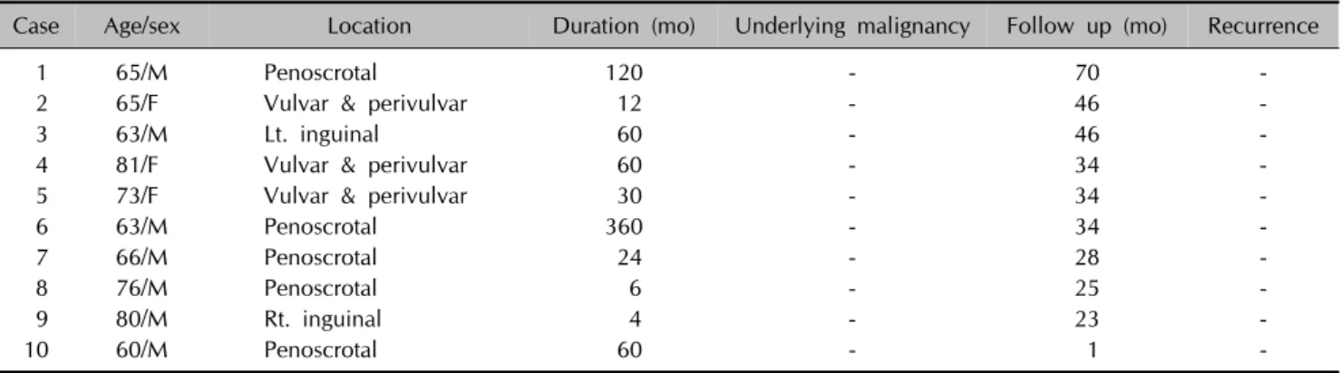
Case Age/sex Location Duration (mo) Underlying malignancy Follow up (mo) Recurrence
1 65/M Penoscrotal 120 - 70 -
2 65/F Vulvar & perivulvar 12 - 46 -
3 63/M Lt. inguinal 60 - 46 -
4 81/F Vulvar & perivulvar 60 - 34 -
5 73/F Vulvar & perivulvar 30 - 34 -
6 63/M Penoscrotal 360 - 34 -
7 66/M Penoscrotal 24 - 28 -
8 76/M Penoscrotal 6 - 25 -
9 80/M Rt. inguinal 4 - 23 -
10 60/M Penoscrotal 60 - 1 -
M: male, F: female, Lt.: left, Rt.: right.
Fig. 1. Preoperative photographs of the patients. (A) Penoscrotal (case 6), (B) vulvar (case 3).
Methods
EMPD was diagnosed based on the patients’ biopsy results. They had been screened for underlying malignan- cies and systemic metastases by clinical examinations and appropriate imaging (positron emission tomography- computed tomography, computed tomography etc.). Se- condary EMPD was ruled out using immunohisto- chemistry against markers such as cytokeratin (CK) 7, CK20 and GCDFP15. Multiple scouting biopsies were done with a 3 mm punch preoperatively. The biopsy sites were located 1 cm lateral to the clinically apparent margin. More than 5 biopsy sites were sampled in average, and the results were used to determine the surgical margins. EMPD lesions were excised with surgical margins of 1 cm lateral to the biopsy site when the biopsy result was negative, and 1.5 cm when the biopsy result was positive. The lesions were excised to the depth of the midsubcutaneous fat layer in the pubic area to the dartos muscle in the scrotum and to the superficial fascia of the penis. Histological control by examination of frozen tissue
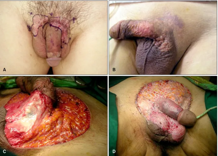
was performed in all the patients. The resultant skin defects were reconstructed with 16/1000 inch split- thickness skin grafts on the penile shaft and mesh grafts on the other sites (Fig. 2). Postoperatively, topical imiquimod was applied every other night, 3 times a week, over the margin and adjacent normal skin for a period of 6 months.
RESULTS
The most frequent location of the EMPD lesions was in the penoscrotal area for male patients and in the vulva for the female patients. None of the patients had underlying malignancies or systemic metastases (Table 1).
The pathologic analysis of the lesions revealed that the epidermis was extensively infiltrated by Paget cells, which are cells found exclusively in the epidermis without dermal invasion, in all the specimens. Immunohistochemi- cal examination of sections using CK7 and CK20 mono- clonal antibodies, PAS, D-PAS, and GCDFP15 and Alcian blue PH 2.5, revealed primary EMPD in all the patients (Table 2).
Fig. 2. (A) Location of scouting biopsies marked 1 cm lateral to the clinically apparent margin. (B) Preoperative preparation. (C) Deep to the midsubcutaneous fat layer with a negative margin on a frozen pathological tissue section. (D) A 16/1000 inch split-thickness skin graft on the penile shaft and mesh grafts on the other sites.
Table 2. Histochemical and immunohistochemical findings of 10 patients
Case Dermal invasion PAS D-PAS CK7 CK20 GCDFP15 Alcian blue pH 2.5
1 - + + + - + +
2 - Focal positive + + - + +
3 - + + + - + +
4 - + + + - + +
5 - + + + - + +
6 - Focal positive + + - + +
7 - Focal positive + + - + +
8 - + + + - + +
9 - + + + - + +
10 - + + + - + +
Intraoperative frozen sections were examined to confirm negative margin status.
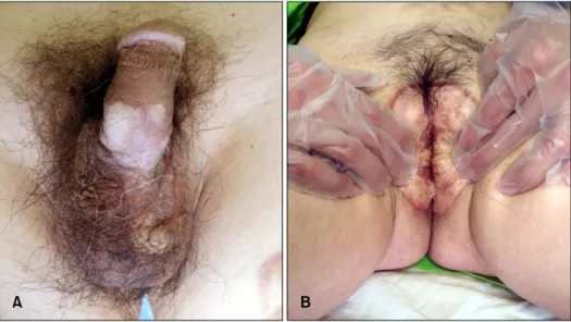
The patients returned for postoperative follow-up exami- nation every 3 months to allow for the evaluation of local or systemic recurrence and genitourinary complications (Fig. 3). All the 10 patients who were followed-up for 1 to 70 months (mean: 34.1 months) remained free of local
recurrence and metastases (Table 1).
DISCUSSION
In Korea, EMPD is more common in males than in females (male-female ratio; 3.9:1), which contrasts with findings from studies in Western populations. As a result of this
Fig. 3. Postoperative photographs of lesional sites. (A) Case 6, (B) case 3.
pattern of male predominance, the most common sites of disease are the scrotum, penile shaft, and pubic area5. The patients in the present study also showed a pattern of male predominance and involvement of predilection sites in the penoscrotal area.
Diagnosis of EMPD is confirmed by biopsy. Paget cells are usually identified by their abundant pale-staining cyto- plasm and large nuclei, with prominent nucleoli, and typical expression of simple epithelia-type CKs (CK7), sweat gland antigens (EMA, CEA, and GCDFP-15), and mucins (MUC1, and MUC5AC). On the basis of the immunophenotypic heterogeneity of EMPD, 2 distinct types of EMPD have been recognized. Type 1 (endoder- mal) EMPD cells express an endodermal phenotype (keratin 7+/keratin 20+/GCDFP-15-) and are associated with distant carcinomas. Type 2 (cutaneous and ectoder- mal) EMPD cells, alternatively, express sweat gland markers (keratin 7+/keratin 20-/GCDFP-15+) and are of cutaneous origin6. According to the immunohistochemical staining results, the patients described in this report showed type 2 cells, leading to the presumption that all of the cases involved primary EMPD. Therefore, extensive examinations for distant carcinomas were not conducted.
There is no consensus on the gold standard treatment for EMPD. Treatment options include topical 5-fluorouracil and topical bleomycin sulfate, radiation therapy, cryo- therapy, chemotherapy, CO2 laser therapy and photo- dynamic therapy7. The histologic extension of EMPD is usually much greater than the clinically apparent margin.
Furthermore, EMPD almost exclusively occurs in the anogenital area. Therefore, a paradox confronts surgeons:
potentially extensive surgery should be performed in an area where tissue preservation is vital. The most reco- mmended therapeutic mode is complete removal of the
lesion by wide excision and complete margin control, using MMS when available. MMS has theoretical advan- tages for this type of tumor. Lee et al.8 reported that MMS had a recurrence rate of 18.2% versus 36.4% in patients who underwent wide excision. However, compared with surgical excision, the total treatment costs of MMS are significantly higher, mainly owing to the longer surgical time and higher costs of pathological examination9,10. MMS also requires an appropriately equipped procedure room, well-trained nursing staff, and a laboratory with a histotechnician who specializes in MMS tissue processing.
In addition, MMS is a highly technical procedure that requires careful attention to detail along with considerable training and experience to achieve the expected high cure rate. Wilson et al.11 reported that MMS incurs higher fees than does wide excision for primary nonmelanoma skin cancer even after adjusting for associated risk factors. In EMPD, treatment cost for MMS is particularly higher than that for wide excision because of the size and complexity of the lesion. Moreover, many EMPD patients are treated by surgeons trained in urology, gynecology, plastic sur- gery or general surgery without training in MMS. There- fore, the determination of an adequate surgical margin for wide excision is important when MMS is unavailable.
Surgical margins are quite variable in the literature because of the rarity of the lesion and the resultant limited experience of authors. Resection margin status is very important because EMPD lesions are irregular and multi- centric3. Traditionally, the recommended surgical margin in wide excision is 5 cm12; however, such a large margin may cause serious functional and aesthetic impairment and may not lower the risk of recurrence. Recently, a safe resection margin of 2 cm has been recommended, even though a margin of 1 cm may be sufficient for lesions with
clinically clear margins4. In our institution, multiple scouting biopsies 1cm lateral to the clinically evident lesion, have started to be routinely performed before lesional excision13. The purpose of biopsies is to increase preoperative knowledge of the subclinical extent of the tumor. In the present study, the surgical margin was 1 cm lateral to the biopsy site if the biopsy result was negative and 1.5 cm lateral if it was positive. All the specimens were found to be free of tumor cells in the intraoperative frozen specimen examination14.
Imiquimod cream has been thought to be a useful postoperative adjuvant therapy. Imiquimod is a biological response modifier that binds to Toll-like receptor 7 on the cell surface of dendritic cells, macrophages, and mono- cytes, stimulating both innate and acquired immune function. Some reports about the efficacy of imiquimod in the treatment of EMPD have been recently published15. The aim of postoperative use of EMPD in our study was to improve the chances of cure, reduce patient morbidity, and enhance surgical efficacy. However, considering the high cost of imiquimod and short duration of application, the cost-benefit of imiquimod is not conclusive yet, thus requiring further studies.
The limitation of our study lies in the small number of patients due to the rarity of EMPD in Korea.
We suggest minimal surgical therapy combined with multiple preoperative scouting biopsies and postoperative topical imiquimod as a cost-effective EMPD treatment modality alternative to MMS.
REFERENCES
1. Yoon SN, Park IJ, Kim HC, Yu CS, Lee MW, Koh JK, et al.
Extramammary Paget's disease in Korea: its association with gastrointestinal neoplasms. Int J Colorectal Dis 2008;23:
1125-1130.
2. Dionyssopoulos A, Mandekou-Lefaki I, Vakirlis E, Manoli- adis I, Nikolis A, Dionyssopoulou E. Extramammary Paget's disease: isolated localization on the groin--wide local excision with immediate reconstruction. Ann Surg Oncol 2006;13:1235-1240.
3. Yang WJ, Kim DS, Im YJ, Cho KS, Rha KH, Cho NH, et al.
Extramammary Paget's disease of penis and scrotum.
Urology 2005;65:972-975.
4. Murata Y, Kumano K. Extramammary Paget's disease of the genitalia with clinically clear margins can be adequately resected with 1 cm margin. Eur J Dermatol 2005;15:
168-170.
5. Lee SJ, Choe YS, Jung HD, Ahn SK, Cha YC, Cho KH, et al;
Korean Society for Skin Cancer and Korean Dermato- pathology Research Group. A multicenter study on extrama- mmary Paget's disease in Korea. Int J Dermatol 2011;50:
508-515.
6. Kanitakis J. Mammary and extramammary Paget's disease. J Eur Acad Dermatol Venereol 2007;21:581-590.
7. O'Connor WJ, Lim KK, Zalla MJ, Gagnot M, Otley CC, Nguyen TH, et al. Comparison of mohs micrographic surgery and wide excision for extramammary Paget's disease.
Dermatol Surg 2003;29:723-727.
8. Lee KY, Roh MR, Chung WG, Chung KY. Comparison of Mohs micrographic surgery and wide excision for extrama- mmary Paget's disease: Korean experience. Dermatol Surg 2009;35:34-40.
9. Essers BA, Dirksen CD, Nieman FH, Smeets NW, Krekels GA, Prins MH, et al. Cost-effectiveness of mohs micro- graphic surgery vs surgical excision for basal cell carcinoma of the face. Arch Dermatol 2006;142:187-194.
10. Rogers HW, Coldiron BM. A relative value unit-based cost comparison of treatment modalities for nonmelanoma skin cancer: effect of the loss of the Mohs multiple surgery reduction exemption. J Am Acad Dermatol 2009;61:96-103.
11. Wilson LS, Pregenzer M, Basu R, Bertenthal D, Torres J, Asgari M, et al. Fee comparisons of treatments for nonmela- noma skin cancer in a private practice academic setting.
Dermatol Surg 2012;38:570-584.
12. Hendi A, Brodland DG, Zitelli JA. Extramammary Paget's disease: surgical treatment with Mohs micrographic surgery.
J Am Acad Dermatol 2004;51:767-773.
13. Appert DL, Otley CC, Phillips PK, Roenigk RK. Role of multiple scouting biopsies before Mohs micrographic surgery for extramammary Paget's disease. Dermatol Surg 2005;31:1417-1422.
14. Xu K, Fang Z, Zheng J, Lu Y, Li B, Sun C, et al. Intraopera- tive frozen biopsy in wide surgical excision of Paget's disease of the scrotum. Urol Oncol 2009;27:483-485.
15. Dias Coelho J, Vale E, Viana I, Martins O. Treatment of primary extramammary Paget's disease of the perineum with topical imiquimod 5% cream. Eur J Dermatol 2010;20:
532-533.