서 론
1. 연구의 필요성
우리 몸의 정신적 혹은 신체적 위협이나 스트레스 요인에 대하여 즉각적인 생리적 기능의 변화들에 대한 반응을 스트레스라고 한다.
스트레스는 뇌의 활동과 다양한 신경시스템의 장·단기적 변화를 나타낸다. 우리가 스트레스 요인에 노출될 때, 우리 뇌는 항상성을
유지하기 위하여 다양한 전달물질(transmitters), 펩타이드(peptides) 그리고 호르몬을 방출한다[1]. 신체는 스트레스에 대하여 교감신경 계와 시상하부-뇌하수체-부신 축(hypothalamic-pituitary-adrenal, HPA axis)의 작용으로 반응한다[2]. 스트레스는 HPA axis를 활성화 시키고 글루코코르티코이드(glucocorticoids)를 부신 피질로부터 분 비시킨다[3]. 스트레스는 카테콜라민과 글루코코르티코이드를 분 비시켜 감정, 인지기능 등에 많은 영향을 미친다고 알려져 있다[4].
구속 스트레스 쥐 모델에서 스트레스 반응 감소에 대한 사카린 섭취의 효과
박종민1·송민경2·김윤주2·김연정3
1
경북보건대학교 간호학과,
2경희대학교 간호과학대학,
3경희대학교 간호과학대학·동서간호학 연구소
Effect of Saccharin Intake in Restraint-induced Stress Response Reduction in Rats
Jong Min Park
1, Min Kyung Song
2, Yoon Ju Kim
2, Youn Jung Kim
31
Department of Nursing, Gyeongbuk College of Health, Gimcheon;
2College of Nursing Science, Kyung Hee University, Seoul;
3College of Nursing Science· East West Nursing Institute, Kyung Hee University, Seoul, Korea
Purpose: Stress activates the sympathetic nervous system and hypothalamic–pituitary–adrenal (HPA) axis and induces the release of
glucocorticoids. Saccharin is 300 times sweeter than sucrose, but does not increase blood insulin levels. Thus, this study was designed to evaluate the effect of saccharin intake in restraint-induced stress response reduction in rats. Methods: Adult male Sprague-Dawley (SD) rats had stress induced by restraint for 2 hours/day for 1 week. Saccharin was provided in sufficient amounts to allow them to in- take it voluntarily at 0.1% diluted in water. The Y-maze test and forced swim test (FST) were performed to evaluate cognitive function and the depressive behavior of the rats. The protein expression of the glucocorticoid receptor (GR) in hippocampal cornu ammonis (CA) 1 was investigated by using immunohistochemistry. Results: It was found that, the percentage of alternation in the Y-maze test was significantly (p< .01) higher in the Stress + saccharin group than in the Stress group. Immobility time in the FST was significantly (p< .01) lower in the Stress + saccharin group than in the Stress group. Also, the positive cells of GR in hippocampus CA1 were signifi- cantly (p< .05) lower in the Stress + saccharin group than in the Stress group. Conclusion: This study showed that there was an effect of saccharin intake in restraint-induced stress response reduction in rats.Key Words: Stress; Saccharin; Glucocorticoid receptor 국문주요어: 스트레스, 사카린, 글루코코르티코이드 수용체
Corresponding author: Youn Jung Kim
College of Nursing Science·East West Nursing Institute, Kyung Hee University, Kyung Hee Dae-ro, Dongdaemun-gu, Seoul 02447, Korea Tel: +82-2-961-0311 Fax: +82-2-961-9398 E-mail: [email protected]
* 이 논문은 2011년 교육과학기술부의 재원으로 한국연구재단의 지원을 받아 수행된 연구사업임(NRF-2010-0013256).
* This research was supported by the National Research Foundation of Korea (NRF) (NRF-2010-0013256).
Received: January 31, 2016 Revised: February 5, 2016 Accepted: February 10, 2016
This is an Open Access article distributed under the terms of the Creative Commons Attribution Non-Commercial License (http://creativecommons.org/licenses/by-nc/3.0) which permits
unrestricted non-commercial use, distribution, and reproduction in any medium, provided the original work is properly cited.
해마(hippocampus)는 대뇌 변연계의 양쪽 측두엽에 존재하며 학습 과 기억을 담당하는 부위로, 글루코코르티코이드 수용체(glucocor- ticoid receptor, GR)를 많이 가지고 있고, 글루코코르티코이드에 정 교하게 반응한다[5]. 또한 학습 전에 스트레스에 노출되면, 그 이후 의 기억 과정이 손상되는 것을 볼 수 있다[6].
스트레스가 유발될 때, 인체에서는 코티솔을 분비하여 대사율을 떨어뜨리고, 대뇌의 식욕중추를 자극하여 허기가 지지 않아도 음식 물 섭취를 하도록 작용한다. 대부분의 사람들은 스트레스를 받을 때 개인의 식습관이 바뀌는 것을 경험한다. 음식 섭취가 늘어나거나 [7], 음식 섭취가 평소보다 감소한다[8]. 게다가, 스트레스를 받을 동 안에 대부분의 사람들은 음식섭취량의 증감과 무관하게 단 음식에 대한 섭취량이 증가되는 모습을 보인다[9]. 동물실험에서 사람이 단 음식을 먹는 습관처럼 쥐에게 소량의 설탕 또는 사카린을 섞은 음료 를 하루 2회 4 주간 제공했을 때, 혈중 코르티코스테론이 감소하였으 며, 시상하부의 뇌실곁핵(paraventricular nucleus)에서 코르티코트로 핀분비호르몬(corticotropin-releasing hormone, CRH) messenger RNA (ribo nucleic acid)의 발현이 감소되었다[10]. 그러나 이러한 신체적 반 응으로 인한 고 칼로리 음식의 섭취는 비만이라는 또 다른 문제를 낳게 된다. 이러한 점에서 단맛은 가지나 열량을 가지지 않는 사카린 의 적용은 또 다른 스트레스 완화방법의 제시가 될 수 있다.
사카린은 설탕과 비슷하다는 의미로, 1879년에 Remsen과 Fahl- berg에 의하여 합성되었다. 사카린은 단맛을 내는 감미료로 사용되 며 설탕보다 300배 이상의 단맛을 내며, 단/다당류와는 다르게 열 량을 가지고 있지 않다[11]. 1960년대에 동물실험에서 사카린이 방 광암을 유발시킨다는 결과가 발표된 이후로 부정적인 인식을 갖게 되었으나 추후의 연구 결과 쥐에서 방광암을 일으키는 기전이 사 람에게는 발견되지 않았다는 것을 확인하였다[12]. 2010년에 미국 환경보호청(Environmental Protection Agency, EPA)은 사카린은 유 해우려 물질목록에서도 삭제했다. 현재 당뇨환자나 비만환자에 설 탕 대용품으로 사용되고 있다[13].
이에 본 연구는 스트레스에 대한 사카린 섭취의 효과를 입증하 기 위하여 구속 스트레스 동물모델에서 사카린 섭취에 대한 신경 과학·행동학적 변화를 확인하고자 한다.
2. 연구 목적
본 연구의 목적은 구속 스트레스 동물모델에서 사카린의 스트레 스 반응 감소 효과를 증명하여, 스트레스 관리 중재방안의 근거를 마련하고자 한다. 구체적인 목적은 다음과 같다.
1) 구속 스트레스 동물모델에 사카린을 자유로이 섭취하도록 중 재하여 스트레스로 인한 인지기능 회복과 우울행동의 극복 효과
를 행동실험을 통해 분석 및 확인한다.
2) 구속 스트레스 동물모델에 사카린을 자유로이 섭취하도록 중 재하여 뇌의 해마 부위에서 면역조직화학법(immunohistochemis- try)을 통한 GR의 발현을 분석 및 확인한다.
연구 방법
1. 연구 설계
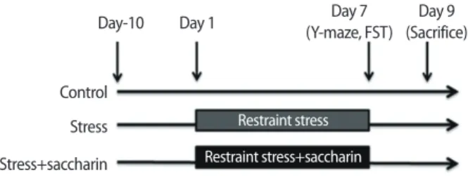
실험동물은 무작위로 선별하여 Control군(n= 4), Stress군(n= 8), Stress+saccharin군(n= 8)으로 분류하였다. 모든 군은 실험환경에 적 응을 위하여 10일간의 적응기간을 두고, 구속 스트레스 유발 실험을 실시하였다. 실험 시작일로부터 7일간 매일 같은 시간에 구속 스트 레스를 가했으며, 사카린은 0.1%로 물에 희석하여 자유롭게 섭취할 수 있도록 하였다. 실험시작 후 7일째에 행동실험을 시행하였다. 모 든 실험이 종료된 후 그 다음날인 9일째에 희생(sacrifice)시켰다(Fig- ure 1).
2. 연구 대상
본 연구는 K대학교의 실험동물윤리 위원회의 승인을 받은 후 규 정에 따라 실행하였다(KHU-12-35). 수컷 Sprague-Dawley (SD) 쥐 (250±10 g, 8 weeks, Daehan bio, Chungcheongbuk-do, Korea)를 실험 기간 동안 평균 온도 23± 2˚C, 습도 50± 2%로 유지하였으며, 밤낮 주 기(12시간 light/12시간 dark, light turn on 6 am)가 조절되는 환경에 수용하였다. 실험 기간 동안 물과 고형사료는 자유롭게 섭취할 수 있도록 제공하였다.
3. 연구 방법
1) 스트레스 유발
본 실험에서는 밑면 지름 40 cm, 높이 35 cm의 크기의 삼각 투명 아크릴(triangular transparent acrylic) 도구를 제작하여 Control군을 제외한 군에 일주일 동안 하루에 2시간(17:00-19:00)씩 restraint를 적 용하였다. 이 도구는 쥐가 호흡이 가능하고, 밖을 볼 수 있도록 설계
Figure 1. Design of experiment.
FST= Forced swim test.
Day-10
Control Stress
Stress+saccharin Restraint stress+saccharin Restraint stress
Day 1 Day 7
(Y-maze, FST)
Day 9
(Sacrifice)
되어 있다(Figure 2A).
2) 사카린 섭취
사카린(Sigma-Aldrich, St. Louis, USA)은 0.1%로 물에 희석하여 자 유롭게 섭취할 수 있도록 충분한 양을 7일간 제공하였고, 하루에 한 번씩 일정한 시간(08:00)에 섭취량을 기록하였다.
3) Y자형 미로 실험
Y자형 미로는 나무로 구성되어 있으며, 각각의 팔은 길이 25 cm, 높이 14 cm, 넓이 5 cm로 동일한 각도로 이루어져 있다. 실험은 각각 의 팔을 A, B, C 영역으로 정한 뒤, 일정한 영역(C 영역)에 쥐를 두고 5분간 측정 및 실시간으로 기록하였다. 각 팔의 출입 기록은 쥐의 뒷다리가 미로의 절반이상을 통과하였을 때만 인정하였다. 교대(al- ternation)는 3개의 팔을 연속으로 통과한 경우를 말한다.
Percentage of alternation (%)= (alternation / total number of arm en- tries-2)×100
4) 강제 수영 검사(Forced swim test, FST)
실험은 일반적으로 첫날 15분간 물에 빠뜨리는 예비실험(pretest)
과 24시간 후에 시행하는 본 시험(test)으로 구분하여 시행한다. 예 비실험은 첫날 유리로 된 투명한 원통(100 cm 깊이×30 cm 직경)에 25˚C 내외의 물을 바닥으로부터 60 cm까지 채우고 쥐를 15분간 물 에 빠뜨리면 초반에는 원통 밖으로 나가려 애를 쓰다가 결국 포기 하고 부동자세(immobility)를 취하는 일종의 행동 좌절(behavioral despair) 양상을 보이게 된다. 그리고 24시간 후에 흰쥐를 물에 빠뜨 려 본 실험(test)을 진행하게 되는데 이때 부동자세를 취하는 시간 (immobility duration)을 비디오 장치를 이용하여 측정하였다[14,15].
5) 뇌 적출 및 조직 처리
쥐를 에틸 에테르(ethyl ether)로 호흡마취를 한 후 흉강을 열어 심 장의 좌심실에 바늘을 꽂아 50 mM 인산염 완충 식염수(phosphate buffered saline, PBS)을 관류시켜 혈관 내 혈액을 제거한 후 4% 파라 포름알데하이드(paraformaldehyde, PFA) in 20 mM 인산염 완충액 (phosphate buffer, PB)를 관류시켜 뇌를 고정시켰다. 뇌를 꺼낸 후 4%
파라포름알데하이드에 담궈 4˚C에서 24시간 고정시킨 후 30% Su- crose 용액으로 옮겨 2-3일간 침전시켜 냉동절편기(cryocut micro- tome, Leica, Germany)로 뇌 조직을 40 μm 두께로 연속 관상 절편하 여 4˚C 냉장 보관하였다.
A
450 400 350 300 250 200
W eigh t (g)
Control Stress Stress+saccharin
Day 1 Day 7
B 700
600 500 400 300 200 100
0 Day 1 Day 2 Day 3 Day 4 Day 5 Day 6 Day 7
In tak e of 0.1% sac charin solution (mL)
C
Figure 2. Restraint stress equipment and set up (A) Rats were placed into restraint tool for 2 hour. (B) Results of weight in rats. (C) Total saccharin
consumption during each drinking day.
Data are expressed as mean± S.E.
6) 면역조직화학염색(Immunohistochemistry, IHC)
해마의 cornu ammonis (CA) 1 부분에서 GR의 발현을 확인하기 위해 절편한 조직을 꺼내어 PBS로 세척한 후 3% H2O2를 상온에서 30분간 반응시키고 기포가 사라질 때까지 충분히 세척하였다. 1차 항체인 GR (1:1,000, Santa Cruz, California, USA)을 4˚C에서 12시간 동안 반응시켰다. 다음날 PBS로 5분간 3회 세척한 후 2차 항체를 상 온에서 1시간 동안 반응시키고 PBS로 세척하고 ABC (Avidin-Biotin Complex, Vector, USA) 용액을 1시간 동안 반응시키고, PBS로 세척 하였다. 이후 발색제인 DAB kit (diaminobenzidine, Vector, USA)를 사용하여 10-30초 정도 반응시킨 후 PBS로 세척하여 조직을 슬라 이드 글라스에 붙여 12-24시간 동안 자연 건조하였다. 건조 후에는 탈수 과정을 위하여 ethanol을, 조직의 투명화를 위해 xylene에 각각 담근 후 permount (SP15-500. Fisher Scientific, Pittsburgh, USA)를 사 용하여 커버 글라스를 덮었다. 모든 과정 후 광학현미경(BX51, Olympus, Japan) 40배에서 촬영을 하였다. GR이 발현된 세포 수는 CA1 중앙에 250× 250 um의 사각형을 만들어, 실험에 참여하지 않 은 3명의 연구원에게 육안으로 측정한 후 평균 값을 구하였다[16].
4. 자료 분석
모든 실험결과는 SPSS 18.0을 이용하여 평균, 표준편차, 표준오 차를 산출하였고, one-way ANOVA로 분석한 후 scheffe로 사후 검 증을 하였으며 통계적 유의수준은 p<.05에서 채택하였다.
연구 결과
1. 체중 변화
모든 실험동물은 실험 시작(Day 1), 실험 시작 7일 후(Day 7)에 체 중을 측정하였다. 체중을 측정한 결과는 Figure 2B와 같으며, 체중 의 변화는 Control군(107 g)에 비해 Stress군(71 g), Stress+saccharin군 (46 g)에서는 체중 증가의 폭이 적었다. 사카린 섭취량은 7일간 측정 하였고, 기간 동안 사카린 섭취량은 통계적으로 유의한 변화 없이 유지되었다(Figure 2C).
2. Y자형 미로 검사에서의 교대의 증가
Y 미로 실험은 공간작업기억(spatial working memory)을 측정하
100
80 60 40 20 0
Per cen tage of alt erna tion (%)
Control Stress Stress+saccharin
#
*
Figure 3. Saccharin intake ameliorated the memory impairment by re-
straint stress. Quantitative analysis of Y-maze alternation test.
Data are expressed as mean± S.E.
#p< .01 versus control group; *p< .01 versus stress group.
60 50 40 30 20 10 0
Immobilit y time (sec)
Control Stress Stress+saccharin
#
*
Figure 4. Saccharin intake reduced the immobility time. Quantitative
analysis of FST.
Data are expressed as mean± S.E.
#p< .01 versus control group; *p< .01 versus stress group. FST= Forced swim test.
Figure 5-1. Saccharin intake reduced positive cells of GR in hippocampus CA1 region.
Immunohistochemical staining of GR (a, Control; b, Stress; c, Stress + saccharin). Scale bar represents 100 um. The tissue was measured by microscope on magnification 40. GR= Glucocorticoid receptor; CA= Cornu ammonis.
A B C
는 실험으로 구속스트레스에서 사카린 섭취가 인지 기능에 미치는 영향을 알아보기 위해 시행하였다. 공간작업기억을 확인할 수 있는 교대는 Control군(Mean=73.13±7.10)이 Stress군(Mean= 61.50± 2.29) 보다 통계적으로 유의하게 높았으며(p <.01), Stress군과 비교하여 Stress+saccharin군(Mean=72.50±3.25)이 통계적으로 유의하게 높 았다(p<.01) (Figure 3).
3. 강제 수영 검사에서 부동시간(immobility time)의 감소 강제 수영 검사는 우울과 관련 행동을 관찰하기 위한 검사로 스트 레스 모델에서 사카린 투여가 우울에 미치는 영향을 측정하기 위해 시행하였다. 우울행동의 지표인 부동시간은 Control군(Mean=20.25
±1.08)이 Stress군(Mean= 49.13±7.20)에서 통계적으로 유의하게 낮았 으며(p<.01), Stress군과 비교하여 Stress+saccharin군(Mean=19.00±
3.26)이 통계적으로 유의하게 낮았다(p<.01) (Figure 4).
4. 해마 CA1 부위에서 glucocorticoid receptor 발현 증가 면역조직화학염색을 이용하여 해마 CA1 부위의 GR 발현을 확 인하였다. Control군(Mean=3.13±1.14)이 Stress군(Mean=32.13±
5.79)보다 통계적으로 유의하게 낮았으며(p<.01), Stress군과 비교하 여 Stress+saccharin군(Mean=16.50±4.40)이 통계적으로 유의하게 낮았다(p<.05) (Figure 5-1, Figure 5-2).
논 의
본 연구에서는 구속 스트레스 동물 모델에서 사카린 섭취를 통 한 스트레스 반응의 감소 효과를 규명하고자 실험을 하였다.
구속 스트레스로 인한 신체적 반응 중의 하나는 체중감소이다.
본 실험에서 실험 시작일에 측정한 체중과 7일 간의 스트레스 적용 후 측정한 체중에서 Control군 이외의 군은 증가의 폭이 적었다. 구 속 스트레스를 3일간 하루 3시간 적용하자 일시적으로 사료 섭취량 이 줄고, 그 이후에 체중은 Control군에 비해 낮게 유지 되었던 이전 의 결과와 일치한다[17]. 구속 스트레스와 사카린 섭취군은 스트레 스군보다도 체중 증가율이 감소하는 양상을 보였는데, 사카린 섭취 로 인해 식욕저하, 저지방혈증, 저콜레스테롤혈증을 야기한다고 하 는 이전의 연구 결과와 일치한다[18].
Y자형 미로 검사에서 정상의 쥐들은 #1 arm에 들어간 다음에는
#2, #3을 방문하는 것이 정상적인 탐색 활동이다. 이 활동은 문제 해 결 능력 및 규칙을 얻는 능력을 나타낸다[19]. 본 연구에서 Y자형 미 로 검사의 결과는 모든 군에서 통계적으로 유의한 차이가 있는 것 으로 나타났다(p<.01). 이는 구속 스트레스가 공간작업기억에 부 정적인 영향을 미친 것으로 보인다. 우리가 일반적으로 알고 있는 만성 스트레스가 장기기억 형성에 필요한 기억의 강화(consolida- tion)에 부정적인 영향을 미치는 것뿐만 아니라, 쥐의 해마에서의 과 립세포(granule cell)의 증식을 감소시키고, 기억 형성에 영향을 미친 다는 선행 연구의 결과와 일치한다[20,21]. 또한 사카린 섭취가 공간 작업기억의 손상을 막아주는 효과가 있음을 확인하였다.
강제수영 검사에서 부동시간이 길어질수록 삶에 대한 희망이 적 은 것으로 판단하여 우울증 지표로 사용되고 있다. 본 연구에서는 구속 스트레스를 주었을 때, 부동시간이 통계적으로 유의미하게 증 가되었다가, 사카린을 섭취한 구속 스트레스군에서 다시 감소하는 양상을 보임으로써, 사카린 섭취가 구속 스트레스로 인한 우울의 정도를 감소시켰음을 확인하였다(p<.01).
다양한 스트레스와 우울 행동과 관련된 선행연구에서 설탕과 사카린의 선호 및 섭취 증가를 확인하였다[22, 23]. 또한 만성적인 신 체 구속으로 인한 스트레스의 결과 고형 사료 섭취량은 변화가 없 지만, 단 음식을 찾는 등의 식욕은 변화되었다[24]. 또한 고지방, 단 음식의 섭취가 스트레스로 인한 우울감을 감소시키고, 신체적인 스트레스 반응을 감소시켜준다는 것을 밝혀냈다[25]. 그러나 설탕 과 같은 단 음식의 과다섭취는 체중 증가를 일으키며, 비만은 대사 증후군, 심혈관 질환, 암과 사망률이 높은 질환을 일으키는 요인으 로 작용한다[26]. 사카린은 앞에서 설명한 것과 같이 열량을 가지고 있지 않아 스트레스의 중재 방안으로 적절하다고 할 수 있다.
스트레스를 경험하게 되면 신체 내에서 아드레날린과 같은 카테 콜아민을 먼저 급격하게 방출한다[27]. HPA axis가 활성화되어 코르 티코트로핀분비호르몬이 시상하부로부터 분비되어 뇌하수체를 자 극하여 부신피질자극호르몬(adrenocorticotrophic hormone, ACTH) 을 분비시켜 최종적으로 부신 수질에서 글루코코르티코이드를 혈
40
30
20
10
0
Positiv e c ells of GR
Control Stress Stress+saccharin
#
*
Figure 5-2. Saccharin intake reduced positive cells of GR in hippocam- pus CA1 region.
Quantitative analysis of GR. Data are expressed as mean± S.E. #p< .01 versus control group; *p< .05 versus stress group. GR= Glucocorticoid receptor;
CA= Cornu ammonis.
류로 보낸다. 증가된 글루코코르티코이드는 주로 해마와 편도체의 GR을 증가시키고, 이 두 영역에서의 GR은 기억 및 두려움, 스트레스 경험뿐 아니라, 스트레스 반응에 대한 HPA axis에 중요한 역할을 한 다[28]. 본 연구에서 해마의 CA1에서의 GR 발현의 결과는 Stress군과 비교하여 Stress + saccharin군이 통계적으로 유의하게 낮았다 (p<.05). 이는 Y자형 미로 검사 결과와도 일치하며, 스트레스와 글루 코코르티코이드는 해마에서의 시냅스 가소성(synaptic plasticity)을 조절하고 시냅스 후의 통로형 글루탐산 수용체(inotropic glutamate receptor)에 영향을 준다는 선행 연구 결과와도 일치한다[29].
스트레스의 간호 중재들이 현재 많은 선행 연구결과 입증되어 왔 다. 본 실험연구는 임상 상황에서 일어날 수 있는 스트레스에서 사 카린이 비만 예방뿐 아니라, 스트레스 반응을 경감시킬 수 있음을 동물실험을 통해 생행동학적, 분자생물학적으로 입증하여, 근거기 반 간호의 적절한 간호 중재의 개발에 기여한다고 사료된다.
결 론
본 연구는 구속 스트레스 쥐 모델에서 사카린 섭취를 이용하여 신체 스트레스 반응의 감소 효과를 확인하였다. 행동 실험 결과, 사 카린 섭취가 공간작업기억의 손상을 줄이고, 강제적 수영 검사에서 부동시간을 감소시켜 스트레스로 인한 우울 행동을 감소시켰고, 해마 CA1에서의 스트레스 호르몬에 대한 반응인 GR의 발현을 감 소시켰다. 이상의 결과로 사카린 섭취가 스트레스로 인한 신체 반 응을 감소시킨다는 결론을 내렸으나, 구속할 당시, 혈중 코르티코 스테론의 양을 측정하지 못해 이상의 연구 결과를 좀 더 완벽하게 지지하지 못한 제한점이 남았다.
REFERENCES