Vol. 17, No. 2, November, 2009 □ 증 례 □
1)
Introduction
Neurofibromatosis type I(NF-1) is an autoso- mal dominant neurocutaneous syndrome with the genetic abnormality localized to the long arm of chromosome 17
1). It is one of the most common genetic disorders, with a prevalence of approxi- mately 1 in 3,500 to 4,000 individuals
2). NF-1 has a predilection for males, with a male-female ratio of 3:1
2). The diagnosis can be made if two or more of the followings are present: (1) six or more café-au-lait spots, (2) two or more neurofi- bromas, (3) one or more plexiform neurofibromas (PNF), (4) freckling in the axilla or inguinal re- gion, (5) optic glioma, (6) two or more Lisch no- dules, (7) a distinctive osseous lesion, or (8) a first-degree relative with NF-1
3). NF-1 has va- riable manifestations and can involve diverse systems: the central and peripheral nervous sys-
: 2009 5 22 , : 2009 10 27
: ,
Tel : 062)220-6646, Fax : 062)222-6103 E-mail : yjwoo@chonnam.ac.kr
tem; the skin; the bone; the endocrine system;
gastrointestinal system; and the vascular sys- tem
4).
Gastrointestinal involvement occurs in 10-25
% of patients with NF-1 and includes solitary or multiple neurofibromas, leiomyomas and rarely PNF
5, 6). However, for children with NF-1, gas- trointestinal involvement is rare, especially the presence of a PNF
5). The most common site of a mesenteric PNF, according to the report by Imamoglu et al., is the terminal ileum, followed by the jejunum and colon
5). We report a rare case of radiologically suspected mesenteric PNF, along the celiac axis, in an 8-year-old boy.
Case Report
An 8-year-old boy was admitted to Chonnam National University Hospital(CNUH) due to severe periumbilical pain that was relieved after defecation. His mother had the diagnosis of NF- 1. On physical examination he had multiple café-
A Case of Radiologically Suspected Mesenteric Plexiform Neurofibromas in a Patient with
Type I Neurofibromatosis
Soo Min Oh, M.D., Young Ok Kim, M.D.
Young Jun Son, M.D. and Young Jong Woo, M.D.
Department of Pediatrics, School of Medicine, Chonnam National University, Gwangju, Korea
= Abstract =
Neurofibromatosis type I(NF-1) is an autosomal dominant neurocutaneous syndrome characterized by café-au-lait spots, optic glioma, skeletal dysplasia, and iris hamartoma.
Mesenteric plexiform neurofibromas(PNF) have been rarely reported in NF-1, especially in children. We report a case of radiologically suspected mesenteric PNF along the celiac axis in an 8-year-old boy who had café-au-lait spots and a family history of maternal NF-1.
Key Words : Neurofibromatosis Type 1, Neurofibroma, Plexiform
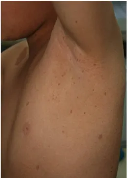
au-lait spots over the upper body with axillary freckling(Fig. 1). Plain films of the abdomen showed severe fecal loading with bowel dilatation from the ascending colon to the rectum. The ab- dominal computed tomography(CT) incidentally revealed a 6.3×2.0 cm, well defined, non-enhan- cing, hypo-attenuated mass along and encasing the celiac axis, suspicious for a PNF(Fig. 2A);
in addition, there was a spindle shaped, non enhancing hypo-attenuated nodule noted in the left pelvic wall. All of the results including a complete blood cell count and serum chemistry (serum total protein and albumin, electrolytes, liver and kidney function and acute phase reac- tants) were normal. Although the abdominal pain was relieved with an enema and constipation was the suspected diagnosis, surgical resection of the mass was recommended for histopatholo- gical confirmation and treatment. However, the patient's guardian declined further treatment.
The patient has remained well since this episode.
Three years after the first visit, the patient was readmitted to CNUH due to recurrent peri- umbilical pain. Basic laboratory studies included a complete blood cell count, serum chemistry (serum total protein and albumin, electrolytes, liver and kidney function, amylase, lipase, muscle enzymes and acute phase reactants) and urine analysis. All of the results were normal except for the lactate dehydrogenase(elevated to 507 U/
L). The tumor markers(CEA, AFP) were within normal limits. The follow-up abdominal CT showed no change(Fig. 2B). The mass did not cause any disturbance as determined by the small bowel study, upper endoscopy and colonoscopy.
Fig. 1. Multiple café-au-lait spots and axillary freckling.
Fig. 2. Abdominal computed tomography on the first visit shows a 6.3×2.0 cm, well defined, non-enhancing, hypo-attenuated mass along and encasing the celiac axis(A). There were no changes in the abdominal CT over 3 years, which showed a well defined, non-enhancing hypoattenuated mass along and encasing the celiac axis(B).
The growth and development was normal but the brain MRI demonstrated ill defined hyperintense lesions at the left cerebellar peduncle and both posterior thalami suggesting hamartomas on T2- weighted image(Fig. 3). Surgical resection was still declined and the patient was lost to follow- up.
Discussion
Gastrointestinal PNFs associated with NF-1 are rare and extremely rare in children
5). In a review by Hochgerg et al.
6), all patients with gastrointestinal involvement were 10 years or older. In the largest series(16 patients) of retro- peritoneal PNFs reported by Bass et al.
7), the youngest patient was 15 years old
7). In the report by Imamoglu et al. reviewing nine children with mesenteric PNF, the average age at diagnosis was 9.2 years and ranged from 6 to 15 years
5). The clinical presentation of gastrointestinal PNFs depends on the location and size of the PNF
5). An intestinal(serosal) PNF can present
with ulceration, bleeding, intestinal obstruction, and, rarely, intussusception, volvulus, perforation, and failure to thrive due to impaired absorption
5,6)
. However, most cases of mesenteric PNFs are asymptomatic or present with non-specific vague symptoms; however, as they become larger they are more likely to be symptomatic
5). In the review by Imamoglu et al, 55%(5 of total 9 pati- ents) of the patients presented with abdominal symptoms, including mild abdominal pain and distension, and occasionally, nausea and vomi- ting
5). By contrast, there were 45% of the pati- ents(4 of 9 patients) that were truly asympto- matic
5). Therefore, because the gastrointestinal PNF is rare and presents with vague or absent clinical symptoms, a high degree of suspicion is needed for a timely diagnosis
5). Delay in the diagnosis of gastrointestinal involvement with a PNF is common. The average interval from on- set of GI symptoms to the diagnosis and detec- tion of GI neoplasms was 2.8 years in one series
8). The term plexiform neurofibroma is derived from the pathological analysis
9). The PNF is
Fig. 3. Brain MRI on T2-weighted image shows ill defined hyperintense le-sions at the left cerebellar peduncle(A) and both posterior thalami(B) sug- gesting hamartomas.
composed of an interdigitating network of finger- like fronds of tumor extending in a serpentine fashion along a nerve and its branches
10). Growth of a PNF, in general, can occur at any time in life
9). Clinical experience, however, sug- gests that PNFs tend to grow at two distinct periods
9). The first is during early childhood and the second is during periods of hormonal change, most notably during puberty or, in women, du- ring pregnancy
9). Appearance of new cutaneous neurofibromas may also occur at these times.
However, there have been no risk factors identi- fied for the growth of PNFs to date
9).
Symptomatic PNFs are usually evaluated by imaging studies, such as magnetic resonance imaging(MRI) or computed tomography(CT) scanning
9). In general, the MRI is the modality of choice. Although their morphology is variable, PNFs display high signal intensity on the T2- weighted MRI often with a central area of low signal intensity
11). The CT findings of PNFs have been described as homogeneous low-atte- nuation masses on post-contrast images
12). These findings have been attributed to entrapment of adipose tissue, cystic degeneration and the pre- sence of a myxoid matrix
13). Imaging plays an important role in the diagnosis, evaluation and follow-up of patients with abdominal manifesta- tions of NF-1. Barium studies may demonstrate intraluminal mass lesions
14). Sometimes, intussu- sception of the bowel may be detected along with intestinal obstruction. Duplex color and spectral Doppler ultrasonography reveal the vascular complications of NF-1 such as aneurysms and stenoses
14). Biliary strictures and bile duct intra- luminal NF are rarely associated with NF-1, but if present, may be visualized by antegrade or retrograde cholangiographic imaging
14).
For treatment, complete surgical resection is
the goal when possible
1). However, aggressive surgical procedures should be avoided with unre- sectable masses
1). In such cases, the optimal approach is close follow-up both clinically and radiologically for the evaluation of signs of ma- lignant transformation after multiple biopsies. A rapidly growing tumor or worsening of symp- toms may indicate a malignancy
15). The rate of malignant degeneration is not known but is esti- mated to be approximately 13%
15). The prognos- tic factors for tumor recurrence, in childhood PNF, have been reported to be an age less than 10 years at the time of the initial surgery, pre- sence of residual tumor after surgery and location of the tumor on the head, neck or face
16). Some medical treatment options including ketotifen fumarate, cis-retinoic acid, interferon- thalido α - mide, and the farnesyl protein transferase inhibi- tor may have a role in the management of sym- ptomatic or progressive benign lesions
17).
In the current case, surgical resection of the abdominal mass was needed for histopathological confirmation and to rule out other tumors such as lymphangioma, neurofibrosarcoma or neuro- blastoma, and as treatment. Although histopatho- logical confirmation of the abdominal mass was not available in this case, the findings were radiologically consistent with a mesenteric PNF.
한 글 요 약
제 형 신경섬유종증에서 발생한 1 방사선학적으로 의심되는 복부 얼기형 신경섬유종 1 례
오수민 김영옥 손영준 우영종․ ․ ․
1
, . 8
café-au-lait ,
,
, .
References
1) Fenton LZ, Foreman N, Wyatt-Ashmead J.
Diffuse, retroperitoneal mesenteric and intra- hepatic periportal plexiform neurofibroma in a 5-year-old boy. Pediatr Radiol 2001;31:637-9.
2) Wilkinson LM, Manson D, Smith CR. Plexi- form neurofibroma of the bladder. Radiogra- phics 2004;24:S237-42.
3) Mulvihill JJ, Parry DM, Sherman JL, Pikus A, Kaiser-Kupfer MI, Eldridge R. NIH conference.
Neurofibromatosis 1 (Recklinghausen disease) and neurofibromatosis 2 (bilateral acoustic neurofibromatosis). An update. Ann Intern Med 1990;113:39-52.
4) Huson SM, Harper PS, Compston DAS. Von Recklinghausen neurofibromatosis: a clinical and population study in south-east Wales.
Brain 1988;111:1355-81.
5) Imamoglu M, Cay A, Yaris N, Yayla S, Sari- han H. Intestinal mesenteric involvement with plexiform neurofibroma in neurofibromatosis type 1. Pediatr Int 2006;48:337-9.
6) Hochberg FH, Dasilva AB, Galdabini J, Rich- ardson EP Jr. Gastrointestinal involvement in von Recklinhausens neurofibromatosis. Neuro- logy 1974;24:1144-51.
7) Bass JC, Korobkin M, Francis IR, Ellis JH, Cohan RH. Retroperitoneal plexiform neurofi- bromas: CT findings. AJR Am J Roentgenol 1994;163:617-20.
8) Bakker JR, Haber MM, Garcia FU. Gastrointe- stinal neurofibromatosis: an unusual cause of gastric outlet obstruction. Am Surg 2005;71:
100-5.
9) Korf BR. Plexiform neurofibromas. Am J Med Genet 1999;89:31-7.
10) Packer RJ, Gutmann DH, Rubenstein A, Visko- chil D, Zimmerman RA, Vezina G, et al. Plexi- form neurofibromas in NF1: toward biologic- based therapy. Neurology 2002;58:1461-70.
11) Filler AG, Kliot M, Howe FA, Hayes CE, Saunders DE, Goodkin R, et al. Application of magnetic resonance neurography in the eva- luation of patients with peripheral nerve pa- thology. J Neurosurg 1996;85:299-309.
12) Rodriquez E, Pombo F, Rodriquez I, Vazquez Iglesias JL, Galed I. Diffuse intrahepatic peri- portal plexiform neurofibroma. Eur J Radiol 1993;16:151-3.
13) Kumar AJ, Kuhajda FP, Martinez CR, Fishman EK, Jezic DV, Siegelman SS. Computed tomo- graphy of extracranial nerve sheath tumors with pathologic correlation. J Comput Assist Tomogr 1983;7:857-65.
14) Rastogi R. Intra-abdominal manifestations of von Recklinghausens neurofibromatosis. Saudi J Gastoentrol 2008;14:80-2.
15) Leslie MD, Cheung KY. Malignant transfor- mation of neurofibromas at multiple sites in a case of neurofibromatosis. Postgrad Med J 1987;63:131-3.
16) Needle MN, Cnaan A, Dattilo J, Chatten J, Phillips PC, Shochat S, et al. Prognostic signs in the surgical management of plexiform neu- rofibroma: the childrens hospital of philadelphia experience, 1974-1994. J Pediatr 1997;131:678- 82.
17) Gupta A, Cohen BH, Ruggieri P, Packer RJ, Phillips PC. Phase I study of thalidomide for the treatment of plexiform neurofibroma in neurofibromatosis 1. Neurology 2003;60:130-2.