VOL. 18, NO. 2, 2019 Case Report
CLINICAL PAIN
88
https://doi.org/10.35827/cp.2019.18.2.88
접수일 : 2019 년 1 월 29 일 , 게재승인일 : 2019 년 7 월 1 일 책임저자 : 임성훈 , 수원시 팔달구 중부대로 93
16247, 가톨릭대학교 의과대학 성빈센트병원 재활 의학과
Tel: 031-249-7650, Fax: 031-251-4481 E-mail: [email protected]
비소세포폐암의 뇌전이로 인한 갑작스런 수부 통증 및 마비
가톨릭대학교 의과대학 성빈센트병원 재활의학과
성원진ㆍ홍보영ㆍ김준성ㆍ유재완ㆍ임성훈
A Man Presenting with Sudden Weakness and Pain of the Right Hand, by Non-Small Cell Lung Cancer with Brain Metastases
Won Jin Sung, M.D., Bo Young Hong, M.D., Ph.D., Joon Sung Kim, M.D., Ph.D., Jae Wan Yoo, M.D. and Seong Hoon Lim, M.D., Ph.D.
Department of Rehabilitation Medicine, St. Vincent’s Hospital, College of Medicine, The Catholic University of Korea, Seoul, Korea
Unexplained pain and weakness, i.e., without obvious predisposing factors, are often encountered by physiatrists and efforts should be made to determine the cause. A 63-year-old male presented with radiating pain in his right arm and mild weakness of the right hand. An electrodiagnostic examination revealed distal symmetric sensory polyneuropathy in the upper and lower extremities, and denervation potentials in the forearm muscles, which were inconsistent with the cervical spine MRI images and symptoms. A predisposing undiscovered disease was revealed, i.e., squamous cell carcinoma in the lung; brain metastasis affecting the left primary motor cortex was also detected. Therefore, we concluded that the pain and weakness were related to paraneoplastic syndrome and brain metastases of the hand knob. The observed denervation potentials were characterized as trans-synaptic changes in the brain metastasis. This case highlights the importance of unexplainable focal pain and weakness in the increasing prevalence of cancer. (Clinical Pain 2019;18:88-91)
Key Words: Lung cancer, Squamous cell carcinoma, Paraneoplastic syndrome
INTRODUCTION
Unexplained pain and weakness are often encountered by physiatrists in the clinic; incidental sensory polyneuro- pathy and incidental denervation potentials are also fre- quently detected. The clinical importance of sensory only polyneuropathy and incidental denervation potentials tend to be overlooked by clinicians due to its typically benign course and the absence of proper treatment. However, pain and weakness are common symptoms of sensory neuro- pathies caused by paraneoplastic syndrome.
1Thus, sensory polyneuropathy, or a subtle change in incidental denerva- tion potentials, may suggest a hidden disease.
This case report describes incidental sensory polyneuro-
pathy caused by paraneoplastic syndrome. The weakness was finally determined to be caused by trans-synaptic de- nervation of brain metastases in the primary motor cortex area corresponding to the hand. This case highlights the importance of checking for incidental sensory polyneuro- pathy and denervation potentials when seeking to deter- mine the cause of unexplained focal pain.
CASE REPORT
A 63-year-old male visited the Department of Neurosur-
gery for radiating pain in his right arm and elbow with
mild weakness of the right hand, especially in the fourth
and fifth fingers. Cervical spine magnetic resonance imag-
ing (MRI) showed minimal central disc protrusion at C6∼7,
mild central disc protrusion, and C6∼7 spondylosis. He was
referred for an electrodiagnostic study to evaluate weakness
inconsistent with the MRI findings. The weakness had been
present for 1 month. Weakness of the right upper limb was
diffuse, but was more severe in the right hand. The degree
of weakness was stable and had not progressed rapidly dur-
성원진 외 4 인: 비소세포폐암의 뇌전이로 인한 갑작스런 수부 통증 및 마비
CLINICAL PAIN
89 Fig. 1. Computed tomography scan showed a 7.2 cm heterogeneously enhancing mass with necrosis in the right lower lobe anterobasal segment. (A, B) Axial images of the lung, (C) coronal image of the lung, (D) sagittal image of the lung.
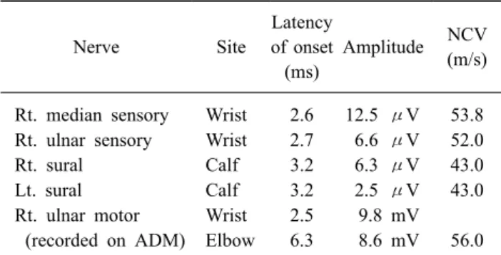
Table 1. Findings of the Nerve Conduction Study
Nerve Site
Latency of onset
(ms)
Amplitude NCV (m/s)
Rt. median sensory Wrist 2.6 12.5 μV 53.8 Rt. ulnar sensory Wrist 2.7 6.6 μV 52.0
Rt. sural Calf 3.2 6.3 μV 43.0
Lt. sural Calf 3.2 2.5 μV 43.0
Rt. ulnar motor Wrist 2.5 9.8 mV (recorded on ADM) Elbow 6.3 8.6 mV 56.0 NCV: nerve conduction velocity, Lt.: left, Rt.: right, ADM: ab- ductor digiti minimi.
ing the past month. Physical and neurological examinations revealed no Hoffmann reflex in either hand. The deep ten- don reflexes of the biceps and triceps brachii muscles were normal. A manual muscle test revealed a fair to fair + grade for the right elbow flexor and wrist extensor, a fair grade for the elbow extensor and finger flexor, and a poor + grade for the finger abductor.
Nerve conduction studies revealed distal symmetric sen- sory polyneuropathy in both the upper and lower extremi- ties (Table 1). Needle electromyography showed a denerva- tion potential of the right pronator teres and extensor carpi radialis longus muscles. The patient was diagnosed with mid to C6 and C7 radiculopathies. However, the findings were inconsistent with his symptoms.
The patient's sensory polyneuropathy could not be ex- plained by his medical or social history, which was charac- terized by the absence of diabetes, vascular disease, heavy
alcohol use, and poisoning. Thus, a physiatrist and a neuro-
surgeon analyzed the abnormal electrodiagnostic findings
VOL. 18, NO. 2, 2019
CLINICAL PAIN
90
Fig. 2. Enhanced magnetic reso- nance imaging revealed a 2.4 × 2.0
× 2.5 cm necrotic mass with peri- tumoral edema in the left hand knob of the primary motor cortex.
(A, B) Axial images of the brain, (C, D) coronal images of the brain.
and referred the patient to the internal medicine department for a work up of the malignancy. Chest computed tomog- raphy (CT) performed by a pulmonologist revealed a 7.2-cm heterogeneously enhancing mass in the right lower lobe anterobasal segment (Fig. 1) that was diagnosed as squamous cell carcinoma on biopsy.
After diagnosing lung cancer, enhanced brain MRI was performed to evaluate the metastasis. Brain MRI revealed a 2.4 × 2.0 × 2.5-cm necrotic mass with vasogenic edema in the left ‘hand knob’ of the primary motor cortex (Fig.
2). The metastatic lesion was consistent with the motor area corresponding to the patient’s weak right arm and hand.
2Thus, we concluded that the weakness in the right upper limb was induced by a brain metastasis in the pri- mary motor area. In addition, peripheral neuropathy and paraneoplastic syndrome may have contributed to the pain.
Despite not testing for the anti-Hu antibody or other para- neoplastic syndrome antibodies, due to a lack of insurance
coverage, we concluded that the sensory polyneuropathy was related to paraneoplastic syndrome, and that the hand weakness was caused by brain metastases in the primary motor cortex corresponding to the hand.
3Indeed, denerva- tion potentials in the right forearm were not explained by MRI images of the cervical spine, nor by any other nerve conduction studies. The physiatrist presumed that the cause of the incidental denervation potentials in the right forearm was trans-synaptic denervation with brain metastasis.
4However, the exact cause of the denervation potentials was not fully revealed. The patient was referred to another hos- pital for further radiotherapy and oncological treatment.
DISCUSSION
Focal radiating pain and mild weakness is commonly
seen by clinicians. Focal radiating pain of an upper limb
and mild weakness may typically present as cervical radi-
성원진 외 4 인: 비소세포폐암의 뇌전이로 인한 갑작스런 수부 통증 및 마비
CLINICAL PAIN