Cell Therapy for Wound Healing
In covering wounds, efforts should include utilization of the safest and least invasive methods with goals of achieving optimal functional and cosmetic outcome. The recent development of advanced wound healing technology has triggered the use of cells to improve wound healing conditions. The purpose of this review is to provide information on clinically available cell-based treatment options for healing of acute and chronic wounds.
Compared with a variety of conventional methods, such as skin grafts and local flaps, the cell therapy technique is simple, less time-consuming, and reduces the surgical burden for patients in the repair of acute wounds. Cell therapy has also been developed for chronic wound healing. By transplanting cells with an excellent wound healing capacity profile to chronic wounds, in which wound healing cannot be achieved successfully, attempts are made to convert the wound bed into the environment where maximum wound healing can be achieved. Fibroblasts, keratinocytes, adipose-derived stromal vascular fraction cells, bone marrow stem cells, and platelets have been used for wound healing in clinical practice. Some formulations are commercially available. To establish the cell therapy as a standard treatment, however, further research is needed.
Keywords: Cell-and Tissue-based Therapy; Wounds and Injuries; Tissue Engineering Hi-Jin You and Seung-Kyu Han
Department of Plastic Surgery, Korea University College of Medicine, Seoul, Korea
Received: 29 October 2013 Accepted: 8 January 2014 Address for Correspondence:
Seung-Kyu Han, MD
Department of Plastic Surgery, Korea University Guro Hospital, 148 Gurodong-ro, Guro-gu, Seoul 152-703, Korea Tel: +82.2-2626-3333, Fax: +82.2-868-6698 E-mail: pshan@kumc.or.kr
http://dx.doi.org/10.3346/jkms.2014.29.3.311 • J Korean Med Sci 2014; 29: 311-319
We all experience our fair share of wounds during the course of our lives. The art and science of wound healing are complex and intriguing. During the past few decades, various wound healing technologies for promoting cell activity or minimizing scar formation have been developed and some of them are be- ing actively used at present. The aim of this paper is to provide information on cell-based treatment options in clinical setting for healing of acute and chronic wounds.
WHAT IS WOUND HEALING?
Wound healing is an intricate process whereby the skin (or an- other organ/tissue) repairs itself after injury. The skin is com- posed of three layers; the most external layer is the epidermis, the next layer is the dermis, and beneath the dermis lies the sub- cutaneous fat layer. In superficial or partial thickness wounds, where the damage is limited to the epidermis or the upper der- mal layer, only the epidermis needs to be regenerated, resulting in quick healing and minimal scar formation. However, in seri- ous wounds that penetrate deeper than the mid-dermal layer with skin avulsion or subcutaneous fat exposure, complications such as infection develop more frequently and scars tend to re- main even after the wound is fully healed.
Wounds heal by repair and/or regeneration. There is a subtle distinction between ‘repair’ and ‘regeneration’. Repair refers to the physiologic adaptation of an organ after injury in an effort to re-establish continuity without regard to the exact replace-
ment of lost/damaged tissue. Regeneration refers to the replace- ment of lost/damaged tissue with an ‘exact’ copy, such that both morphology and functionality of the tissue are completely re- stored. The skin of mammals does not regenerate spontane- ously, but heals with scars (repair).
Wounds heal according to a specific, sequenced process : 1) hemostasis (not considered a phase by some authors), 2) in- flammation, 3) proliferation and 4) remodeling. Upon injury to the skin, a set of complex biochemical events takes place in a closely orchestrated cascade to repair the damage. The dura- tion of the wound healing process in reality tends to vary across individuals and different severities of the wound.
WHAT KINDS OF METHODS ARE USED?
Selecting an appropriate wound healing strategy according to the condition of the wound is crucial for successful wound heal- ing, since it can minimize the risk of complications, enhance the speed of wound healing, and minimize scar formation after the wound has fully healed. A variety of methods have been used for wound healing. These include healing by primary in- tention, secondary intention, tertiary intention, skin grafts, and flaps (1-4).
In primary intention, wounds heal by the process of epitheli- alization. Primary intention healing can be applied for wounds involving the epidermis and dermis without total penetration of the dermis. When wound edges are brought adjacent to each
other with sutures (stitches), staples, or adhesive tape (approxi- mated wound), the wound also heals by primary intention. Well- repaired lacerations and most surgical wounds also heal by pri- mary intention healing. Primary intention can minimize scar- ring, but the role of primary closure is limited because of the size and shape of the defect. Only small defects with elliptical shapes yield satisfactory results after primary closure.
In secondary intention, wounds heal by granulation forma- tion (fibrosis), contraction, and epithelialization. Wound care must be performed to prevent infection and to encourage gran- ulation tissue formation. Unrepaired full thickness open wounds heal by secondary intention. Healing by secondary intention usually leaves significantly conspicuous and unfavorable scars.
Darker skin is more prone to hypertrophic scarring or keloid formation (5).
In tertiary intention (delayed primary closure or secondary suture), wounds are initially left open and closed after several days (typically 4 or 5 days) by approximation or by the use of tissue grafts (skin grafts or flaps). During the first 4 to 5 days, the wound is cleaned, debrided, and observed. This type of healing may be desired in cases of contaminated wounds. By the 4th or 5th day, phagocytosis of contaminated tissues takes place and the wound enters the proliferation phase. Usually, the wound is closed surgically at this juncture.
Skin grafting is a type of graft surgery involving the transplan- tation of skin. Skin graft does not have an intact blood supply and therefore relies on the growth of new blood vessels from the wound bed. Skin grafts are often employed to treat extensive wounds caused by trauma, burn, removal of skin cancer, and infection. Skin grafts serve two purposes: reduce the course of required treatment (and time in the hospital), and improve the function and appearance of the area of the body which receives the skin graft. Skin grafting is generally straightforward with a rel- atively low risk of complications. However, two major concerns regarding skin grafting are poor color matching in the recipient site and donor site morbidity, including pain, discomfort, and hypertrophic scarring. Major pigment mismatches are particu- larly common in dark-skinned patients, including Asians (6).
Skin and soft tissue injuries can be also treated using various types of flaps, including local, distant, island, and free flaps. Lo- cal flap can be a useful strategy for entire thickness skin and soft tissue defect. However, in certain cases, the use of a local flap is not feasible, primarily because of limitations of size and rota- tion arc. In such cases, an island or a free flap may also be ap- plied (7-9). However, this surgical procedure often needs micro- surgery facilities and techniques, takes longer, and is frequently associated with donor site morbidity (10, 11). Skin grafting or flap coverage can be also a burden to patients due to the involv- ed surgical procedures, especially for elderly people in whom skin cancer is fairly common.
WHAT IS ADVANTAGE OF CELL THERAPY?
Each conventional repair method has advantages and limita- tions. Recently, the development of advanced wound healing technology has triggered the use of cells to overcome limita- tions of the conventional methods. Cell therapy has a potential to improve wound healing conditions without major surgical procedures and donor-site morbidity. Cell therapy can be ap- plied for both acute and chronic wounds.
In the treatment of acute wounds, cell therapy can increase wound healing rate, reduce scar contracture, and minimize do- nor-site morbidity. This method also provides a tissue that is similar to the surrounding skin, leaving minimal scars and col- or mismatch. Since the epidermal portion can be restored by epithelialization induced by the migration and proliferation of adjacent epidermal cells including melanocytes, the density and activity of melanocytes as well as precursor melanocytes of the epidermis of the graft become similar to those observed in the adjacent skin. In addition, this technique is simple, less time- consuming, and reduces the surgical burden for patients.
In the treatment of chronic wounds, attempts are made to convert the wound bed into the environment where maximum wound healing can be achieved by transplanting cells with an excellent wound healing capacity to the wound bed.
WHAT DISEASES ARE TARGET OF CELL THERAPY?
Cell therapy can be used for the treatment of various types of skin defects including trauma, burn wounds, scar excision, leg ulcer, donor sites of split-thickness skin, and excision of skin tu- mor (12, 13). In particular, cell therapy can be useful for elderly patients in whom skin grafting or flap coverage can be a burden due to surgical procedures or patients who do not want to un- dergo major surgery. In addition, this method can be a good option in covering defects involving the face and upper extrem- ities, in which efforts should include choosing the safest and least invasive method with a goal of achieving optimal func- tional and cosmetic outcomes.
Cell therapy can be also effective in the treatments of chronic wounds including diabetic ulcers. In cases of chronic wounds, wound healing cannot often be achieved successfully because of the involvement of multiple factors (14, 15). One of main con- tributing factors for the non- or delayed healing wound is de- creased function of the key cells for the wound healing includ- ing fibroblasts and keratinocytes. In chronic wound patients these cells have notably decreased mitotic activity compared with normal healthy controls. Besides, there is an insufficient degree of synthesis of extracellular matrices and growth factors that are essential for wound healing. Conversely, secretion of matrix metalloproteinase which destroys tissue proteins becomes activated. The degree of cell migration is also decreased. There-
fore, the wound remains open without being cured for long pe- riods of time. This eventually leads to infections. It would there- fore be mandatory to activate these cells during treatment. By transplanting cells with an excellent profile of wound healing capacity to the wound bed (the term may sound grand, but this method actually refers to the attachment or spraying the cells to the wound bed), attempts are made to convert the wound bed into the environment where maximum wound healing can be achieved.
BASIC MECHANISM AND PROCEDURE OF CELL THERAPY
Cell therapy using autologous cells accelerates the wound heal- ing process by reducing the time needed for the host cells to in- vade the wound tissue and by early synthesis of new skin (16- 18). Based on previous research, cell-only treatment accelerates the rate of wound healing, but does not show a beneficial effect on wound contraction (19, 20). Therefore, cells are usually used with artificial dermis as a tissue-engineered dermis to optimize wound healing. Cell-seeded artificial dermis graft minimizes wound contraction without delaying wound healing for the treat- ment of skin and soft tissue defects. Artificial dermis can be used alone without cells for wound coverage. However, in the absence of cell components, wounds treated with artificial dermis often show delayed healing and heal with altered patterns of scarring, resulting in scar contractures (21, 22).
Cell therapy using allogeneic cells has also impressive thera- peutic value in wound healing (23, 24). Initially there was some confusion as to how the wound-healing effects of allogeneic cells were exerted. It became clear that allogeneic cells do not attach and cover the wound permanently and are soon replac- ed by host cells. However, allogeneic cells promote migration and proliferation of host cells from the wound beds and edges because allogeneic cells release not only growth factors, but also extracellular matrices and basement membrane compo- nents. Eventually, the role of allogeneic cells in the wound heal- ing process is to accelerate epithelialization from the wound edges and promote granulation formation from wound beds.
Procedure of cell therapy is different depending on cells. At present, keratinocytes, fibroblasts, and adipose-derived stromal vascular fraction (SVF) cells are actively used in the clinical set- ting. To culture fibroblasts and keratinocytes for clinical use, a skin biopsy of approximately 1 cm2 is taken from a patient and sent to a commercial laboratory. In Korea, the Korean Food and Drug Administration (FDA) have approved two commercial laboratories for fibroblast culture : S Biomedics (Seoul, Korea) and ChaBio & Diostec (Seoul, Korea) and one for keratinocyte culture : Tego Science (Seoul, Korea). Briefly, cells are isolated using enzyme and propagated. SVF cells, which can be obtain- ed from adipose tissue, are relatively easy to harvest in large quan-
tities without cell cultures. Briefly, abdominal adipose tissues are harvested from a patient by liposuction. The obtained sam- ples are rinsed and then incubated in cell culture medium con- taining collagenase. The top lipid layer is removed and the re- maining liquid portion is centrifuged. The resulting pellet is treat- ed with NH4Cl to lyse red blood cells. The remaining cells are then washed and filtered through a 100 μm nylon mesh. Cell density and viability is assessed. Based on the authors’ experi- ence, 6.3 × 10⁴ to 2.2 × 10⁵ viable SVF cells can be isolated per mL of aspirated adipose tissue.
The cultured or isolated cells are then seeded on scaffolds in- cluding artificial dermis. The artificial dermis should have opti- mal physical and biochemical factors for its successful use in regenerative medicine. Collagen sponge and hyaluronic acid sheet are two main biomedical materials that are frequently used in clinical medicine. These materials accelerate tissue gran- ulation. Collagen and hyaluronic acid matrices promote migra- tion of host cells and blood vessels into the structure, allowing rapid replacement by host tissue. The cell-scaffold complex is grafted onto the defect area and covered with a polyurethane foam dressing.
APPLICATION OF CELLS Keratinocytes
Rheinwald and Green et al. (25) succeeded in culturing kerati- nocytes in 1975. Treatment using cultured autologous keratino- cytes was first applied to a burn patient in 1981. Over the past three decades, cultured keratinocytes have been used as auto- grafts or allografts to cover various wounds (26).
Autologous keratinocytes should become an integral part of the management of patients with large burn wounds and pa- tients with limited donor site availability. This treatment can minimize donor-site morbidity when the patient needs a skin graft because it is possible to culture a large sheet of the patient’s epidermis from only a small piece of skin. For example, a com- mercialized product (Holoderm®; Tego Science, Seoul, Korea) is composed of approximately 1 billion keratinocytes. A skin bi- opsy as small as 1 to 3 cm2 can be expanded 10,000 times in about 14 to 18 days to cover an entire adult body.
Many investigators studied the use of allogeneic keratino- cytes in healing partial thickness burns and other wounds (23, 24). They demonstrated that the strategy had impressive thera- peutic value.
Previous studies have also shown that cultured allogeneic keratinocytes are effective in the treatment of chronic leg ulcers.
Recently, a biological dressing material derived from cultured keratinocytes of a Korean infant’s foreskin (Kaloderm®; Tego Science) has newly been developed. A sheet of the cultured al- logeneic keratinocytes contains > 2 × 10⁷ cells backed by vase- line gauze. A randomized, controlled, multicenter study has
been carried out involving 59 patients with diabetic foot ulcers.
Results showed that complete wound healing occurred in all patients (100%) in the keratinocyte-treated group and 69% in the control group (P < 0.05) after 12 weeks. The Kaplan-Meier median time to complete ulcer healing was 35 days for the treat- ment group and 57 days for the control group (Fig. 1) (26).
Fibroblasts
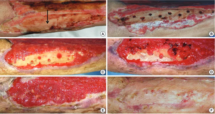
The application of autologous cultured fibroblasts is one of the representative treatments in the wound coverage. Fibroblasts are the most important mesenchymal cells involved in wound healing (27). The authors have analyzed 16 clinical cases of the tissue-engineered dermis comprising of autologous cultured dermal fibroblasts seeded on a hyaluronic-acid sheet for the coverage of facial defects created by the resection of skin can- cer. Based on the authors’ experiences, the graft was well taken by all patients. The entire tissue-engineered dermis graft reepi- thelialized after grafting within 21 to 42 days. All patients had satisfactory results in both functional and cosmetic matters with high quality skin characteristics. Regarding scar contracture, no patient required an additional operation for further improve- ment of the graft areas. There were no significant complications
or recurrences during the follow-up. Patient satisfaction with the tissue-engineered dermis graft was also excellent (Fig. 2).
There is considerable interest in the treatment of chronic wo- unds with grafts of cultured fibroblasts. Most fibroblast therapy products are composed of cryopreserved allogeneic cells. How- ever, the effect has generally shown to be not very dramatic. One of the possible reasons for the limited effect of these products is that it is difficult for grafted frozen cells to recover, colonize, and persist in chronic wound beds which are deficient in oxygen and nutrients. Moreover, cell activities are impaired due to cryopre- servation. Generally, cells in cryopreserved cultures show about 50% viability compared to non-frozen cells, and protein synthe- sis is inhibited by 70%-98%. In terms of growth factor expression and angiogenesis, there is an even lower rate of recovery (28-30).
To overcome the limitations of such cryopreserved cell allo- graft techniques, the authors have developed a non-cryopre- served fresh fibroblast allograft method and reported promis- ing results. Based on the authors’ clinical study for the treatment of diabetic foot ulcers, the efficacy, safety, and tolerability of fi- broblast allograft treatment were excellent. Within 8 weeks, com- plete wound closure was achieved in 84% of the treatment group and 50% of the control group (P < 0.05). There was no adverse
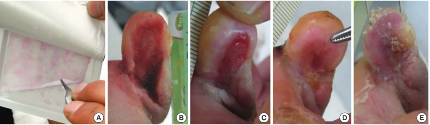
Fig. 1. Treatment of a diabetic foot ulcer using an allogeneic keratinocyte sheet. A woman (aged 63 yr) with a chronic non-healing diabetic ulcer over 10 weeks old on the medi- al side of the right fifth toe. After debridement, freshly bleeding wound bed was prepared. (A) View of the cultured allogeneic keratinocyte sheet. (B) Preoperative view. (C-E) Wound appearance was taken every week. Re-epithelization occurred progressively from the periphery to the center of the wound. The wound was completely healed after 3 weeks.
A B C D E
Fig. 2. Cheek defect created by removal of a basal cell carcinoma was reconstructed by autologous fibroblasts seeded on a hyaluronic acid sheet. The entire wound re-epitheli- alized 21 days after grafting. (A and B) Fibroblast pellet and non-woven hyaluronic acid sheet. (C) After wide excision of the tumor. (D) Immediately after the graft of the tissue- engineered dermis. (E) One-year after the graft. The result demonstrates excellent color match with minimal scar contracture.
A B C D E
effect related to fibroblast allograft treatment (Fig. 3) (28-30).
In recent years, a new sheet comprising of autologous cultur- ed fibroblasts and a scaffold made from the benzyl ester of hy- aluronic acid (Hyalograft 3D; ChaBio & Diostec) has been de- veloped in Korea for the treatment of diabetic foot ulcers. Hyal- uronic acid is a naturally occurring biopolymer whose molecu- lar structure is highly conserved between mammalian species.
First described in 1934, it has since been used across a wide va- riety of medical fields as diverse as neurosurgery and cutane- ous wound healing. The benzyl ester of hyaluronic acid is an ideal material for wound healing, as it is biocompatible and re- sorbable and integrates with ulcer tissues. Based on the authors’
clinical trial study with 12 week follow-up, complete wound healing occurred in 84% in the autologous fibroblast-hyaluron- ic acid complex-treated group and in 34% in the control group (P < 0.01). The safety and tolerability were also excellent.
Adipose-derived SVF cells
As mentioned earlier, utilizing cultured cells for clinical pur- poses requires FDA-approved facilities and techniques and a lengthy culture period. Adipose-derived SVF cells can substi- tute for fibroblasts. The authors have fairly extensive clinical cases of the tissue-engineered dermis using the SVF cells for the coverage of defects on the face, hand, and foot. Based on the authors’ experience, the results were favorable (Fig. 4 and 5) (31-35).
The SVF cells can be also applied for chronic wounds. An in vitro study has been performed to determine the effect of cell therapy using uncultured SVF cells on cell proliferation and col- lagen synthesis of diabetic fibroblasts, which are the major con- tributing factors in wound healing (36). The results showed that cell proliferation and collagen synthesis in the SVF cell treat-
ment group were 28% and 44% higher than those in the control group, respectively. In addition, a synergic effect of the SVF cell autograft was observed possibly due to mutual stimulation of the diabetic fibroblasts and SVF cells by growth factors secreted from them. A clinical trial study, which included 54 patients with diabetic foot ulcers, also demonstrated that SVF cell therapy ac- celerate diabetic wound healing. After 8 weeks of treatments, complete wound healing occurred in all patients (100%) in the SVF cell treated group and in 16 patients (62%) in the control group (P < 0.05) (36).
Bone marrow stromal stem cells
Bone marrow stroma is the source of mesenchymal stem cells which may serve as long-lasting precursors for bones, cartilag- es, muscles, and connective tissues. Mesenchymal stem cells also have a low immunity-assisted rejection rate, and they have a greater ability to divide without apoptosis than differentiated cells. Therefore, they have been drawing intense attention in the field of bioengineering.
At present, however, it is difficult for a clinician to utilize the cultured bone marrow stromal stem cells (BSCs) for the pur-
Fig. 3. A woman (aged 50 yr) with 6 week old chronic non-healing diabetic ulcers on the left second and third toe tips. The second toe with the larger wound was treated with the fibroblast allograft, and the third toe with the smaller wound was conserva- tively treated. (A) Preoperative view. (B) Immediate after treatments. (C) Two weeks after treatments. (D) Twenty days after treatments, the second toe wound completely epithelized, but the third toe wound (control) had a raw surface. (E) Three months af- ter treatments, the second toe was good with a healthy texture, but the third toe had not yet completely epithelized.
A
C D E
B
Fig. 4. Reconstruction using stromal vascular fraction (SVF) cells. (A) A squamous cell carcinoma on the hand. (B) After wide excision of the tumor, flap surgery was re- quired since the first metacarpal bone and tendons were exposed. However, a proce- dure which needs a general anesthesia or a long operation time could not be per- formed due to the poor general health condition of the patient. (C and D) To prepare the wound bed to be allowed for a skin graft, stromal vascular cells were isolated and grafted on the wound bed. (E) Three weeks after the graft, a healthy granulation tis- sue was formed. (F) The wound was finally closed by a split-thickness skin graft un- der local anesthesia.
A
C
E
B
D
F
A
D
B
E
C
F Fig. 5. Results of a stromal vascular fraction (SVF) cell graft applied to a thumb. (A) A bone and pulp defect on the thumb caused by trauma. Black and yellow arrows indicate the cortex and medulla of the distal phalanx, respectively. (B) SVF cells suspended in fibrin glue were transplanted one day after trauma. (C-F) Two-weeks, 4-weeks, 3-months, and 1-yr after the graft. The wound was completely closed 4 weeks after the graft.
pose of wound healing due to issues regarding the approval by the FDA. The authors have used uncultured BSCs in selected cases with exposed bone on the wound bed, especially on the tibial area. The BSCs in the tibia can be taken out by drilling in the bone and then applied on the wound bed of the exposed bony area. The results were favorable (Fig. 6).
Platelets
Attempts to deliver growth factors to wounds utilizing platelets have been developed and have shown beneficial efficacy on chronic wound healing (32, 37). The usefulness of autologous platelets in chronic wound healing has been demonstrated by many studies. Moreover, some authors have also tried homolo- gous platelets to treat chronic wounds, and have concluded that homologous platelets are as effective as autologous equivalents.
However, in the case of the autologous platelets, patient blood has to be aspirated and processed to yield the required platelet products. Moreover, patients with chronic wounds usually have poor general health and anemia, and thus the large volume blood withdrawal required to produce autologous platelet-rich plasma or platelet gel may have detrimental effects on hemo-
dynamic stability. In addition, equipment and techniques are used to separate platelets, and although homologous platelet gels have been used, healthy donors are required for its produc- tion, and testing of blood samples must be done for a history of infectious diseases.
To overcome these limitations, the authors performed serial studies to determine the potential of a straightforward method using a blood bank platelet concentrate (BBPC). BBPC is a trans- fusable blood product that is used to restore low platelet counts.
A BBPC is readily available and 1 unit contains approximately 5
× 1010 platelets in 50 mL of plasma. The results demonstrated that the use of a BBPC can provide a simple, safe, and effective means of treating diabetic foot ulcers (Fig. 7) (38, 39).
DIFFERENCES OF CELLS USED FOR THERAPY Keratinocytes mainly release growth factors (TGF-α, PDGF, bFGF, VEGF, and TGF-β) and cytokines (IL-1, -6, -8, and -10), but lack the ability to secrete extracellular matrices. Fibroblasts trans- planted onto the wound secrete various cytokines and growth factors that control cell proliferation, induce angiogenesis, and
Fig. 7. A 67-yr-old man with diabetes mellitus had a non-healing ulcer on the lateral border of his right foot at the metatarsophalangeal joint level. Before participating in the study, the patient had been treated for 12 weeks. (A) Preoperative view. (B) After surgical debridement, the metatarsal and proximal phalangeal bones were exposed.
(C) Three days after the first application of blood bank platelet concentrate (BBPC) (immediate before the second application of the BBPC). (D) Three weeks after the first application of BBPC. (E) Five weeks after the first application of BBPC. (F) Seven weeks after treatment, the wound was completely epithelialized. Two applications were performed for this patient.
A
D
B
E
C
F
Fig. 6. A 73-yr-old man with diabetes mellitus had a non-healing ulcer over 8 weeks old on the anterior tibial area of the lower leg. (A) A large wound on the lower leg with ex- posed tibial bone (an arrow). (B) Drilling in the bone to take out bone marrow stromal stem cells (BSCs). (C-E) Two, 3, and 4 weeks after drilling. Granulation tissue was formed from the drilling sites. (F) The wound was closed by a skin graft.
A
C
E
B
D
F
modify the inflammatory process. They also produce three-di- mensional extracellular matrices comprising collagens, proteo-
glycans, and other proteins. Therefore, the overall environment of wounds can be improved by controlling cell proliferation, in- ducing angiogenesis, and stimulating production of the three- dimensional extracellular matrices.
SVF cells can be obtained large quantities without cell cul- ture. They contain heterogenous cell populations, including fi- broblasts, stem cells, endothelial cells, mast cells, pericytes, prea- dipocytes, smooth muscle cells, and progenitor cells, which are well-known to accelerate wound healing. Based on the results of Suga et al. (40), SVF cells contain blood-derived cells (37%), adipose-derived stem/stromal cells (37%), endothelial cells (15%), and other cells (11%). All these SVF cells grafted on the wound bed could not only stimulate host cells around the wound, but also be taken on the wound bed and function independently by providing growth factors and extracellular matrices due to the autologous nature of the cells.
Bone marrow stromal stem cells (BSCs) are superior to fibro- blasts in wound healing activities. BSCs produce collagen and growth factors much higher than fibroblasts. In particular, the VEGF synthesis of the BSCs is 12 times higher than that of fibro- blasts. These results suggest that BSCs may possibly be used as a replacement for fibroblasts, which are commonly being used currently for wound healing (41-44).
Platelets contain at least seven locally acting growth factors from alpha-granules, i.e., three isomers of platelet derived grow- th factor (PDGF-AA, PDGF-AB, and PDGF-BB), two isomers of transforming growth factor-β (TGF-β1 and TGF-β2), vascular
endothelial growth factor (VEGF), and epidermal growth factor (EGF). Platelets applied on chronic wounds secrete various cy- tokines and growth factors, which are absent in chronic wounds.
They can compensate for impaired activities of cells crucial for wound healing.
PERSPECTIVES OF CELL THERAPY
As the populations of industrialized countries age and become more sedentary and increasing number of patients seek a pro- cedure of the least degree of invasiveness in wound repair, cell therapy for wound healing should be increasing dramatically.
To meet these demands, various commercially available cell- scaffold complexes have been developed and will be widely used. More and more, cell therapy methods are replacing the conventional tissue transfer.
Stem cells hold great promise for addressing the need for via- ble cell sources because they share the advantages of both allo- geneic and autologous cells. Mesenchymal stem cells (MSCs) have the ability to proliferate, form extensive colonies of healthy newly differentiated fibroblasts, demonstrate low levels of im- munity-assisted rejection, and divide without apoptosis than differentiated cells. In addition, it was demonstrated that even after 20 or 30 cycles of cell doubling in culture, they still retain stem cell properties. Accordingly, MSCs have attracted much attention in the bioengineering field. In the meantime, bone marrow stromal MSCs and umbilical cord blood-derived MSCs are on the way of research for this purpose.
WHAT ARE LIMITATIONS FOR PRESENT APPLICATION?
While a lot of reports regarding cell therapy for wound healing have been published, clinically available treatments are still lim- ited. There appears to be sufficient evidence to support the use of cell therapy to facilitate wound healing in chronic wounds.
However, clinical research regarding the cell therapy for acute wounds is limited. Further controlled studies are required be- fore this modality is considered a standard treatment for heal- ing of acute wounds.
There is limited information of “take” rates of the grafted cells.
Inconsistent “take” rates have been reported. These reports at- tribute their engraftment failure to the sensitivity of the cultured cells to bacterial colonization and inappropriate wound beds.
In applying cell therapy for healing of chronic wounds, the most important point is to identify good responders to cell ther- apy. Before cells are applied, the wound condition should be assessed for appropriateness. The wound bed should be healthy and clean with or without granulation tissue. There should not be any accompanying devitalized tissue, increased drainage, increase in size or odor. Wound edges should be open to allow
for epithelial migration. Periwound tissue should not be macer- ated or indurated. It is important to emphasize that these treat- ment modalities must be used along with other standard prin- ciples of chronic wound management, including debridement, infection control, pressure off-loading, and revascularization.
Without adhering to these important principles, addition of an active adjunctive modality is unlikely to result in improved heal- ing in chronic wound patients. Lastly, it must be emphasized that non-commercialized cell therapies need to be approved by the FDA.
WHAT KINDS OF RESEARCH ARE REQUIRED?
Future research should be focused on improving wound bed preparation and infection control to maximize cell engraftment, expediting cell culturing, and looking at long-term patient fol- low-ups both functional and aesthetic. Accurate identification of the causes and the appropriate cell therapy methods are there- fore mandatory to obtain the best treatment outcomes.
To establish cell therapy as a standard treatment, more inves- tigation with a larger number of patients is necessary. In addition, further studies are needed to determine the fate of transplanted cells and the number of cells required to show definitive effects.
Further studies on the length of time for cells to be maintained after harvesting and still retain viability are also required.
DISCLOSURE
The authors have no conflicts of interest to disclose.
ORCID
Hi-Jin You http://orcid.org/0000-0002-9997-2736 Seung-Kyu Han http://orcid.org/0000-0002-2875-9276
REFERENCES
1. Moon HS, Burm JS, Yang WY, Kang SY. Prognosis of full-thickness skin defects in premature infants. Arch Plast Surg 2012; 39: 463-8.
2. Park YS, Lee JW, Huh GY, Koh JH, Seo DK, Choi JK, Jang YC. Algorithm for primary full-thickness skin grafting in pediatric hand burns. Arch Plast Surg 2012; 39: 483-8.
3. Yun MJ, Park JU, Kwon ST. Surgical options for malignant skin tumors of the hand. Arch Plast Surg 2013; 40: 238-43.
4. Bae SH, Bae YC, Nam SB, Choi SJ. A skin fixation method for decreasing the influence of wound contraction on wound healing in a rat model.
Arch Plast Surg 2012; 39: 457-62.
5. Han SK, Yoon WY, Jeong SH, Kim WK. Facial dermis grafts after remov- al of basal cell carcinomas. J Craniofac Surg 2012; 23: 1895-7.
6. Han SK, You HJ. Wound coverage using advanced technology in Korea. J Korean Med Assoc 2011; 54: 594-603.
7. Gu JH, Han SK, Jeong SH, Kim WK. Hand coverage using venous island
flaps. J Plast Reconstr Aesthet Surg 2012; 65: e366-7.
8. Han SK, Lee BI, Kim WK. The reverse digital artery island flap: an up- date. Plast Reconstr Surg 2004; 113: 1753-5.
9. Han SK, Lee BI, Kim WK. The reverse digital artery island flap: clinical experience in 120 fingers. Plast Reconstr Surg 1998; 101: 1006-11.
10. Ghanem AM, Hachach-Haram N, Leung CC, Myers SR. A systematic review of evidence for education and training interventions in microsur- gery. Arch Plast Surg 2013; 40: 312-9.
11. Myers SR, Froschauer S, Akelina Y, Tos P, Kim JT, Ghanem AM. Micro- surgery training for the twenty-first century. Arch Plast Surg 2013; 40:
302-3.
12. Whang KK, Kim MJ, Song WK, Cho S. Comparative treatment of giant congenital melanocytic nevi with curettage or Er: YAG laser ablation alone versus with cultured epithelial autografts. Dermatol Surg 2005; 31:
1660-7.
13. Sood R, Roggy D, Zieger M, Balledux J, Chaudhari S, Koumanis DJ, Mir HS, Cohen A, Knipe C, Gabehart K, et al. Cultured epithelial autografts for coverage of large burn wounds in eighty-eight patients: the Indiana University experience. J Burn Care Res 2010; 31: 559-68.
14. Kim HR, Han SK, Rha SW, Kim HS, Kim WK. Effect of percutaneous trans- luminal angioplasty on tissue oxygenation in ischemic diabetic feet. Wound Repair Regen 2011; 19: 19-24.
15. Park DJ, Han SK, Kim WK. Is the foot elevation the optimal position for wound healing of a diabetic foot? J Plast Reconstr Aesthet Surg 2010; 63:
561-4.
16. Seo YK, Song KY, Kim YJ, Park JK. Wound healing effect of acellular arti- ficial dermis containing extracellular matrix secreted by human skin fi- broblasts. Artif Organs 2007; 31: 509-20.
17. Erdag G, Sheridan RL. Fibroblasts improve performance of cultured com- posite skin substitutes on athymic mice. Burns 2004; 30: 322-8.
18. Morimoto N, Saso Y, Tomihata K, Taira T, Takahashi Y, Ohta M, Suzuki S.
Viability and function of autologous and allogeneic fibroblasts seeded in dermal substitutes after implantation. J Surg Res 2005; 125: 56-67.
19. Yates CC, Whaley D, Wells A. Transplanted fibroblasts prevents dysfunc- tional repair in a murine CXCR3-deficient scarring model. Cell Trans- plant 2012; 21: 919-31.
20. El-Ghalbzouri A, Gibbs S, Lamme E, Van Blitterswijk CA, Ponec M. Ef- fect of fibroblasts on epidermal regeneration. Br J Dermatol 2002; 147:
230-43.
21. Kang BS, Na YC, Jin YW. Comparison of the wound healing effect of cel- lulose and gelatin: an in vivo study. Arch Plast Surg 2012; 39: 317-21.
22. Kim H, Son D, Choi TH, Jung S, Kwon S, Kim J, Han K. Evaluation of an amniotic membrane-collagen dermal substitute in the management of full-thickness skin defects in a pig. Arch Plast Surg 2013; 40: 11-8.
23. Gallego L, Junquera L, Villarreal P, Peña I, Meana A. Use of cultured hu- man epithelium for coverage: a defect of radial forearm free flap donor site. Med Oral Patol Oral Cir Bucal 2010; 15: e58-60.
24. Yanaga H, Udoh Y, Yamauchi T, Yamamoto M, Kiyokawa K, Inoue Y, Tai Y. Cryopreserved cultured epidermal allografts achieved early closure of wounds and reduced scar formation in deep partial-thickness burn wo- unds (DDB) and split-thickness skin donor sites of pediatric patients. Burns 2001; 27: 689-98.
25. Rheinwald JG, Green H. Serial cultivation of strains of human epidermal keratinocytes: the formation of keratinizing colonies from single cells. Cell 1975; 6: 331-43.
26. You HJ, Han SK, Lee JW, Chang H. Treatment of diabetic foot ulcers us- ing cultured allogeneic keratinocytes: a pilot study. Wound Repair Regen 2012; 20: 491-9.
27. Zou SB, Yoon WY, Han SK, Jeong SH, Cui ZJ, Kim WK. Cytotoxicity of silver dressings on diabetic fibroblasts. Int Wound J 2013; 10: 306-12.
28. Han SK, Choi KJ, Kim WK. Clinical application of fresh fibroblast allo- grafts for the treatment of diabetic foot ulcers: a pilot study. Plast Recon- str Surg 2004; 114: 1783-9.
29. Han SK, Kim WK. Revisiting fresh fibroblast allograft as a treatment for diabetic foot ulcers. Plast Reconstr Surg 2009; 123: 88e-9e.
30. Han SK, Kim HS, Kim WK. Efficacy and safety of fresh fibroblast allografts in the treatment of diabetic foot ulcers. Dermatol Surg 2009; 35: 1342-8.
31. Castro-Govea Y, De La Garza-Pineda O, Lara-Arias J, Chacón-Martínez H, Mecott-Rivera G, Salazar-Lozano A, Valdes-Flores E. Cell-assisted li- potransfer for the treatment of parry-romberg syndrome. Arch Plast Surg 2012; 39: 659-62.
32. Choi J, Minn KW, Chang H. The efficacy and safety of platelet-rich plas- ma and adipose-derived stem cells: an update. Arch Plast Surg 2012; 39:
585-92.
33. Lee JH, Lee KH, Kim MH, Kim JP, Lee SJ, Yoon J. Possibility of undiffer- entiated human thigh adipose stem cells differentiating into functional hepatocytes. Arch Plast Surg 2012; 39: 593-9.
34. Lee SK, Kim DW, Dhong ES, Park SH, Yoon ES. Facial soft tissue aug- mentation using autologous fat mixed with stromal vascular fraction.
Arch Plast Surg 2012; 39: 534-9.
35. Sung HM, Suh IS, Lee HB, Tak KS, Moon KM, Jung MS. Case reports of adipose-derived stem cell therapy for nasal skin necrosis after filler injec- tion. Arch Plast Surg 2012; 39: 51-4.
36. Han SK, Kim HR, Kim WK. The treatment of diabetic foot ulcers with uncultured, processed lipoaspirate cells: a pilot study. Wound Repair Re- gen 2010; 18: 342-8.
37. Shin HS, Oh HY. The effect of platelet-rich plasma on wounds of OLETF rats using expression of matrix metalloproteinase-2 and -9 mRNA. Arch Plast Surg 2012; 39: 106-12.
38. Jeong SH, Han SK, Kim WK. Treatment of diabetic foot ulcers using a blood bank platelet concentrate. Plast Reconstr Surg 2010; 125: 944-52.
39. Han SK, Kim DW, Jeong SH, Hong YT, Woo HS, Kim WK, Gottrup F. Po- tential use of blood bank platelet concentrates to accelerate wound heal- ing of diabetic ulcers. Ann Plast Surg 2007; 59: 532-7.
40. Suga H, Matsumoto D, Inoue K, Shigeura T, Eto H, Aoi N, Kato H, Abe H, Yoshimura K. Numerical measurement of viable and nonviable adipo- cytes and other cellular components in aspirated fat tissue. Plast Recon- str Surg 2008; 122: 103-14.
41. Kim JB, Chun KW, Han SK, Kim WK. Effect of human bone marrow stro- mal cell allograft on proliferation and collagen synthesis of diabetic fi- broblasts in vitro. J Plast Reconstr Aesthet Surg 2010; 63: 1030-5.
42. Lee CH, Han SK, Choi WI, Kim WK. Effect of human bone marrow stro- mal cells and dermal fibroblasts on collagen synthesis and epithelization.
Ann Plast Surg 2007; 59: 713-9.
43. Han SK, Chun KW, Gye MS, Kim WK. The effect of human bone marrow stromal cells and dermal fibroblasts on angiogenesis. Plast Reconstr Surg 2006; 117: 829-35.
44. Han SK, Yoon TH, Lee DG, Lee MA, Kim WK. Potential of human bone marrow stromal cells to accelerate wound healing in vitro. Ann Plast Surg 2005; 55: 414-9.