https://doi.org/10.22643/JRMP.2018.4.1.26
Molecular imaging of atherosclerosis using reporter gene system
Ran Ji Yoo1,2, Kyochul Lee1, Joo Hyun Kang1, Yong Jin Lee1,*
1 Department of RI-Convergence Research, Korea Institute of Radiological and Medical Sciences, Seoul, Korea
2 Korea Drug Development Platform using radio-isotope, Korea Institute of Radiological and Medical Sciences, Seoul, Korea
Macrophages play a key role in atherosclerotic plaque formation, but their participation has been discerned largely via ex vivo analyses of atherosclerotic lesions. Therefore, we aimed to identify atherosclerosis on noninvasive in vivo imaging using reporter gene system. This study demonstrated that recruitment of macrophages could be detected in atherosclerotic plaques of Apolipoprotein E knockout (ApoE-/-) mice with a sodium iodide symporter (NIS) gene imaging system using 99mTc-SPECT. This novel approach to tracking macrophages to atherosclerotic plaques in vivo could have applications in studies of arteriosclerotic vascular disease.
ABSTRACT
Key Word: Atherosclerosis, Click chemistry, Macrophage, Reporter gene, SPECT imaging, PET imaging
Atherosclerosis
Atherosclerotic diseases are a major cause of death and disability worldwide. Preventive strategies currently focus on controlling risk factors, such as smoking, blood pressure, serum glucose and lipid levels (1). Despite the success of these preventive measures, substantial residual risk remains even when treatment goals are fully met (2). In patients that suffered a myocardial infarction, the recurrence risk of an acute coronary syndrome is high, particularly within the first year when recurrence rates are up to 17.4% (3).
A recent study explained this phenomenon by showing that a systemic response to ischemic injury aggravates inflammation in atherosclerotic plaques at a distance, due to increased monocyte recruitment (4). Monocytes that infiltrate the plaque differentiate into macrophages, which produce proteolytic enzymes
that digest extracellular matrix causing plaque rupture (5). The immediate site of plaque rupture contains a high concentration of inflammatory cells (6). Plaque inflammation is therefore pursued as a therapeutic target to prevent atherothrombotic events (7) (Figure 1, 2).
The study of atherosclerosis progression in humans is hindered by the complexity and chronicity of the disease process, by the difficulty to longitudinally monitor the changes of the plaques in an individual patient and by deficiency of the noninvasive detection modalities that provide limited information on the composition of the lesions. The investigation of pathological changes in the arteries of humans is restricted to studies in the cross-sections of autoptic or surgical samples.
Therefore, there has been a reliance on animal models, and, since 1992, the mouse has become an excellent system for the study of atherosclerosis, progressively
Received: June 18, 2018 / Revised: June 27, 2018 / Accepted: June 29, 2018
Correspnding Author : Yong Jin Lee, PhD. Department of RI-Convergence Research, Korea Institute of Radiological and Medical Sciences (KIRAMS), 75 Nowon-gil, Gongneung-Dong, Nowon-Gu, Seoul, 139-706, Republic of Korea Tel: +82-2-970-1364, Fax:
+82-2-970-1341, E-mail: [email protected]
replacing the use of large animals (8), in particular due to the ability to easily over- or under-express specific genes such as leukocyte adhesion receptors, indicators of macrophage infiltration and angiogenesis.
Molecular imaging using an atherosclerosis model
Recently, studies targeting a specifically expressed gene in atherosclerotic lesions have been actively conducted in mouse models using molecular imaging (9,10). Molecular imaging has become an indispensable tool both in cardiovascular research and clinical care within the last decades. Molecular imaging relies on diverse imaging techniques, which include contrast-
enhanced ultrasound (CEUS), magnetic resonance imaging (MRI), positron emission tomography (PET), single-photon emission computed tomography (SPECT), computed tomography (CT), fluorescence molecular tomography (FMT) and photoacoustic imaging (PAI). Various imaging technologies are now available and they have their own strengths and weaknesses (Table 1). Imaging is mostly restricted to depicting anatomy and quantifying the degree of vessel stenosis.
Studies in an atherosclerosis model using nuclear
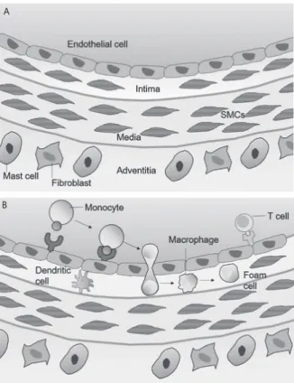
Figure 1. The development of atherosclerotic lesions. (A) There are three layers in the artery, and the inner membrane consists of a monolayer of endothelial cells in contact with blood. (B) In the early stages of atherosclerosis, blood leukocytes accumulate on the activated endothelial layer, and bound white blood cells migrate to the endothelium. When mononuclear cells are macrophage-maturated and lipid-uptake, foam cells are produced.
Figure 2. The progress of atherosclerotic lesions. (A) Progression of the lesion is accompanied by proliferation of smooth muscle cells (SMCs) and medium derived SMCs by migration of SMC from the medium to the intima.
And increased synthesis of extracellular matrix macromolecules such as collagen, elastin, and proteoglycans. Increased plaque macrophages and SMCs can accumulate in the central region of plaques marked with lipid or necrotic cores. A fibrous cap is formed in the vessel wall and the fat layer in the vessel wall becomes atheroma along with the necrosis process.
(B) Thus, the plaque formed causes bleeding, and these processes are repeated, leading to atherosclerosis in which the vessel wall becomes narrower and narrower. The rupture of the fibrous cap of the plaque may cause the blood clotting component to contact the tissue factor inside the plaque and expand into the lumen of the vessel to cause thrombosis, a complication of atherosclerosis
techniques such as PET and SPECT potentially provide detection sensitivities in the nanomolar- picomolar range. Such functional imaging enables the investigation of biological events that lead to plaque rupture with high specificity and offers relevant potential results of clinical translatability from basic research to identify high-risk patients. Furthermore, the combination of the nuclear medicine images with the morphological information provided by CT in hybrid scanners, or with the high soft tissue contrast obtained
through MRI, has the potential to map molecular signals with precise anatomic details (11–13). Also, more recently, the association with optical imaging was shown to be particularly helpful for testing innovative probes and for in vivo tracking of targeted cells (14).
Reporter gene in molecular imaging
In a previous study, it was shown that differentiated
Imaging
modality Spatial resolution Sensitivity (mol/L) Contrast agent /
probe concentration Advantage limitation
Ultrasound
imaging 50~500 μm Not well characterized yet
Microbubbles / μM to nM
real-time, low cost high temporal resolution (0.1- 100 s) no ionizing radiation
operator-dependent
Magnetic resonance imaging
10~100 μm 10-3~10-5 USPIOa), SPIOb) / mN to nM
high tissue contrast and functional parameters no ionizing radiation
high cost, operator- dependent
Nuclear imaging
PET: 1~2 mm SPECT: 0.5~2 mm
PET: 10-11~10-12 SPECT: 10-10~10-11
Positron or Y ray emitting radionuclides / pM
molecular and functional parameters high sensitivity
ionizing radiation limited spatial resolution (mm) high-medium cost
Computed tomography imaging
30~400 μm 10-2~10-3
Iodinated particles, gold nanorods / mM to nM
fast acquisition time high temporal resolution (1-300 s)
provides molecular and structural information
ionizing radiation, medium cost low soft tissue contrast resolution
Fluorescence tomographic imaging
1~2 mm 10-10~10-11 NIR fluorophores
/ nM to pM
high sensitivity, low cost no ionizing radiation
limited depth of penetration (1-20 mm)
limited spatial resolution (mm)
Photoacoustic
imaging <100 μm <10-12 NIR fluorophores
/ nM to pM
high sensitivity, low cost, no ionizing radiation high depth of penetration (<5 cm)
data post-processing and acquisition procedures still being optimized Table 1. Noninvasive molecular imaging in mouse models of vulnerable atherosclerotic plaques
a) Gd-based contrast agents, b) Iron oxide and other superparamagnetic nanoparticles
thyroid cancers and all anaplastic thyroid cancers fail to concentrate radioiodine due to reduced NIS expression, which prevents the use of radioiodine in metastatic thyroid cancer therapy (15). The recent cloning and characterization of the NIS gene could lead to the development of a novel gene strategy for radioiodine therapy in thyroid cancer (16,17). The transfer of NIS gene and the functional expression of NIS protein in cancer cells would enable these cells to concentrate iodide from plasma and enable radioiodine therapy (18). Previous studies have reported the result for the production of a recombinant NIS (rNIS) (15), and many researchers are interested in the basic and clinical applicability of rNIS. On expression of the NIS protein in the cell membrane following NIS gene transfection, it is possible to perform imaging using
99m Tc-pertechnetate, 131I and 123I, and it is possible to perform therapy using therapeutic radionuclides such as 131I, 186Re, 188Re (19).
Mutant dopamine D2 receptor (D2R) and Herpes simplex virus type 1 thymidine kinase (HSV1-tk) are the most widely used among the reporter genes besides NIS, which was developed for nuclear medicine and molecular imaging (20). Recently, interest has concentrated on the complex molecular image using a multi-reporter system that provides composite gene expression (21).
Especially, the dual reporter gene imaging system expressing a nuclear medical reporter gene (HSV1- TK or NIS) and a reporter gene at the same time for use in optical molecular imaging (luciferase or green fluorescence protein) can be used extensively to assess the progress, challenges of cancer and tracking the treatment progress in vitro and in vivo. Also, an optical imaging reporter gene as a monitoring gene showed that
131I, 186Re or 188Re in the transfected NIS will be able to evaluate the effect of treatment with radionuclides (15,16) (Figure 3).
Detection of atherosclerotic lesion with reporter gene system
Macrophages accumulate in the blood vessel walls where atherosclerosis has developed. Therefore, atherosclerotic plaque can be identified by molecular imaging using macrophages (22).
To detection atherosclerotic lesion with reporter gene system, we sought to establish a noninvasive technique to determine macrophage tracking to atherosclerotic lesions in apolipoprotein E-/-(ApoE-/-) mice with an imaging system based on sodium iodide symporter (NIS) gene coupled with 99mTc-SPECT imaging. In this study, RAW 264.7 macrophage cells were stably transduced with retrovirus containing NIS gene (RAW-NIS). In RAW-NIS cells, the atherosclerotic ApoE-/- mice were divided into four study groups (Saline, RAW cells, RAW-NIS cells and RAW-NIS cells after atorvastatin treatment). Through 99mTc-SPECT/CT imaging, in the RAW-NIS cell injected group, the 99mTc uptake in aorta was higher than other groups, but the injected RAW-
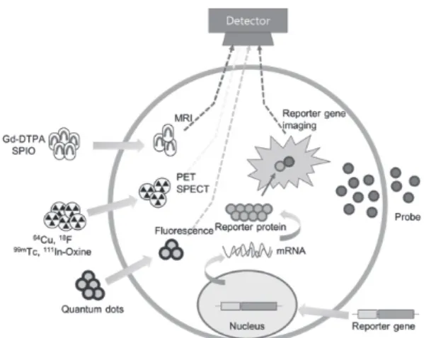
Figure 3. Molecular imaging using reporter gene. A vector containing a DNA reporter construct with the reporter gene(s) driven by a specific promoter.
Transcription and translation lead to the expression of mRNA and reporter protein, respectively. After administration of a corresponding reporter probe systemically, the reporter probe will be catalyzed by specific cells that have the reporter proteins. The signals occurred in this amplification process can be detected by a sensitive imaging device.
NIS cell for the atorvastatin treated group demonstrated reduced recruitment to the aorta (Figure 4). This study confirmed a positive correlation between cell imaging intensity and macrophage marker histological staining of the aorta. Areas of high 99mTc-pertechnetate uptake correlated with higher immunohistochemistry staining using CD68 antibody.
Conclusion
Generally, atherosclerosis is characterized by intimal plaques of the arterial vessels that develop slowly.
Therefore, noninvasive tools for early diagnosis are necessary to screen atherosclerotic lesions at high risk of acute complications.
In the current study, macrophages, known to be activated at the onset of atherosclerosis, trafficking the atherosclerotic lesions of the ApoE-/- model was performed using noninvasive imaging, the NIS reporter gene system. In study, the macrophage cell line which allowed NIS to be stably expressed, the novel approach showed that migration of macrophages to the inflammatory atherosclerotic lesions was effectively monitored by SPECT imaging. In addition, the efficacy of statin could be evaluated by imaging.
An attempt to identify atherosclerosis by molecular imaging using NIS reporter gene was the first in this study. This potential novel approach including the NIS reporter gene system may provide new insights into the role of macrophage tracking monitoring in a living body for making the diagnosis of atherosclerosis and may contribute importantly to enhanced risk assessment.
Acknowledgments
This work was supported by a grant of Korea Institute of Radiological and Medical Sciences(KIRAMS) and National Reseach Foundation of Korea(NRF) grant, by Ministry of Science and ICT(MSIT), Republic of Korea. (Ministry of Science, ICT)(No.50536-2018 and NRF-2013M2C2A1074238)
References
1. Grundy SM. Atherosclerosis imaging and the future of lipid management. Circulation 2004;110: 3509–3511.
2. Libby P. The forgotten majority: unfinished business in cardiovascular risk reduction. J Am Coll Cardiol.
2005;46:1225–1228.
3. Milonas C, Jernberg T, Lindbäck J, Agewall S, Wallentin L, Stenestrand U. Effect of angiotensin- converting enzyme inhibition on one-year mortality and frequency of repeat acute myocardial infarction in patients with acute myocardial infarction. Am J Cardiol 2010;105:1229–1234.
4. Dutta P, Courties G, Wei Y, Leuschner F, Gorbatov R, Robbins CS, Iwamoto Y, Thompson B, Carlson AL, Heidt T, Majmudar MD, Lasitschka F, Etzrodt M, Waterman P, Waring MT, Chicoine AT, van der Laan AM, Niessen HWM, Piek JJ, Rubin BB, Butany J, Stone JR, Katus HA, Murphy SA, Morrow DA, Sabatine MS, Vinegoni C, Moskowitz MA, Pittet MJ, Figure 4. SPECT/CT images of atherosclerotic lesions in ApoE-/- mice on
Western diet for 30 weeks. SPECT/CT images of atherosclerotic lesions in ApoE-/- mice confirmed that 99mTc-pertechnetate uptake of injected saline, RAW264.7 cell, RAW-NIS and atorvastatin-treated RAW-NIS group. Arrows indicate the 99mTc-pertechnetate uptake in heart and aorta. CT imaging was acquired only on day 5.
Libby P, Lin CP, Swirski FK, Weissleder R, Nahrendorf M. Myocardial infarction accelerates atherosclerosis.
Nature 2012;487:325–329.
5. Libby P, DiCarli M, Weissleder R. The vascular biology of atherosclerosis and imaging targets. J Nucl Med 2010;51:33S–37S.
6. Van der Wal AC, Becker AE, Van der Loos CM, Tigges AJ, Das PK. Fibrous and lipid-rich atherosclerotic plaques are part of interchangeable morphologies related to inflammation: a concept. Coron Artery Dis 1994;5:463–469.
7. Weber M, Bhatt DL, Brennan DM, Hankey GJ, Steinhubl SR, Johnston SC, Montalescot G, Mak K-H, Fox KAA, Easton DJ, Topol EJ, Hamm CW. High-sensitivity C-reactive protein and clopidogrel treatment in patients at high risk of cardiovascular events: a substudy from the CHARISMA trial. Heart 2011;97:626–631.
8. Moghadasian MH. Experimental atherosclerosis: A historical overview. Life Sci. 2002;70:855–865.
9. Whitman SC. A practical approach to using mice in atherosclerosis research. Clin Biochem Rev 2004;25:81–
93.
10. Li X, Liu Y, Zhang H, Ren L, Li Q, Li N. Animal models for the atherosclerosis research: a review.
Protein Cell. 2011;2:189–201.
11. Nahrendorf M, Zhang H, Hembrador S, Panizzi P, Sosnovik DE, Aikawa E, Libby P, Swirski FK, Weissleder R. Nanoparticle PET-CT imaging of macrophages in inflammatory atherosclerosis.
Circulation 2008;117:379–387.
12. Seo JW, Baek H, Mahakian LM, Kusunose J, Hamzah J, Ruoslahti E, Ferrara KW. (64)Cu-labeled LyP-1- dendrimer for PET-CT imaging of atherosclerotic plaque. Bioconjug Chem 2014;25:231–239.
13. Foss CA, Bedja D, Mease RC, Wang H, Kass DA, Chatterjee S, Pomper MG. Molecular imaging of inflammation in the ApoE -/- mouse model of atherosclerosis with IodoDPA. Biochem Biophys Res Commun 2015;461:70–75.
14. Larmann J, Frenzel T, Schmitz M, Hahnenkamp A, Demmer P, Immenschuh S, Tietge UJF, Bremer C, Theilmeier G. In vivo fluorescence-mediated tomography imaging demonstrates atorvastatin- mediated reduction of lesion macrophages in ApoE-/- mice. Anesthesiology 2013;119:129–141.
15. Kang JH, Chung J-K, Lee YJ, Shin JH, Jeong JM, Lee DS, Lee MC. Establishment of a human hepatocellular carcinoma cell line highly expressing sodium iodide symporter for radionuclide gene therapy. J Nucl Med 2004;45:1571–1576.
16. Dai G, Levy O, Carrasco N. Cloning and characterization of the thyroid iodide transporter.
Nature. 1996;379:458–460.
17. Smanik PA, Liu Q, Furminger TL, Ryu K, Xing S, Mazzaferri EL, Jhiang SM. Cloning of the human sodium lodide symporter. Biochem Biophys Res Commun 1996;226:339–345.
18. Spitzweg C, Morris JC. Approaches to gene therapy with sodium/iodide symporter. Exp Clin Endocrinol Diabetes. 2001;109:56–59.
19. Haberkorn U, Henze M, Altmann A, Jiang S, Morr I, Mahmut M, Peschke P, Kübler W, Debus J, Eisenhut M. Transfer of the human NaI symporter gene enhances iodide uptake in hepatoma cells. J Nucl Med 2001;42:317–325.
20. Gambhir SS, Barrio JR, Herschman HR, Phelps ME.
Assays for noninvasive imaging of reporter gene expression. Nucl Med Biol 1999;26:481–490.
21. Shu CJ, Guo S, Kim YJ, Shelly SM, Nijagal A, Ray P, Gambhir SS, Radu CG, Witte ON. Visualization of a primary anti-tumor immune response by positron emission tomography. Proc Natl Acad Sci 2005;102:17412–17417.
22. Yoo RJ, Kim MH, Woo SK, Kim K Il, Lee TS, Choi YK, Kang JH, Lim SM, Lee YJ. Monitoring of macrophage accumulation in statin-treated atherosclerotic mouse model using sodium iodide symporter imaging system.
Nucl Med Biol 2017;48:45–51.