INTRODUCTION
With the rapid increase in the number of upper and lower gastrointestinal endoscopic procedures as a part of the na- tional cancer screening project, early detection of various gas- trointestinal precancerous or cancerous lesions have been incr- eased, recently. These early detection of the lesions in early st- age has been greatly helpful for the enhancement of the quali- ty of life or life prolongation and have also allowed the signi- ficant reduction of medical costs through the endoscopic sur- gery without cutting the abdomen or chest.1-3 Regardless of such rapid development in the endoscopic field, laryngopha- ryngeal lesions are generally considered as a field of otolar- yngology, and thus it is thought that not a lot of gastrointesti- nal endoscopists commonly take interest in these lesions dur- ing the upper gastrointestinal endoscopic examinations. Th- erefore, most laryngopharyngeal lesions are difficult to be detected by gastrointestinal endoscopists. The laryngophar- ynx is a structure that must be passed inevitably during upper
gastrointestinal endoscopic insertion; hence various diseases can be detected if the area is examined in detail during endo- scopy. According to the reports which have studied laryngo- pharyngeal lesions that can be observed during upper gas- trointestinal endoscopic procedures, 0.9% to 3.5% of all tests have displayed abnormalities in the laryngopharynx.4-10 Fur- thermore, the field of endoscopic treatment is expanding to include laryngopharynx lesions.11 Accordingly, this thesis will review the availability of upper gastrointestinal endoscopy in laryngopharyngeal area, normal structures of laryngopharynx, and the lesions that can be observed with the standard upper gastrointestinal endoscopic procedure.
STRUCTURE OF THE LARYNGOPHARYNX AND NORMAL FINDINGS
The pharynx is divided into three parts: nasopharynx, oro- pharynx, and hypopharynx. From the soft palate, the top part is the nasopharynx, and from the soft palate to the epiglottis is the oropharynx, and from the bottom of the epiglottis to the entrance of the esophagus is the hypopharynx. The hypoph- arynx applies to the back of the larynx.12 The larynx is the vo- cal organ that includes the vocal cords, and the oropharynx is located on the top, with the trachea located on the bottom and the hypopharynx on the back. The oropharynx and lar- ynx can be observed in a standard upper gastrointestinal en- doscopy, and the nasopharynx can be observed with the re-
Observable Laryngopharyngeal Lesions during the Upper Gastrointestinal Endoscopy
Kyung Sik Park
Department of Internal Medicine, Keimyung University School of Medicine, Daegu, Korea
Regardless of outstanding developments in the endoscopic field, laryngopharyngeal lesions are generally considered as a field of otolaryn- gology, and thus it is thought that not a lot of gastrointestinal endoscopists commonly take interest in these lesions during the upper gas- trointestinal endoscopic examinations. Therefore, here in this thesis, I reviewed the availability of upper gastrointestinal endoscopy in laryn- gopharyngeal area, normal structures of laryngopharynx, and the lesions that can be observed with the standard upper gastrointestinal endoscopic procedure.
Key Words: Laryngopharynx; Endoscopy; Larynx; Pharynx Open Access
Received: March 24, 2013 Revised: March 29, 2013 Accepted: March 29, 2013
Correspondence: Kyung Sik Park
Department of Internal Medicine, Keimyung University Dongsan Medical Cen- ter, Keimyung University School of Medicine, 56 Dalseong-ro, Jung-gu, Daegu 700-712, Korea
Tel: +82-53-250-7088, Fax: +82-53-250-7088, E-mail: [email protected]
cc This is an Open Access article distributed under the terms of the Creative Commons Attribution Non-Commercial License (http://creativecommons.org/
licenses/by-nc/3.0) which permits unrestricted non-commercial use, distribution, and reproduction in any medium, provided the original work is properly cited.
cently developed endoscopy, although its use is not popular.
There are differences based on the type of mouth-piece used during endoscopic procedures, but when the endoscope is inserted using the most commonly used tongue depressor type mouth-piece, observations can be conducted from the soft palate. After the uvula and both tonsils connected to the soft palate are observed, and when the tube is progressed fur- ther, the epiglottis and lymph nodes near the root of the ton- gue are seen, followed by most of the larynx and parts of the hypopharynx can be seen. Here, the vocal cords, insides of the epiglottis, the corniculate cartilage, arytenoids cartilage, and pyriform sinus can be observed (Fig. 1). However, when air is released while these parts are observed, severe coughing can occur due to the inflow of secretion through the airway, thus air must be inflowed as less as possible, and the parts must be observed within a short period of time. If necessary, the area should be reobserved when removing the endoscope after ob- serving the esophagus, stomach, and duodenum.
OBSERVABLE LESIONS
Oral cavityA part of the mouth can sometimes be observed—there are not many parts that can be observed thoroughly due to the mouthpiece used during the endoscopic procedure; however, soft palate lesions can sometimes be observed quite easily, with greater care. The diseases that can occur within the oral cavity, such as the lips, gums, teeth, hard palate, and soft pal- ate have been shown on Table 1. It includes various diseases based on various causes; oral ulcers, connective tissue diseas- es, various infectious diseases, and benign or malignant tu-
mors (Fig. 2). Even if lesions do exist, it is difficult to discover them during the actual endoscopic procedure because the parts covered by the mouth-piece are commonly affected. How- ever, when suspicious lesions are discovered, biopsy should be taken or consulted to the laryngologist, if available.
Inflammatory lesions
Laryngopharyngeal reflux (LPR) diseases
The gastroesophageal reflux disease does not only display esophageal symptoms but also includes various extraesopha- geal symptoms or complications, including coughing, laryn- gitis, asthma, dental caries, pharyngitis, sinusitis, pulmonary fibrosis, otitis media, etc.13 LPR refers to the effect of gastric contents, such as gastric acid or pepsin passing through the upper esophageal sphincter to the laryngopharynx, and may display symptoms such as abnormal vocalization, hoarse vo- ice, globus, dysphagia, and chronic coughing.14,15
Meanwhile, laryngitis occurs due to various causes, but most Table 1. Oral Lesions Observable during Endoscopy
Tumors or lesions that mimic Squamous cell carcinoma Leukoplakia
Melanoma Amalgam tattoos Fordyce spots Mucoceles Infections Candidiasis Herpes simplex virus Varicella-zoster virus Coxsackie virus
Human immunodeficiency virus infection Syphilis
Aphthous oral ulcer (aphthae) Oral and genital ulcers Behcet’s disease Complex aphthosis Lichen planus
Connective tissue and autoimmune disease Systeminc lupus erythematosus Bullous pemphigoid
Cicatrical pemphigoid Pemphigus vulgaris Erythema multiforme Miscellaneous lesions Black hairy tongue Geographic tongue
Contact and irritant stomatitis Epidermolysis bullosa Fig. 1. Normal laryngopharyngeal structure under upper gastroin-
testinal endoscopic procedure. The vocal cords, insides of the epiglottis, the corniculate cartilage, arytenoids cartilage, and pyri- form sinus can be observed.
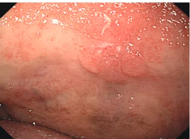
of them display similar appearances by the naked eye with no relation to the causes; LPR can also become an important cau- se, and laryngitis caused by LPR is referred to as reflux laryn- gitis. There may be controversy on standard diagnostic meth- ods that can easily be conducted concerning the diagnosis of LPR. If abnormal findings are found with the aforementioned symptoms during the endoscopy or laryngoscopy, the diag- nosis of LPR can be considered with priority. These abnor- mal finding may include various appearances, such as eryth- ema, edema, granuloma, and stenosis. Erythema and edema which occur in the mucous membrane between the arytenoid and back of the arytenoid are the most common findings of reflux laryngitis, considering the positional relationship with the upper esophageal sphincter (Fig. 3).16 However, there is diagnostic limitation in that these abnormalities can occur in as high as 70% of general people who do not display specific symptoms, and in that standards of dividing normality and abnormality are not clear.17-19
There have been many studies conducted under the pre- sumption that the 24-hour pH monitoring is the most supe-
rior of all diagnostic tools for LPR.20 However, there is no cl- earcut standard, and there are also reports which show large amount of false positive results, displaying a positive rate of 10% to 60% in healthy people with no symptoms. Thus, this test is not recommended to all patients.21
Recently, reflux finding score, which consists of eight cate- gories observed through the laryngoscope, has been proposed in the otolaryngologic field to standardize the diagnosis of LPR (Table 2).22 This system displayed a highly significant con- sistency within and between the observers, as well as repro- ducibility, and was reported to help the judgment of improve- ment after the treatment of reflux laryngitis. Furthermore, this
Fig. 2. Soft palate cancer. In a 75-year-old asymptomatic male patient, ill demarcated friable lesion is found on the soft palate.
Fig. 3. Reflux laryngitis. (A) In a 63-year-old male patient who presents severe globus and chronic coughing, diffuse erythema is found in the vocal folds and arytenoids wall. (B) In a 59-year-old male patient who presents globus, severe edema is found in the posterior larynx.
A B
Table 2. Reflux Finding Score Subglottic edema Ventricular
Erythema/hyperemia Vocal fold edema
Diffuse laryngeal edema
Posterior commissure hypertrophy
Granuloma/granulation tissue Thick endolaryngeal mucus
0=absent 1=present 2=partial 4=complete 2=arytenoids only 4=diffuse 1=mild 2=moderate 3=severe 4=polypoid 1=mild 2=moderate 3=severe 4=obstructing 1=mild 2=moderate 3=severe 4=obstructing 0=absent 2=present 0=absent 2=present
scoring system and a survey that grades reflux symptoms have been used in reports that have successfully researched the re- action to treatments, thus is considered as being an element worth considering for use in the gastroenterologic field.23-25 Caustic damage
Other than in special cases, such as having no consciousness or being unable to communicate, caustic damage can gener- ally be predicted through medical history taking. Various fi- ndings can be shown based on the causing material. Erythe- ma, edema, and bleeding can be seen during the acute phase, and as time progresses, edema, and erythema can decrease, with change in the surrounding tissues due to fibrosis. During the chronic phase, stenosis due to deformity becomes the gr- eatest problem.26
Candidiasis
White membranous substances similar to esophageal can- didiasis can be seen in the oral membrane or the laryngophar- ynx, and can commonly occur in situations with suppressed immunity, such as with chronic diseases or use of steroids.
Membranous substances can be diagnosed by dying potassi- um hydroxide.27
Neoplasic lesions Laryngeal cancer
There is a need to suspect laryngeal cancer in patients with changes in their voice. The larynx is located directly above the vocal cord, so laryngeal cancer is relatively easy to detect.
However, if the cancer is located in the dented area between the epiglottis and vocal cord, it may be difficult to diagnose.
It is observed in various shapes, and can easily bleed on con- tact with the endoscope, and displays a tendency of easy fria- bility during biopsies.28 Caution must be taken during biopsies so that pulmonary aspiration due to bleeding does not occur.
Hypopharyngeal cancer
During endoscopic procedure, the scope enters through the left pyriform sinus, thus hypopharyngeal cancer located on the back of the left arytenoid is occasionally found. If the mass is large, it can easily be found, but without care, it can easily be missed even after several procedures. The shape is very diverse and is almost undistinguishable with the naked eye, but it displays easy bleeding on contact with the scope, and thus can be diagnosed by biopsies. The surface of oral ca- vity, larynx, and hypopharynx is composed of squamous epi- thelium, just as the esophagus. Thus, laryngeal or hypopha- ryngeal cancer can occur synchronously or metachronously with esophageal cancer.29 Therefore, the hypopharynx or lar-
ynx must thoroughly be examined in esophageal cancer pa- tients,30 and inversely, it is necessary to thoroughly examine the esophagus in hypopharynx or laryngeal cancer patients.
Recently, new video imaging methods such as narrow band imaging are contributing to the early detection of these dis- eases.31
Other benign tumors
Papillomas, cysts, vocal fold polyps, lipomas, hymangiomas, and other various benign tumors can be discovered in the larynx, hypopharynx, epiglottis, and oral cavity.32 Sometimes local leukoplakia is found in various parts. This is a precan- cerous lesion and it is recommended to be removed, if possible.
Other diseases Intubation granuloma
As side effects of tracheal intubation in patients treated with mechanical ventilation or general anesthesia, tracheosteno- sis, organ-esophageal fistula, and granulomas may occur due to the fibrosis of the stimulated area.33 It is known that the in- tubation period or early laryngeal damage cannot predict the occurrence of granulomas. If a hoarse voice continues 7 to 10 days after extubation, the formation of a granuloma should be suspected and a diagnostic evaluation is recommended. Many cases require surgical removal.
Edema
Laryngopharyngeal edemas may occur after forceful insert- ing of the scope or during tracheal intubation, thus caution is needed. It also may occur after repetitive procedures due to hematemesis, or after difficult procedure due to severe vom- iting.
Postoperative deformity
Various deformities may be observed after the operation of laryngeal or hypopharyngeal cancer. In those cases, there is a need to confirm the surgical history of the patient through detailed medical history taking before the procedure. If one side of the pyriform sinus is closed after surgery, the scope must be entered through the pyriform sinus located on the op- posite side. And thorough observation for synchronous or metachronous lesions is needed.
Behcet’s disease
Unlike simple aphthous ulcers, oral ulcers based on Behcet’s disease are not easily healed, and frequently relapse. If ulcers also occur in other areas, such as the genital area, this disease may be suspected. Most ulcers occur in the mouth, but also can occur in the pharynx or hypopharynx. Deformity of the sur-
rounding areas can also be developed due to deep ulcers.
Discoloration of the mucous membrane
Local discoloration may occur on the mucous membrane due to angiodysplasia or infections, and it can also be ob- served before or after the formation of ulcers.
Vocal cord paralysis
Vocal cord paralysis can occur due to a large variety of causes, such as gastric acid reflux, exercise, stress, postopera- tion, neurological disorders, etc. It is commonly accompanied by the change in voice. Although, recently, endoscopic proce- dures under conscious sedation may restrict observation of the vocal cords, vocal cord paralysis can also be confirmed by allowing the patient to use the vocal cord during the diagnosis.34 Tonsillar hypertrophy
Congenital or infection-based tonsillar hypertrophy can be observed relatively easily during the insertion of the endo- scope.
CONCLUSIONS
As mentioned, oral and laryngopharyngeal lesions which can easily be observed with careful upper gastrointestinal en- doscopic procedure were reviewed. On the other hands, these lesions may easily be missed if careful examination is not per- formed. Considering the current medical circumstances in Korea where a large number of patients must be examined wi- thin a short period of time, it is not easy to diagnose the le- sions which exist in these oropharyngolaryngeal areas. How- ever, with thorough examinations conducted with great care within the possible area, various diseases can be detected in early stage and it can be a great help to the patients. These ca- reful examinations are also expected to increase the effective- ness of the endoscopic procedures.
Conflicts of Interest
The author has no financial conflicts of interest.
REFERENCES
1. Chang DK. Current status of colorectal endoscopic submucosal dis- section in Korea. Clin Endosc 2012;45:288-289.
2. Goh PG, Jeong HY, Kim MJ, et al. Clinical outcomes of endoscopic sub- mucosal dissection for undifferentiated or submucosal invasive early gastric cancer. Clin Endosc 2011;44:116-122.
3. Kim YS, Cho WY, Cho JY, Jin SY. Successful treatment of early gastric cancer adjacent to a fundal varix by endoscopic submucosal dissection and endoscopic cyanoacrylate therapy. Clin Endosc 2012;45:169-173.
4. Katsinelos P, Kountouras J, Chatzimavroudis G, et al. Should inspection of the laryngopharyngeal area be part of routine upper gastrointestinal endoscopy? A prospective study. Dig Liver Dis 2009;41:283-288.
5. Cammarota G, Galli J, Agostino S, et al. Accuracy of laryngeal examina-
tion during upper gastrointestinal endoscopy for premalignancy screen- ing: prospective study in patients with and without reflux symptoms.
Endoscopy 2006;38:376-381.
6. Raju GS. Value of screening the laryngopharyngeal area during routine upper gastrointestinal endoscopy. Nat Clin Pract Gastroenterol Hepatol 2005;2:22-23.
7. Mullhaupt B, Jenny D, Albert S, Schmid S, Fried M. Controlled prospec- tive evaluation of the diagnostic yield of a laryngopharyngeal screening examination during upper gastrointestinal endoscopy. Gut 2004;53:
1232-1234.
8. Yarze JC, Chase MP, Herlihy KJ. Laryngopharyngeal examination: an important but not-so-new role of upper gastrointestinal endoscopy. Ann Intern Med 2000;133:314-315.
9. Kozarek RA. Evaluation of the larynx, hypopharynx, and nasopharynx at the time of diagnostic upper gastrointestinal endoscopy. Gastroin- test Endosc 1985;31:271-273.
10. Lehman G, Compton M, Meadows J, Elmore M. Screening examination of the larynx and pharynx during upper gastrointestinal panendoscopy.
Gastrointest Endosc 1982;28:176-178.
11. Choi JH, Park JJ, Jee JB, et al. Endoscopic treatment of benign hypo- pharyngeal tumors. Korean J Gastrointest Endosc 2005;31:306-310.
12. Matsuo K, Palmer JB. Anatomy and physiology of feeding and swallow- ing: normal and abnormal. Phys Med Rehabil Clin N Am 2008;19:691- 13. Vakil N, van Zanten SV, Kahrilas P, Dent J, Jones R; Global Consensus 707.
Group. The Montreal definition and classification of gastroesophageal reflux disease: a global evidence-based consensus. Am J Gastroenterol 2006;101:1900-1920.
14. Ford CN. Evaluation and management of laryngopharyngeal reflux.
JAMA 2005;294:1534-1540.
15. Koufman JA. The otolaryngologic manifestations of gastroesophageal reflux disease (GERD): a clinical investigation of 225 patients using am- bulatory 24-hour pH monitoring and an experimental investigation of the role of acid and pepsin in the development of laryngeal injury. La- ryngoscope 1991;101(4 Pt 2 Suppl 53):1-78.
16. Hickson C, Simpson CB, Falcon R. Laryngeal pseudosulcus as a pre- dictor of laryngopharyngeal reflux. Laryngoscope 2001;111:1742-1745.
17. Qadeer MA, Swoger J, Milstein C, et al. Correlation between symptoms and laryngeal signs in laryngopharyngeal reflux. Laryngoscope 2005;
115:1947-1952.
18. Hicks DM, Ours TM, Abelson TI, Vaezi MF, Richter JE. The prevalence of hypopharynx findings associated with gastroesophageal reflux in normal volunteers. J Voice 2002;16:564-579.
19. Branski RC, Bhattacharyya N, Shapiro J. The reliability of the assessment of endoscopic laryngeal findings associated with laryngopharyngeal re- flux disease. Laryngoscope 2002;112:1019-1024.
20. Merati AL, Lim HJ, Ulualp SO, Toohill RJ. Meta-analysis of upper probe measurements in normal subjects and patients with laryngopharyngeal reflux. Ann Otol Rhinol Laryngol 2005;114:177-182.
21. Noordzij JP, Khidr A, Desper E, Meek RB, Reibel JF, Levine PA. Corre- lation of pH probe-measured laryngopharyngeal reflux with symp- toms and signs of reflux laryngitis. Laryngoscope 2002;112:2192-2195.
22. Belafsky PC, Postma GN, Koufman JA. The validity and reliability of the reflux finding score (RFS). Laryngoscope 2001;111:1313-1317.
23. Lien HC, Wang CC, Hsu JY, et al. Classical reflux symptoms, hiatus her- nia and overweight independently predict pharyngeal acid exposure in patients with suspected reflux laryngitis. Aliment Pharmacol Ther 2011;
33:89-98.
24. Reichel O, Dressel H, Wiederänders K, Issing WJ. Double-blind, place- bo-controlled trial with esomeprazole for symptoms and signs associat- ed with laryngopharyngeal reflux. Otolaryngol Head Neck Surg 2008;
139:414-420.
25. Belafsky PC, Postma GN, Koufman JA. Laryngopharyngeal reflux symptoms improve before changes in physical findings. Laryngoscope 2001;111:979-981.
26. Scott JC, Jones B, Eisele DW, Ravich WJ. Caustic ingestion injuries of the upper aerodigestive tract. Laryngoscope 1992;102:1-8.
27. Giannini PJ, Shetty KV. Diagnosis and management of oral candidia- sis. Otolaryngol Clin North Am 2011;44:231-240.
28. Chu EA, Kim YJ. Laryngeal cancer: diagnosis and preoperative work- up. Otolaryngol Clin North Am 2008;41:673-695.
29. Kim JH, Park JE, Nam JH, et al. A case of synchronous esophageal ba- saloid squamous carcinoma and cancer of the base of tongue. Korean J Gastrointest Endosc 2005;31:383-386.
30. Chung KW, Sun HS, Park DH, et al. A case of esophageal cancer with metastasis to the pharynx. Korean J Gastrointest Endosc 1996;16:63-67.
31. Yoshimura N, Goda K, Tajiri H, et al. Diagnostic utility of narrow-band imaging endoscopy for pharyngeal superficial carcinoma. World J Gastroenterol 2011;17:4999-5006.
32. Tanaka S, Morita Y, Fujita T, et al. Clinicopathological characteristics of abnormal micro-lesions at the oro-hypopharynx detected by a mag- nifying narrow band imaging system. Dig Endosc 2012;24:100-109.
33. Sue RD, Susanto I. Long-term complications of artificial airways. Clin Chest Med 2003;24:457-471.
34. Jung SW. Two cases of vocal cord paralysis complicated by upper gas- trointestinal endoscopy. Korean J Gastrointest Endosc 2006;33:32-36.