서 언
밀은 세계적으로 매년 약 억톤이 생산되는 육배체7 (2n=6x=42, 작물로 빵이나 국수를 포함한 다양한 가공 제품으로 AABBDD)
인간에게 영양분을 제공한다(Shewry & Hey 2015).밀의 가공적 성은 종자 저장단백질인 글루테닌과 글리아딘의 이황화결합 과 비공유성 수소결합으로 이루어진 글루텐의 (disulfide bond)
양과 질에 의해 결정된다(Gilbert et al. 2000).글루테닌은 단백질 분자량에 따라 고분자 글루테닌(high-molecular-weight glutenin
과 저분자 글루테닌
subunits, HMW-GS) (low-molecular-weight 으로 분류되며 밀가루 반죽의 탄성 glutenin subunits, LMW-GS)
과 강도 를 결정한다
(elasticity) (strength) (Ortolan & Steel 2017).
유전자좌는 각 번 염색체 장완에 있으며
H MW-GS A, B, D 1 ,
분자량이 큰x-type과 분자량이 작은y-type의 유전자로 나뉘며, 대부분의 품종에서 Glu-A1 유전자좌의y-type유전자는 비활성 화되어 단백질이 합성되지 않는다(Payne & Lawrence 1983).
그룹의 초기 보고에서는
Payne Glu-A1 유전자좌에는 개의3
대립유전자가 보고되었고, Glu-B1 유전자좌에는 개11 , Glu-D1 유전자좌에 개의 대립유전자가 알려 졌다 최근 단백질 동정7 . 기술과 유전체 정보 분석 기술의 발달로 현재까지 Grain Genes
에는
2.2 Database Glu-A1과 Glu-B1, Glu-D1 유전자좌에 각각 개 개 개의 대립유전자가 보고되었다
22 , 52 , 36 (Peng et al. 2015, 그러나 세계적으로 대부분 육성 품종에는 Roy et al. 2018).
특정 Glu-1 대립유전자의 빈도가 높았는데, Glu-A1 유전자좌에 서는 Glu-A1b 대립유전자가55%로 가장 높았고, Glu-B1 유전자 좌에서는 Glu-B1b, Glu-B1c와 Glu-B1i의 대립유전자 빈도가 높았으며, Glu-D1 유전자좌에서는 Glu-D1a와 Glu-D1d 대립유 전자의 빈도가 높았다(Table 1, Yasmeen et al. 2015). 또한, 빵에 적합한 글루텐 특성을 지닌 품종은 대부분 Glu-A1과 Glu-D1 유전자좌에서 각각 Glu-A1a나 Glu-A1b 대립유전자와 Glu-D1d 대립유전자를 지녔으며, Glu-B1 유전자좌에서는 Glu-B1i과 Glu-B1al 대립유전자를 지닌 품종의 제빵 적성이
K J B S Print ISSN: 0250-3360
Korean J. Breed. Sci. 52(3):235-243(2020. 9) https://doi.org/10.9787/KJBS.2020.52.3.235
밀 고분자 글루테닌 조성 분석을 위한유전자 특이 분자표지 선발
신동진*⋅차진경 이소명 고종민 이종희⋅ ⋅ ⋅ 국립식량과학원 남부작물부
Validation and Selection of Functional Allele-specific Molecular Markers to Analyze High-Molecular-Weight Glutenin Subunit Composition in Wheat
Dongjin Shin*, Jin-Kyung Cha, So-Myeong Lee, Jong-Min Ko, and Jong-Hee Lee
Department of Southern Area Crop Science, National Institute of Crop Science, RDA, Miryang, 50424, Republic of Korea
Abstract The high-molecular-weight glutenin subunit (HMW-GS) composition of wheat is the main factor controlling gluten strength related to bread baking quality. Reported molecular markers for HMW-GS were validated and selected for improved breeding efficiency in South Korean wheat breeding programs. Sodium dodecyl sulfate polyacrylamide gel electrophoresis, lab-on-a-chip electrophoresis, sequence-tagged site (STS) markers, and Kompetitive Allele-Specific PCR were performed to re-evaluate the known HMW-GS of 14 wheat cultivars. Glu-A1b and Glu-A1c alleles were separated by the STS marker, UMN19, and KASP marker, namely Glu-Ax1/2*_SNP, at Glu-1 loci. At the Glu-B1 locus, Glu1-By8 and Glu1-By9 could be distinguished from Glu-B1b and Glu-B1c alleles by two STS markers, namely ZSBy8 and ZSBy9a, respectively. Glu1-Bx17 and Glu1-7
OEcould respectively be separated from non-Glu-B1i and non-Glu-B1al alleles by cauBx642 and BX7
OE_866_SNP. The Glu-D1d allele, used to determine bread baking quality, could easily be distinguished from other alleles by Glu-D1d_SNP at Glu-D1 loci. Validated molecular markers in this study could therefore be used to select wheat lines for good bread baking quality in South Korean wheat breeding programs.
Keywords Wheat, High-molecular-weight glutenin subunit, molecular marker, KASP Received on March 26, 2020. Revised on May 18, 2020. Accepted on June 5, 2020.
* Corresponding Author (E-mail: [email protected], Tel: 82-55-350-1185, Fax: 82-55-352-3059)
우수하였다(Anjum et al. 2007, Gao et al. 2020).
조성 분석을 위하여 분석 방법이 최근
HMW-GS SDS-PAGE
까지 활용되고 있지만분석 시간이 길고 대량 검정에 한계가, 있어DNA분자 표지를 활용한 방법이 많이 이용되고 있다(Ravel 최근에는 형광물질 이용하여 전기영동없이 유전형 et al. 2020).
분석을 쉽고 빠르게 할 수 있는TaqMan Kompetitive allele-과 등을 활용한 분자표지가 개발되어 육종 specific PCR (KASP)
프로그램에서 대량 유전형 검정이 이루어지고 있다(Tan et al.
밀에서도 목표 형질을 도입하고 유전체 2017, Kang et al. 2020).
배경을 분석할 수 있는 방법으로 신뢰도가 높고 가격이 저렴한 방법이 제안되었으며 국내에서도 밀 종실 경도에 연관
rhAMP ,
된 Pinb-D1 유전형 분석에 활용되었다(Ayalew et al. 2019, Choi 조성 분석을 위한
et al. 2020). HMW-GS STS, SNP, KASP 등의 분자표지가 다양하게 개발되었지만국내 밀 육종 프로그램, 에서 빵용 품종 개발을 위한 HMW-GS의 분자 표지 활용은 미흡한 실정이다(Yuan et al. 2014, Rasheed et al. 2016, Ravel 본 연구에서는 기존에 보고된 분자표지를
et al. 2020). HMW-GS
검토하여 국내 육종 프로그램에 활용하기 위한 기초자료를 얻기 위하여 수행 하였다.
재료 및 방법
와 을 이용한 고분자 글루테
SDS-PAGE Lab-on-a-chip 닌 단백질 조성 분석
고분자 글루테닌의 조성이 알려져 있는 조경과 조품 등 국내 육성 품종 종과5 Petrel, Brimstone 등 국외 육성 품종 종을11 사용하였다 종자 립에서 배를 제거하고 배유를 막자사발로. 1 분쇄하고 Singh et al. (1991)의 방법을 변형하여 밀 고분자 글루테닌의 단백질을 추출하였다 분쇄된 종자에. 1,000 ml의 을 extraction buffer (50% 1-propanol, 80 mM TrisHCl pH 8.0) 첨가하고 원심분리, (12,000rpm, 5 )분 후 글리아딘과 알부민 등이 포함되어 있는 상등액을 제거하였다 침전물에. 500 ml의 denaturation buffer (50% 1-propanol, 1% (w/v) dithiothreitol, 을 첨가하고 에서 분간 방치하고 80 mM TrisHCl pH 8.0) 60°C 30
원심분리(12,000 rpm, 10 )분 하여 고분자 글루테닌이 포함된 상 등액을 새 튜브에 옮겨 사용 전까지 초저온냉동고에 보관하였다.
고분자 글루테닌의 단백질 조성 분석을 위하여 3-8% Criterion 미국 을 사용하여
XT Tris-acetate protein gel (Bio-Rad, ) Criterion 미국 에서
vertical electrophoresis cell (BioRad, ) SDS-PAGE 전기영동을 매뉴얼에 따라 수행하였다(Geisslitz et al. 2020).
추출한 10 ml 고분자 글루테닌 단백질에 동량의 Laemmli samplebuffer를 넣고150V에서 시간 동안 전기영동 하였다4 . 으로 염색 후 젤 사진을 Coomassie brilliant blue staining solution
확보하였다.
을 이용한 고분자 글루테닌의 조성 분석은 추출 Lab-on-a-chip
한1 ml고분자 글루테닌 단백질을 제조사 매뉴얼에 따라Protein 미국 칩에 넣고
230(Agilent Technologies, ) 2100 bioanalyzer 미국 기기를 이용하여 전기영동 하였다 (Agilent Technologies, ) . 고분자 글루테닌의 조성은Mondal et al. (2008)의 방법에 따라 판독하였다 고분자 글루테닌의 단백질 정량은. 2100 bioanalyzer
미국 프로그램을 이용하였다 (Agilent Technologies, ) .
분자표지를 이용한 고분자 글루테닌 대립유전자 유전형 분석 추출은 파종 후 일된 유묘의 잎을 채취하여
DNA 21 CTAB
방법으로 추출하였다 (cetyltrimethyl ammonium bromide) (Murray
문헌조사를 통하여 기존 개발된 젤 기반
& Thompson 1980).
분자표지 중 다수의 문헌에서 검토된 분자표지 종과
HMW-GS 12
분자표지 종 선발하였다 젤 기반 분자표지의 KASP 5 (Tables 2, 3).
은 분리된 와 유전자 특이 프라이머
PCR gDNA 50 ng 0.5 nmole,
0.3 ml Taq polymerase, 200 mM dNTP, 1 ×PCR buffer을 첨가하 여 수행하였다. PCR 기기는 Veriti Thermal Cycler (Applied
미국 를 사용하였다 분자표지는
biosystem, Carlsbad, ) . KASP
기존 보고된 염기서열을 사용하였으며, PCR반응은 50 ng의
Locus
name Locus allele Gene allele
(x-type + y-type) SDS-PAGE allele
Glu-A1 Glu-A1a Glu1-Ax1 1
Glu-A1b Glu1-Ax2* 2*
Glu-A1c Glu1-AxNull Null
Glu-B1 Glu-B1a Glu1-Bx7 7
Glu-B1b Glu1-Bx7 + Glu1-By8 7+8
Glu-B1c Glu1-Bx7 + Glu1-By9 7+9
Glu-B1d Glu1-Bx6 + Glu1-By8 6+8
Glu-B1f Glu1-Bx13 + Glu1-By16 13+16
Glu-B1i Glu1-Bx17 + Glu1-By18 17+18
Glu-B1al Glu1-Bx7
OE+ Glu1-By8 7
OE+8
Glu-D1 Glu-D1a Glu1-Dx2 + Glu1-Dy12 2+12
Glu-D1c Glu1-Dx4 + Glu1-Dy12 4+12
Glu-D1d Glu1-Dx5 + Glu1-Dy10 5+10
Glu-D1f Glu1-Dx2.2 + Glu1-Dy12 2.2+12
Table 1. Allelic information of HMW-GS at Glu-1 loci.
와
gDNA 1 ×KASP Master mixture, 0.056 ml의 프라이머를 첨가하여 수행하였다 젤 기반 분자표지와. KASP 분자표지의
은 방법으로 에서 시작하여 각 횟수 마다
PCR Touch down 65°C
씩 온도를 낮추어서 을 회 수행한 후 에서 초
0.6°C PCR 10 56°C 30 ,
에서 초 총 회 반복하고 에서 분간 처리한 후
72°C 45 30 72°C 5
반응을 종료하였다 젤 기반 분자표지의. PCR산물은3% agarose
에서 전기영동하고 을 통하여 사진을
gel Gel documentary system
확보하였다. KASP분자표지는7500 fast Real-Time PCR system 미국 을 통하여 과 필터를 이용 (Applied biosystems, ) FAM HEX
하여 유전형을 분석하였다(Allen et al. 2013).
결과 및 고찰
분자표지 선발을 위한 표준 품종의 고분자 글루테닌 단백질 조성 검정
밀 육성 품종과 재래종에서 다양한 HMW-GS가 보고되어 있으나 육성 품종에는 품질에 영향력이 큰 몇몇HMW-GS가 이용되고 있다(Yasmeen et al. 2015). 품종 육성에 활용되고 있는HMW-GS의 주요 대립유전자를 판독할 수 있는 분자표지 를 검정하기 위하여 본 연구에서는Table 1과 같이 주요 대립유전 자를 대표할 수 있는 고분자 글루테닌 조성이 보고된 국내외 품종 14종을 표준품종으로 수집하고 활용하였다(Jang et al.
본 연구에 이용된 품종의
2017). Glu-A1 대립유전자는 Glu-A1a,
Locus Gene
type Primer
name Marker
type Target
Allele PCR size (bp)
Forward primer sequence (5'-3')
References
zReverse primer sequence (5'-3')
Glu-A1 x-type UMN19 Co-dominant 2* 344 CGAGACAATATGAGCAGCAAG Liu et al. (2008) non-2* 362 CTGCCATGGAGAAGTTGGA
Glu-Ax2* Dominant 2* 1200 ATGACTAAGCGGTTGGTTCTT Ma et al. (2003) non-2* No band ACCTTGCTCCCCTTGTCTTT
Glu-B1 x-type Bx-17 Co-dominant Bx17 669 CGCAACAGCCAGGACAATT Ma et al. (2003) nonBx17 630,766 AGAGTTCTATCACTGCCTGGT
cauBx642 Co-dominant Bx17 534 GGGCAATCGGGGTACTTCC Xu et al. (2008) nonBx17 642 CCCTTGTCTTGGCTGTTGTC
Bx-7
OEleft Dominant Bx7
oe447 ACGTGTCCAAGCTTTGGTTC Ragupathy et al. (2008) non-Bx7
oe(x) GATTGGTGGGTGGATACAGG
Bx-7
OEright Dominant Bx7
oe844 CCACTTCCAAGGTGGGACTA Ragupathy et al. (2008) non-Bx7
oeNo band TGCCAACACAAAAGAAGCTG
y-type ZSBy8 Dominant By8 527 TTAGCGCTAAGTGCCGTCT Liu et al. (2008) nonBy8 (x) TTGTCCTATTTGCTGCCCTT
ZSBy9a Co-dominant By9 662 TTCTCTGCATCAGTCAGGA Liu et al. (2008) nonBy9 707 AGAGAAGCTGTGTAATGCC
Glu-D1 x-type UMN25 Co-dominant Dx2 299 GGGACAATACGAGCAGCAAA Liu et al. (2008) Dx5 281 CTTGTTCCGGTTGTTGCCA
Dx-5-1 Dominant Dx5 478 CGTCCCTATAAAAGCCTAGC Liu et al. (2008) non-Dx5 No band AGTATGAAACCTGCTGCGGAC
Dx-5-2 Dominant Dx5 450 GCCTAGCAACCTTCACAATC Liu et al. (2008) non-Dx5 No band GAAACCTGCTGCGGACAAG
y-type UMN26 Co-dominant Dy10 397 CGCAAGACAATATGAGCAAACT Liu et al. (2008) Dy12 415 TTGCCTTTGTCCTGTGTGC
Dy-10 Co-dominant Dy10 576 GTTGGCCGGTCGGCTGCCATG Ahmad (2000) Dy12 612 TGGAGAAGTTGGATAGTACC
z
Each primer information was obtained from published articles.
Table 2. Primer information of high-molecular-weight glutenin subunits used in the study.
Glu-A1b와 Glu-A1c 종류였고3 , Glu-B1 유전자좌에는 Glu-B1c 와 Glu-B1i 등 6 종류였으며, Glu-D1 유전자좌에는 Glu-D1a와 Glu-D1d 등 4 종류를 포함하고 있었다.
방법과 전기영동 방법을 이용하여
SDS-PAGE Lab-on-a-chip
조성을 확인하였다 방법은 주로
HMW-GS (Fig. 1). SDS-PAGE
15 ×15 cm이상의 사이즈가 큰 젤이 많이 이용되는데 크기가, 크면HMW-GS조성을 명확하게 판독할 수 있는 장점이 있지만, 전기영동 시간이 오래 걸리는 단점이 있다 이를 극복하고자. 젤 크기를 13 ×8.7 cm로 줄이고 농도 구배(3-8%)를 포함하여 젤 농도를 다양하게 처리하여 분석하였다(7.5%, 10%, 12%, 와 는 전기영동의 시간이 시간으로 15%). 7.5%, 10% 12% 3-5
줄어들었지만, Glu-D1 유전자좌의Glu-D1y10 Glu-D1y12과 의 구별이 불가능하였다. 15%농도는 전기영동 시간이 시간 이상8 으로 길었다 농도구배 젤의 경우 전기영동 시간은 시간 이내였. 4 으며, Glu-D1y10 Glu-D1y12과 의 구별이 가능하였다(Fig. 1).
농도구배SDS-PAGE를 이용한 결과 본 연구에 이용된 개, 14 품종의HMW-GS조성은 기본에 보고된 결과와 일치하였다(Fig.
1, Table 4).
에서는 분자량이 약 이상이지만
SDS-PAGE HMW-GS 70ka ,
을 이용하였을 때는 이상으로 나타나며
Lab-on-a-chip 95 kDa ,
결과와 달리 이 나 보다
SDS-PAGE Glu-D1x5 Glu-A1a Glu-A1b 분자량이 크게 나타나는 역전현상이Lab-on-a-chip에는 발생한
Locus Primer Name Target Allele SNP Primer sequence (5'-3')
Glu-A1 Glu-Ax1/2*_SNP Ax1, Ax2* G FAM AAGTGTAACTTCTCCGCAACG
Ax-null A HEX ACCTAAGTGTAACTTCTCCGCAACA
Common CGAAGAAGCTTGGCCTGGATAGTAT Glu-Ax2*_IND Ax1, Ax-null Indel FAM ATTCTTGTTGTCCTTGTCCTGGCT
Ax2* HEX CTTGTTGTCCTTGTCCTGGCC
Common GGTTTCATACTATCCAGGCCAAGCTT
Glu-B1 BX7
OE_866_SNP Bx7
OEC FAM GTGGAATATTAGTGATGGCGTGAC
non-Bx7
OEG HEX GTGGAATATTAGTGATGGCGTGAG
Common TTCTTCTCTCGTTGGCCTTATCGC
Bx13_SNP non-Bx13 - FAM CAACGACCGGGACAAGGGCAAC
Bx13 - HEX CAACGACCGGGACAAGGGCAAT
Common CTGTGGAGAGGTTGGGTAGTACCC
Glu-D1 Glu-D1d_SNP non-Dx5 C FAM ATAGTATGAAACCTGCTGCGGAG
Dx5 G HEX ATAGTATGAAACCTGCTGCGGAC
Common TACTAAAAAGGTATTACCCAAGTGTAACTT
z
All primer information was obtained from Rasheed et al. (2016).
Table 3. Kompetitive allele-specific PCR (KASP) assay information used in the study
z.
Fig. 1. Analysis of high-molecular-weight glutenin subunit composition of standard varieties used in this study. A. HMW-GS was analyzed by SDS-PAGE with 3~8% Criterion XT tris-acetate protein gel.
Each 10m of extracted protein from one seed loaded on the gel.
Photograph was taken after coomassie brilliant blue staining and
destaining. B. Analysis of HMW-GS by Lab-on-a-chip electrophoresis
with Protein 230 kit using 2100 bioanalyzer. HMW-GS composition
was analyzed according to its relative molecular weight.
다(Mondal et al. 2008). 그러나Lab-on-a-chip은 분석 시간이 짧고 단백질 정량을 쉽게 할 수 있는 장점이 있다, . Lab-on-
으로 품종의 를 분석한 결과
a-chip 14 HMW-GS SDS-PAGE
분석과 동일하였다.
Glu-A1 유전자좌 유전형 판별 분자표지 검정
Glu-A1 유전자좌의y-type유전자는 유전자 침묵(gene silencing) 으로 발현이 되지않고x-type대립유전자인Glu1-A1a, Glu1-A1b 와 Glu1-A1c만이 발현된다(De Bustos et al. 2000). 3개의Glu-A1 대립유전자 분자표지 탐색을 위하여 아가로스 젤 기반의 STS 분자표지 종2 (UMN19, Ax2*)과 형광 기반의KASP분자표지
종 와 을 검정하였다
2 (Glu-Ax1/Ax2*_SNP GLu-Ax2*_IND)
공우성 (Ma et al. 2003, Liu et al. 2008, Rasheed et al. 2016).
분자표지인 는
(co-dominant) STS UMN19 Glu1-A1b (344 bp) 를 Glu1-A1a와 Glu1-A1c (362 bp)로부터 구별할 수 있으며, 우성(dominant) STS 분자표지인Ax2* Glu1-A1b를 특이적도 으로 구별할 수 있다(Ma et al. 2003, Liu et al. 2008).이들 분자표지는 UMN19는 국내 품종 중에서 Glu1-A1b를 지닌 금강과 Glu1-A1a를 지닌 조경과 Glu1-A1c인 중모2008의 구 분은 가능하였지만조경과 중모, 2008을 구분할 수 는 없었다 이러한 문제를 해결하기 위해 분자표지인
(Fig. 2A). KASP
와 를 적용한 결과
Glu-Ax1/x2*_SNP Glu-Ax2*_IND , Glu1-A1c 를 지닌 품종은 SNP “A” allele로 나타났으며, Glu1-A1a와
Cultivar Glu-A1 Locus Glu-B1 Locus Glu-D1 Locus
Reported SDS-PAGE DNA Marker Reported SDS-PAGE DNA Marker Reported SDS-PAGE DNA Marker
Petrel null null null 7 7 - 5+10 5+10 5+10
Brimstone null null null 6+8 6+8 6+8 2+12 2+12 -
Junggye5336 null null null 7+8 7+8 7+8 2.2+12 2.2+12 -
Chukoku122 null null null 7+9 7+9 7+9 2.2+12 2.2+12 -
Jopoom null null null 13+16 13+16 - 2.2+12 2.2+12 -
Joongmo2008 null null null 17+18 17+18 17+18 5+10 5+10 5+10
Jokyoung 1 1 1 7+8 7+8 7+8 5+10 5+10 5+10
Cajeme 1 1 1 17+18 17+18 17+18 5+10 5+10 5+10
Kenya-5 1 1 1 13+16 13+16 - 5+10 5+10 5+10
Joongmo2012 2* 2* 2* 7+8 7+8 7+8 5+10 5+10 5+10
Norin 61 2* 2* 2* 7+8 7+8 7+8 2.2+12 2.2+12 -
Sukwang 2* 2* 2* 13+16 13+16 - 2.2+12 2.2+12 -
Younbaek 2* 2* 2* 13+16 13+16 - 2.2+12 2.2+12 -
Bl1102 2* 2* 2* 17+18 17+18 17+18 5+10 5+10 5+10
Table 4. High-molecular-weight glutenin subunit of varieties used in the study.
Fig. 2. Evaluation of molecular markers for genotype analysis of
Glu-A1 locus. A. Evaluation of molecular marker UMN19 for
Glu1-Ax2*. PCR with UMN19 was performed by touch-down
method from 65°C for 10 cycles and then followed more 30 PCR
cycles at 55 °C for annealing. Gel photograph was taken after
electrophoresis on 3% agarose gel for 1 hour. B and C. Scatter
plots for Glu-Ax1/2*_SNP and Glu-Ax2*_IND assay showing
clustering of varieties on the X-(FAM) and Y-(HEX) axes. B. Red
color indicates Glu1-Ax1 and Glu1-Ax2* allele and blue color
shows Glu1-AxNull allele of each varieties. C. Red color indicates
Glu1-Ax2* allele and blue color shows Glu1-Ax1 and Glu1-AxNull
allele of each varieties. All experiments were repeated three times
independently.
Glu1-A1b를 지닌 품종은SNP “G” allele로 나타났다(Table 3).
Glu-A1b를 구분하는 것으로 알려진Glu-Ax2*_IND분자표지는 본 연구에서 사용한 실험 방법에서FAM HEX과 의 형광 반응이 축과 축으로 명확히 구분되지 않았다 위의 연구 결과를 종합하
X Y .
면 국내 밀 육종에서, Glu-A1 유전자형을 구분하기 위해서는 STS 분자표지인UMN19 KASP와 분자표지인Glu-Ax1/x2*_SNP를 병행 사용이 효율적인 것으로 보인다.
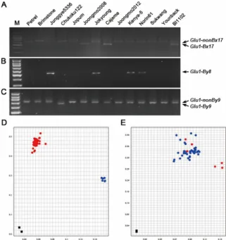
Glu-B1 유전자좌 유전형 판별 분자표지 검정
Glu-B1 유전자좌에는 종류 이상 대립유전자가 보고되었지50 만 빵용에 적합한 재배 품종은 대부분, Glu-B1b, Glu-B1i와 Glu-B1al 등을 지닌 것으로 알려져 있다(Lei et al. 2006).국내 품종에서는 조경과 금강은 Glu-B1b를 중모, 2008 Glu-B1i 대이 립유전자를 지니고 있으며 연백과 조품은, Glu-B1f를 가지고 있다 조경의. Glu-B1b 대립유전자 판별하기 위하여STS분자표 지인ZSBy8 ZSBy9a와 를 적용하였으며 기존의, Glu1-Bx7 대립 유전자 판별 분자표지는2,000 bp이상으로 육종 프로그램에 활용 가능성이 낮아 배제하였다(Anderson & Greene 1989, Rai
우성 분자표지인 을 이용하여
et al. 2018). ZSBy8 Glu-B1b을 보유 한Junggye5336, Kenya-5 Norin 61와 품종에서만특이적으로 가 증폭된 것을 확인하였으며 공우성인 분자표지
DNA , ZSBy9a
를 이용하여 Glu-B1c 대립유전자를 보유한Chukoku122에서 밴드를 확인하였고 다른 대립유전자를 보유한 품종에서
662 bp ,
는 707 bp의 밴드를 확인하였다(Figs. 3A, 3B).
Glu-B1i 대립유전자의 확인은 개의2 STS분자표지를 이용하 였는데, cauBx642로 유전형을 분석한 결과Glu-B1i를 지닌 중모
과 는 의 밴드가 나타났고
2008 Cajeme, Bl1102 534 bp , Glu1-Bx6 을 지닌 Brimstone 660 bp ,는 를 다른 품종은 642 bp밴드를 나타내었다(Fig. 3C).제빵력이 우수한 품종이Glu-B1al을 지닌 것으로 알려지면서 Glu-B1al 대립유전자에 대한 관심이 높아지 고 있는데 분자표지 검정을 위하여 본 연구에서 평가한 품종과,
국내 육성 품종에는 Glu-B1al 대립유전자를 보유한 자원이 없었 다. Glu-B1al 자원 선발을 위하여Lab-on-a-chip방법 통하여 국립식량과학원 남부작물부에서 보유하고 있는 유전자원의
조성을 분석하였다 분석 결과
HMW-GS . Lab-on-a-chip Glu-B1b 의 조경보다 단백질 발현이 배 높은2 Vesna Yecora f-70와 를 이용하여(Fig. 4), Glu-B1al 대립유전자의 x-type 유전자인
Fig. 4. Quantity of each HMW-GS composition. HMW-GS protein was separated on Protein 230 chip with 2100 bioanalyzer and quantity of HMW-GS was calculate with 2100 expert program (Agilent Technologies, CA, USA). Each content was measured as relative content.
Fig. 3. Evaluation of molecular markers for genotype analysis of Glu-B1 locus. A-C. Molecular markers, cauBx642 (A), ZSBy8 (B), and ZSBy9a (C), were examined after agarose gel electrophoresis.
All of markers were applied with touch-down method from 65°C for 10 cycles and then followed more 30 PCR cycles at 55 °C for annealing. Gel photograph was taken after electrophoresis on 3%
agarose gel for 1 hour. D and E. Scatter plots for Bx7
OE_866_SNP
and Bx13_SNP assay, respectively, showing clustering of varieties
on the X-(FAM) and Y-(HEX) axes. D. Red color indicates
Glu1-nonBx17
OEallele and blue color shows Glu1-Bx17
OEallele of
each varieties. E. Red color indicates Glu1-Bx13 allele and blue
color shows Glu1-nonBx13 allele of each varieties. All experiments
were repeated three times independently.
Glu1-Bx7OE대립유전자를 판별할 수 있는STS분자표지 종2 (Bx-7OE left, Bx-7OE right) KASP과 분자표지 종1 (BX7OE_866_SNP)을 검정하였다(Ragupathy et al. 2008). STS 분자표지인Bx-7OE
와
left Bx-7OE right는 우성 분자표지로서Vesna Yecora f-70와 품종에서 각각 특이적으로DNA가 증폭되었다(Data not shown).
분자표지인
KASP BX7OE_866_SNP을 이용한 결과, Vesna와 은
Yecora f-70 BX7OE_866_SNP분자표지의“C” SNP의 를 가지 고 있었으며 다른 대립유전자를 보유한 품종에서는, “G” SNP의 를 가지고 있었다(Fig. 3D).이러한 결과를 통하여BX7OE_866_SNP 분자표지와STS분자표지를 활용하여Glu-B1al 대립유전자의 국내 품종으로 도입을 적극적으로 검토해야 할 것이다.
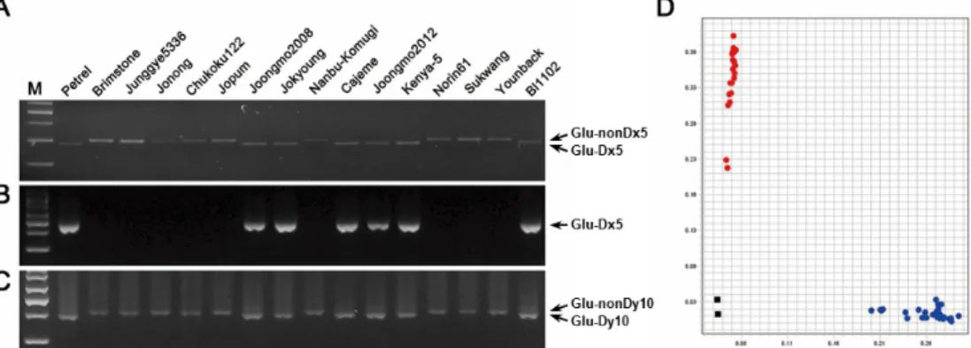
Glu-D1 유전자좌 유전형 판별 분자표지 검정
국내 품종의 Glu-D1 유전자좌에는 Glu-D1a, Glu-D1d와 Glu-D1f 대립유전자가 존재하는데, Glu-D1d는 Glu-D1x5와 Glu-D1y10을 지니고 제빵 적성에 적합한 품종이 반드시 지니고 있어야 하는 대립유전자로 알려져 있다(Zhang et al. 2014).국내 품종 중에는 금강 백강 조경 탑동 황금과 중모, , , , 2008 Glu-D1d이 대립유전자를 지니고 있으며 대부분의 품종은, Glu-D1f 대립유 전자를 가지고 있고 일부 품종이 Glu-D1a 대립유전자를 지니고 있다(Jang el al. 2017). Glu-D1d 대립유전자의 Glu1-Dx5와 Glu1-Dy10을 판별할 수 있는 종과 종을 각각 검정하였다4 2 . Glu1-Dx5 구별을 위해STS 분자표지 종3 (UMN25, Dx5-1과
과 분자표지 종 을 이용하였다
Dx5-2) KASP 1 (Glu-D1d_SNP) . 아가로스 젤 기반 분자분자표지인 공우성 분자표지인UMN25로 Glu1-Dx5를 지닌 중모2008 Petrel과 등은281 bp가 증폭되었으
며 다른 유전형에서는, 299 bp가 증폭되어Glu1-Dx5를 판별 할 수 있었다(Fig. 5A).우성 분자표지인Dx5-1 Dx5-2 Glu1-Dx5과 는 를 지닌 품종은 각각478 bp 450 bp와 의 단편이 증폭되어 판별이 가능하였지만 공우성 분자표지에 비해서 적용의 한계가 있었다 (Fig. 5B). Glu1-Dx5 판별을 위한KASP분자표지Glu-D1d_SNP 검정 결과, Glu1-Dx5를 지닌 품종은“G” (HEX) SNP를 나타내었 고, Glu1-Da와 Glu1-D1f 대립유전자를 지닌 품종은 “C” (FAM)
를 나타내었다
SNP (Fig. 5C).
Glu-D1 대립유전자의 y-type인Glu1-Dy10와 Glu1-Dy12를 판별하기 위해, STS분자표지 종2 (UMN26, Dy-10)을 검정하였 다 공우성 분자표지. UMN26의 경우, Glu1-Dy10을 지닌 품종 은 397 bp DNA의 단편이 증폭되었으며, Glu1-Dy12를 지닌 품종은 415 bp의 DNA 단편을 보였다(Fig. 5D). Ahmad M
이
(2000) 보고한 Glu1-Dy10 판별 분자표지는 본 연구에서는 멀티 밴드 패턴으로 나타나 판별에 이용하기 어려웠다. UMN25 와UMN26을 이용하여Glu-D1d 대립유전자를 판별이 가능하 지만증폭된DNA단편의 차이가 작아 육종 프로그램에서 대량 검정에 활용하였을 때 어려움이 예상되어KASP기반의 분자표 지Glu-D1d_SNP를 함께 이용하는 것을 제안한다 국내 품종이. 많이 보유하고 있는 Glu-D1f의 판별을 위한 추가적인 분석도 필요하다.
적 요
조성은 밀의 품질을 결정하는 주요 요인으로 종자 HMW-GS
단백질을 추출하여SDS-PAGE를 이용한 분석이 주로 이루어졌
Fig. 5. Evaluation of molecular markers for genotype analysis of Glu-D1 locus. A-C. Molecular markers, UMN25 (A), Dx5-1 (B) and
UMN26 (C), were examined after agarose gel electrophoresis. All of markers were applied with touch-down method from 65°C for 10
cycles and then followed more 30 PCR cycles at 55 °C for annealing. Gel photograph was taken after electrophoresis on 3% agarose gel
for 1 hour. D. Scatter plots for Glu-D1d_SNP. D. Red color indicates Glu1-Dx5 allele and blue color shows Glu1-nonDx5 allele of each
varieties. All experiments were repeated three times independently.
으나 분석 시간이 오래 걸리는 단점이 있다 육종프로그램에서, . 대량 검정을 위하여 본 연구에서는STS KASP와 표지인자 적용 을 검토하였다. Glu-A1 유전자좌의 Glu-A1b 대립유전자를 판독 에는STS분자표지인UMN19 Glu-A1c 대립유전자를 판독할와 수 있는 KASP 분자표지 Glu-Ax1/2*_SNP이 적합하였다. Glu-B1 유전자좌의 Glu-B1b, Glu-B1c Glu-B1i 대립유전자와 판별을 위한STS분자표지로는ZSBy8, ZSBy9a cauBx642과 가 각각 적합하였다. Glu-B1al 대립유전자 판별을 위해서는 KASP 분자표지 BX7OE_866_SNP가 가능하였다. Glu-D1 유전자좌의 Glu-D1d 대립유전자를 판별할 수 있는 KASP 분자표지로
이 가능하였다 Glu-D1d_SNP .
사 사
본 논문은 농촌진흥청 연구사업 세부과제명 밀 고분자 글루( : 테닌의 유전적 조성과 품질 연관성 구명, PJ013564022020)의 지원에 의해 이루어진 것임.
REFERENCES