The Effects of Either Chrysin or Moderate Exercise on Inflammasome and Thermogenic Markers in High Fat Fed Mice
Young-Ran Lee1, Hee-Geun Park2 and Wang-Lok Lee2*
1Center for Sport Science in Jeonbuk, Jeonju 54894, Korea
2Department of Sport Science, College of Natural Science, Chungnam National University, Daejeon 34134, Korea Received March 25, 2019 /Revised April 23, 2019 /Accepted April 25, 2019
The purpose of this study was to investigate the effects of either chrysin or exercise on the inflammasome and thermogenic markers in the livers of high-fat fed mice. C57BL/6 mice were randomly assigned to four groups: normal diet control (NC; n=5), high-fat diet control (HC; n=5), high-fat diet with chrys- in (Hch; n=5), and high-fat diet with moderate exercise (HME; n=5). The mice were fed a high-fat diet (60% of calories from fat) or normal diet (18% of calories from fat). Chrysin was supplemented orally as 50mg/kg/day dissolved in a 0.1ml solution of dimethyl sulfoxide. The exercised mice ran on a treadmill at 12-20 m/min for 30-60 min/day, 5 times/week, for 16 weeks. After the intervention, the epididymal fat and liver weights were significantly decreased in the HME group compared with HC and Hch groups. The adipocyte size was effectively decreased in the Hch and HME groups compared with the HC group. The inflammasome markers NLRP3, IL-1β, and caspase1 were significantly de- creased in the Hch and HME groups compared with the HC group. The thermogenic markers PGC-1α and BMP7 were significantly lower in the HC than in the NC group. However, the HME group showed an increase in the thermogenic markers. In conclusion, chrysin and moderate exercise have positive effects on obese metabolic complications induced by high-fat diets by reducing inflammasome genes.
However, chrysin supplementation had no effect on thermogenic gene expression. Moderate exercise would therefore seem to be more effective in controlling obesity-induced metabolic deregulation.
Key words : Browning marker, chrysin, exercise, inflammasome, obese
*Corresponding author
*Tel : +82-42-821-6458, Fax : +82-42-823-0387
*E-mail : [email protected]
This is an Open-Access article distributed under the terms of the Creative Commons Attribution Non-Commercial License (http://creativecommons.org/licenses/by-nc/3.0) which permits unrestricted non-commercial use, distribution, and reproduction in any medium, provided the original work is properly cited.
Journal of Life Science 2019 Vol. 29. No. 5. 607~613 DOI : https://doi.org/10.5352/JLS.2019.29.5.607
Introduction
Chronic inflammation state is a pivotal mark in obesity.
In obesity, inflammatory cytokines released from adipose tis- sue reach out the liver and can directly interfered liver func- tions [30]. Recently, interleukin-1 (IL-1) is one of the regu- lators in inflammation-related diseases, and the IL-1 secre- tion is called an inflammasome [19]. Among inflammasomes, the most studied in the area of metabolic disorder is the NOD-like receptor family, pryin domain containing 3 (NLRP3) inflammasome consisting of NLRP3, apoptosis-as- sociated speck-like protein containing a caspase activation recruitment domain (ASC) and caspase 1 (CASP1) [5]. Previ- ous report has demonstrated that NLRP3 inflammasome may be responsible for the progression of metabolic disorders.
Moreover, NLRP3 inflammasome is implicated in obesity, hepatic inflammation [32]. Vandanmagsar et al. [30] have suggested that obese caused the inflammasome marker in- creased significantly in liver.
Alleviating hepatic inflammation through regular ex- ercise, and diet intervention may help treat obesity. Moder- ate exercise can prevent against of chronic inflammation [18]
and reduces state of fibrosis as a marker of hepatic injury [19]. Our previous study has been shown the positive effect of treadmill exercise and resveratrol supplementation on in- flammatory cytokines, mitochondrial biogenesis, adipo- genesis in adipose tissue of obese animal [9, 11, 15]. Also, recent study reported that resveratrol reduced activation of NLRP3 inflammasome in the subcutaneous fat cell [16].
Many researchers use variety way to treat obese induced metabolic complication such as functional nutrition diet.
Nutritional approaches to reduce hepatic inflammation us- ing functional foods, including blueberry and resveratrol, have been introduced [3]. However the underlying mecha- nisms have not been clearly identified. Chrysin, (5, 7-dihy- droxyflavone), is a natural flavonoids present in many plant extracts, honey and propolis [10]. Several, studies have
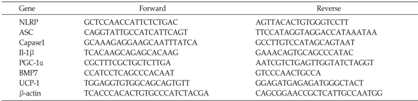
Table 1. Primer sequences used for RT-PCR
Gene Forward Reverse
NLRP ASC Capase1 Il-1β PGC-1α BMP7 UCP-1 β-actin
GCTCCAACCATTCTCTGAC CAGGTATTGCCATCATTCAGT GCAAAGAGGAAGCAATTTATCA TCACAAGCAGAGCACAAG CGCTTTCGCTGCTCTTGA CCATCCTCAGCCCACAAT TGGAGGTGTGGCAGCAGTGTT TCACCCACACTGTGCCCATCTACGA
AGTTACACTGTGGGTCCTT TTCCATAGGTAGGACCATAAATAA GCCTTGTCCATAGCAGTAAT GAAACAGTGCAGCCCATAC AATCGTCTGAGTTGGTATCTAGGT GTCCCAACTGCCA
GGAGATGAGAGATGGGCTACT CAGCGGAACCGCTCATTGCCAATGG shown that chrysin has multiple biological activities, such
as anti-inflammation, anti-oxidation [2, 3, 8]. Although most previous studies reported the gainful effects of chrysin and exercise on high-fat-induced metabolic disturbances, there were few results, comparing the effectiveness among them.
Therefore, the purpose of this study was to compare the ef- fectiveness either chrysin or moderate exercise training on inflammasome, and browning markers in liver of high fat diet mice.
Material and Methods
Animals and diet
Male C57BL/6 (Central Experiment Animal, Korea, N=20) mice were housed in cages in a standard experimental labo- ratory, at temperature 22±2℃, with 60±5% of humidity.
After a one-week acclimatization period, the mice were fed a high fat diet (60% of calories from fat, 20% from carbohy- drate, 20% from protein, Orient Bio Inc., #D12492) or a nor- mal diet (18% calories from fat, 58% from carbohydrate, 24%
from protein, Orient Bio Inc., #2018) ad for 16 weeks. The mice were divided into 4 groups: normal diet control (NC;
n=5), high fat diet control (HC; n=5), high fat with chrysin (Hch; n=5), high fat with moderate exercise (HME; n=5).
Liver tissues were dissected, weighed and immediately frozen. All experiments were approved by the Animal Care and Use Committee at the Chungnam National University (CNU-00494).
Exercise protocol and chrysin supplements In the early phase of moderate exercise, all the mice of exercise ran at 8-10 m/min for 10 minutes on the automati- cally operated rodent treadmill. After that, the speed and time of exercise were gradually increased up to 10-16 m/min for 40-60 minutes. The volume of this exercise training proto- col represents 60-75% of the maximum oxygen consumption
of the rodents as moderate intensity exercise program [23].
A foam sponge was placed in the back of each treadmill lane to prevent the animal from being injured. Electrical shock was not used during the treadmill run. The control mice were exposed to environmental stress as well as noise from the treadmill. Chrysin supplemented mice were orally gavaged with chrysin (50 mg/kg/day) dissolved in a 0.1ml solution of Dimethyl Sulfoxide [10]. The other group re- ceived the vehicle. Each treatment was administrated once a day, five times a week, for 16 weeks before tissue collection.
Histology of adipose tissue size
White adipose tissue samples were taken from character- istic depots identified. Flash-frozen adipose tissues were fixed in 4% buffered paraformaldehyde and sections were stained with hematoxylin and eosin (H&E). To measure adi- pocyte cross-sectional area, four microscope fields in H&E- stained adipose tissue from 5 mice per group were digitally recorded. Images were analyzed using NIS-Elements imag- ing software (Nikon Instruments Inc., Melville, NY).
Gene expression
Total RNA was extracted from 20 mg liver tissue homoge- nate using 1ml Trizol reagent (Ambion, Carlsbad, CA, USA).
The total RNA concentration was calculated by measuring the absorbance at 260 nm and 280 nm using an ultraviolet spectrophotometer. For cDNA synthesis, Maxi RT PreMix kit (iNtRON, Korea) was used according to the manu- facturer’s instructions. The PCR was set using the following program: 95℃ for 2 min, 95℃ for 30 sec 38-40 cycle, the appropriate annealing temperature between 55-57℃ for 30 sec and 72℃ for 2 min. After loaded onto 1% of the agarose gel containing ethidium bromide then measured the PCR band density with Image Lab 4.0 (Bio-Rad, USA). The PCR primer sequences for each studied gene are shown in Table 1.
Fig. 1. The change of (A)epididymal fat ;(B) liver weight of high-fat fed mice. Values are expressed as means ± SEM. NC: normal diet control; HC: high fat diet control; Hch: high fat diet with chrysin; HME: high fat diet with moderate exercise. *p<0.05 difference from NC group, #p<0.05 difference from HC group.
Statistical Analysis
Statistical analysis of the data was performed by SPSS Version 22.0 using one-way ANOVA with LSD post-hoc tests. Statistical significance was defined as α=.05.
Results
The change of white adipose tissue and liver weight Fig. 1 shows the epididymal fat and liver weight of high- fat fed mice. The epididymal fat weight was significantly increased in HD and Hch group compared with NC group (p<0.01). However, the weight was most effectively decreased in HME group compared with HD and Hch group (p<0.05).
The liver weight was significantly increased in HD group compared with NC group (p<0.05). However, the weight was effectively decreased in HME and Hch group compared with HD group (p<0.05).
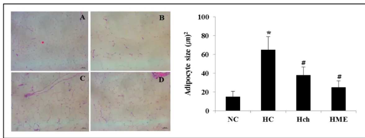
The change of fat cell size in white adipose tissue Fig. 2 shows the fat cell size of high-fat fed mice. The epididymal fat cell size was significantly increased in HD group compared with NC group (p<0.05). However, epi- didymal fat cell size was significantly decreased in Hch and HME group compared with HD group (p<0.05).
The change of inflammasome genes in liver Fig. 3 shows the effect of chrysin or exercise inflamma- some genes in liver.
The NLRP3, caspase1, and IL-1β mRNA expression of HC group significantly increased compared to NC group. How- ever, the NLRP3, caspase1, and IL-1β mRNA expression of Hch and HME group were significantly reduced compared
with HC group (p<0.05). The ASC level was no significant difference among all groups.
The change of thermogenic genes in liver
Fig. 4 shows the effect of chrysin or exercise thermogenic genes in liver.
The PGC-1α mRNA expression was significantly different between NC and HC group, but, HME group was sig- nificantly increased in those. The BMP7 mRNA expression was also significantly decreased in HC and Hch group com- pared with NC group. However, HME group was effectively increased for BMP7 mRNA expression. The UCP-1 mRNA expression was no significant difference among all groups.
Discussion
It is well known that moderate exercise has beneficial ef- fects on the metabolic disorder [4]. Thus, physical activity has been recommended in the treatment of obesity [6]. Also, recent studies demonstrated that Chrysin has many func- tional roles, such as anti-inflammatory and antioxidant [8].
Nevertheless, information concerning the effects of chrysin in animal models is rather scant. Thus, it was meaningful to compare the effectiveness of moderate exercise and chrys- in supplementation on inflammasome and thermogenic genes in high fat fed mice.
After 16 weeks of treatments, the epididymal fat, liver weight, and adipocyte size of HC group were significantly increased in comparison with NC group (Fig. 1, Fig. 2). But, HME group was significantly decreased in that’s comparison with HC group. These research results are consistent with the previous studies that the aerobic exercise allowed to pos-
Fig. 2. The change of adipocyte size of high-fat fed mice. Values are expressed as means ± SEM. NC: normal diet control; HC:
high fat diet control; Hch: high fat diet with chrysin; HME: high fat diet with moderate exercise. *p<0.05 difference from NC group, #p<0.05 difference from HC group.
Fig 3. The change of inflammasome markers of high-fat fed mice. (A) NLRP3; (B) ASC; (C) Cappase1;(D)IL-1β. Values are expressed as means ± SEM. NC: normal diet control; HC: high fat diet control; Hch: high fat diet with chrysin; HME: high fat diet with moderate exercise. *p<0.05 difference from NC group, #p<0.05 difference from HC group.
itively reduce the body and fat weight by increasing the en- ergy consumption [9, 11]. It is suggesting that the moderate exercise can suppress the obesity induced weight gain.
In this study, we found that liver weight and adipocyte size of Hch group were effectively reduced comparison with HC group. But, no significant effect on fat pad weight com- parison with HC group. The previous study reported that resveratrol supplements significantly reduced the epididy- mal fat pad weight [1]. In contrast, in another study this effect was not observed [1, 22]. Thus, in anti-agent studies, the results were still controversial and depended on the dos- age and duration of the supplement. Thus, further studies
are needed in the future.
The NLRP3 inflammasome is a key regulator linking met- abolic dysfunction [5]. Once the NLRP3 inflammasome is ac- tivated, activated caspase1 cleaves a variety of protein pre- cursors [21]. We found that induction of high–fat fed in- duced NLRP3, Il-1β, and Caspase1 expression in liver. On the contrary to this, NLRP3, IL-1β, and Caspase-1 mRNA expressions were significantly decreased in Hch and HME group comparison with HC group. These results have sug- gested that the chrysin supplements or moderate exercise have the function to suppress the expression of inflamma- some related genes in hepatocytes. It seems that the effec-
Fig. 4. The change of thermogenic markers in liver of high-fat fed mice. (A) PGC-1a; (B) BMP7; (C) UCP 1. Values are expressed as means ± SEM. NC: normal diet control; HC: high fat diet control; Hch: high fat diet with chrysin; HME: high fat diet with moderate exercise. *p<0.05 difference from NC group, #p<0.05 difference from HC group.
tiveness on the regulation of inflammasome molecules de- pends on whether there is a significant liver weight loss after intervention. The future study measuring the tissue using the inhibitors of NLRP3 can determine the direct roles of inflammasome in obesity-associated metabolic dysfunction.
Peroxisome proliferator activated receptor gamma co- activator 1 α (PGC-1α), have been identified as the main reg- ulators of mitochondrial biogenesis and thermogenesis [7].
Also, BMP7 inhibits fibrosis in animal models of chronic liv- er disease [17]. Previous experiment results indicated that BMP7 promotes hepatocyte regeneration and inhibits liver fibrosis [20].
In our results, the PGC-1α and BMP7 were significantly decreased in high-fat fed group compared to the normal diet group. However, PGC-1α and BMP7 were significantly in- creased only in moderate exercise group.
Regarding the improve effect of moderate exercise on PGC-1α and BMP7 activation in high-fat fed, we suggest sev- eral pathways. First, in the exercise-induced danger signals, such as free fatty acids, cholesterol, etc., that promotes the NLRP inflammasome activation [32]. In our results, demon- strated that moderate exercise reduce inflammasome related gene in liver of high-fat fed mice (Fig. 3). Second, previous study suggested that aerobic exercise potently stimulates fat- ty-acid oxidation in variety tissue by inhibiting the activity of lipid synthesis related markers [15]. Although, our study
did not measure this metabolite factors in this study. It will be of interest for further researchers to include. In con- clusion, chrysin and moderate exercise have a positive effect on high-fat-diet-induced obese metabolic complication by re- ducing inflammasome genes. However, there was no effect of chrysin supplementation on the thermogenic gene ex- pression. It seems that moderate exercise is more effective to obese-induced metabolic deregulation.
Acknowledgment
This work was supported by the National Research Foundation of Korea Grant funded by the Korean Govern- ment (NRF-2014S1A5A2A01016657).
References
1. Baur, J. A., Pearson, K. J., Price, N. L., Jamieson, N. L., Lerin, C., Kalra, A., Prabhu, V. V., Allard, J. S., Lopez-Lluch, G., Lewis, K., Pistell, P. J., Poosala, S., Becker, K. G., Boss, O., Gwinn, D., Wang, M., Ramaswamy, S., Fishbein, K. W., Spencer, R. G., Lakatta, E. G., Le Couteur, D., Shaw, R. J., Navas, P., Puigserver, P., Ingram, D. K., Cabo, R. and Sinclair, D. A. 2006. Resveratrol improves health and surviv- al of mice on high-calorie diet. Nature 444, 337-342.
2. Cho, H., Yun, C. W., Park, W. K., Kong, J. Y., Kim, K. S., Park, Y., Leem, S. and Kim, B. K. 2004. Modulation of the activity of pro-inflammatory enzymes, COX-2 and iNOS, by
chrysin derivatives. Pharmacol. Res. 49. 37-43.
3. Duarte, J., Jimenez, R., Villar, I. C., Perez-Vizcaino, F., Jimenez, J. and Tamargo, J. 2001. Vasorelaxant effects of the bioflavonoid chrysin in isolated rat aorta. Planta Med. 67, 567-569.
4. Gleeson, M., Bishop, N. C., Stensel, D. J., Lindley, M. R., Mastana, S. S. and Nimmo, M. A. 2011. The anti-inflamma- tory effects of exercise: mechanisms and implications for the prevention and treatment of disease Nat. Rev. Immunol. 5, 607-615.
5. Haneklaus, M. and O’Neill, L. A. J. 2015. NLRP3 at the inter- face of metabolism and inflammation. Immunol. Rev. 265, 53-62.
6. Haram, P. M., Kemi, O. J., Lee, S. J., Bendheim. M. Ø., Al-Share, Q. Y., Waldum, H. L, Gilligan, L. J., Koch, L. G., Britton, S. L., Najjar, S. M. and Wisløff, U. 2009. Aerobic interval training vs. continuous moderate exercise in the metabolic syndrome of rats artificially selected for low aero- bic capacity. Cardiovasc. Res. 81, 723-732.
7. Hondares, E., Rosell, M., Diaz-Delfin, J., Olmos, Y., Monsalve, M., Iglesias, R., Villarroya, F. and Giralt, M. 2011.
Peroxisome proliferator activated receptor-alpha (PPAR α ) induces PPAR γ-coactivator 1 α (PGC-1 α ) gene expression and contributes to thermogenic activation of brown fat: in- volvement of PRDM16. J. Biol. Chem. 286, 43112-43122.
8. Izuta, H., Shimazama, M., Tazawa, S., Araki, Y., Mishima, S. and Hara, H. 2008. Protective effects of Chinese propolis and its component, chrysin, against neuronal cell death via inhibition of mitochondrial apoptosis pathway in SH-SY5Y cells. J. Agric. Food. Chem. 56. 8944-8953.
9. Jeong, J. H., Lee, Y. R., Park, H. G. and Lee, W. L. 2015.
Moderate exercise training is more effective than resveratrol supplementation for ameliorating lipid metabolic complica- tion in skeletal muscle of high fat diet-induced obese mice.
J. Exerc. Nutr. Biochem. 19, 131-137.
10. Juan José, R. E., Johann, S. R., Sara, G. J., Rafael, V. M., Gabriela, A. V., Angélica N. R. O., Germán, B. F. and Samuel, E. S. 2017. Chrysin induces antidiabetic, anti- dyslipidemic and anti-inflammatory effects in athymic nude diabetic mice. Molecules 23, pii:E67.
11. Jun, J. K., Lee, W. L., Park, H. G., Lee, S. K., Jeong, S. H.
and Lee, Y. R. 2014. Moderate intensity exercise inhibits macrophage infiltration and attenuates adipocyte inflamma- tion in ovariectomized rats. J. Exerc. Nutr. Biochem. 18, 119- 127.
12. Kawanishi, N., Yano, H., Yokogawa, Y. and Suzuki, K. 2010.
Exercise training inhibits inflammation in adipose tissue via both suppression of macrophage infiltration and accel- eration of phenotypic switching from M1 to M2 macro- phages in high fat-diet-induced obese mice. Exer. Immunol.
Rev. 16, 105-118.
13. King, G. A., Fitzhugh, E. C., Bassett, Jr. D. R., McLaughlin, J. E., Strath, S. J., Swartz, A. M. and Thompson, D. L. 2001.
Relationship of leisure-time physical activity and occupa- tional activity to the prevalence of obesity. Int. J. Obes. Relat.
Metab. Disord. 25, 606-612.
14. Lagouge, M., Argmann, C., Grhart-Hines, Z., Meziane, H., Lerin, C., Daussin, F., Messadeq, N., Milne, J., Lambert, P., Elliott, P., Geny, B., Laakso, M., Puigserver, P. and Auwerx, J. 2006. Resveratrol improves mitochondrial function and protects against metabolic disease by activating SIRT1 and PGC-1a. Cell 127, 1-14.
15. Lee, Y. R., Jeong, S. H., Park, H. G., Jeong. J. H. and Lee, W. L. 2013. The effects of either resveratrol supplementation or aerobic exercise training combined with a low fat diet on the molecules of adipogenesis and adipocyte inflamma- tion in high fat diet induced obese mice. J. Exerc. Nutr.
Biochem. 17, 15-20.
16. Lee, Y. R., Pipit, P., Park, H. G. and Lee, W. L. Resveratrol ameliorates high-fat-induced metabolic complications by changing the expression of inflammasome markers and macrophage M1 and M2 markers in obese mice. J. Life. Sci.
27, 1462-1469.
17. Li, R. X., Yiu, W. H. and Tang, S. C. 2015. Role of bone morphogenetic protein-7 in renal fibrosis. Front. Physiol. 6, 114.
18. Mardare, C., Kruger, K., Liebisch, G., Seimetz, M., Coutu- rier, A., Ringseis, R., Wilhelm, J., Weissmann, N., Eder, K.
and Mooren, F. C. 2016. Endurance and resistance training affect high fat diet-induced increase of ceramides, in- flammasome expression, and systemic inflammation in mice.
J. Diabetes Res. 2016, 4536470.
19. McCarthy, E. M. and Rinella, M. E. 2012. The role of diet and nutrient composition in nonalcoholic fatty liver disease.
J. Acad. Nutr. Diet. 112, 401-409.
20. Promrat, K, Kleiner, D. E., Niemeier, H. M., Jackvony, E., Kearns, M., Wands, J. R., Fava, J. L. and Wing, R. R. 2010.
Randomized controlled trial testing the effects of weight loss on nonalcoholic steatohepatitis. Hepatology 51, 121-129.
21. Ringseis, R., Eder, K., Mooren, F. C. and Kruger, K. 2015.
Metabolic signals and innate immune activation in obesity and exercise. Exerc. Immunol. Rev. 21, 58-68.
22. Rocha, K. K., Souza, G. A., Ebaid, G. X., Seiva, F. R., Cataneo, A. C. and Novelli, E. L. 2009. Resveratrol toxicity: effects on risk factors for atherosclerosis and hepatic oxidative stress in standard and high-fat diets. Food. Chem. Toxicol.
47, 1362-1367.
23. Schefer, V. and Talan, M. I. 1996. Oxygen consumption in adult and aged C57BL/6J mice during acute treadmill ex- ercise of different intensity. Exp. Gerontol. 31, 387-392.
24. Schroder, K. T. and Schopp, J. 2010. The inflammasomes.
Cell 140, 821-832
25. Schroder, K. Zhou, R. T. and Schopp, J. 2010. The NLRP3 inflammasome: a sensor for metabolic danger? Science 327, 296-300.
26. Skeldon, A. M., Faraj, M. and Saleh, M. 2014. Caspases and inflammasomes in metabolic inflammation. Immunol. Cell Biol. 92, 304-313.
27. Sobocanec, S., Sverko, V., Balog, T., Saric, A., Rusak, G., Likic, S., Kusic, B., Katalinic, V., Radic, S. and Marotti, T.
2006. Oxidant/antioxidant properties of Croatian native propolis. J. Agric. Food. Chem. 54, 8018-8026.
초록:고지방식이 동물의 간 조직에서 크리신 투여 또는 중강도 운동이 Inflammasome과 열 발생 유전 자발현에 미치는 효과
이영란1․박희근2․이왕록2*
(1전북스포츠과학센터, 2충남대학교 스포츠과학과)
본 연구 목적은 고지방식이 동물의 간 조직에서 크리신 투여 또는 중강도운동이 Inflammasome과 thermo- genesis 유전자 발현의 차이를 규명하고자 시도되었다. 본 연구를 위해 정상식이군, 고지방식이군, 고지방식이+크 리신 투여군, 고지방식이+중강도 운동군으로 분류한 후, 크리신 투여군은 16주간 50 mg/kg 농도로 투여하였으며, 운동군은 최대산소섭취량의 60-75%의 중강도 운동으로 실시되었다. 연구결과 크리신 그리고 중강도운동군은 지 방조직, 간조직 무게 그리고 지방세포 크기가 고지방식이 군과 비교해 유의하게 감소하였다. Inflammasome 유전 자 변화는 크리신 투여군 그리고 중강도 운동군에서 NLRP3. ASC, Casepase1 mRNA 발현이 고지방식이 군과 비교해 유의하게 감소하였다. 열발생마커로 알려진 PGC-1a, BMP7 mRNA 발현은 중강도 운동군에서만 고지방식 이군과 비교해 유의하게 증가했다. 결론적으로 중강도 운동은 고지방식이 동물에서 지방무게, Inflammasome, 그 리고 열발생 유전자들의 발현을 비만을 억제하는데 긍정적인 영향을 미치는 것으로 보여진다. 하지만 크리신 투 여는 열발생 유전자 발현에는 유의한 차이를 나타내지 못하였다. 향후 연구에서는 크리신의 비만억제 효과를 규 명하기 위해 투여농도 기간을 고려한 다양한 연구가 진행되어야 할 것이다.
28. Sugimoto, H., Yang, C., LeBleu, V. S., Soubasakos, M. A., Giraldo, M., Zeisberg, M. and Kalluri, R. 2007. BMP-7 func- tions as a novel hormone to facilitate liver regeneration.
FASEB. J. 21, 256-264.
29. Unger, R. H. and Orci, L. 2000. Lipotoxic diseases of non- adipose tissues in obesity. Int. J. Obes. Relat. Metab. Disord.
24, S28-32.
30. Vandanmagsar, B., Youm, Y. H., Ravussin, A., Galgani, J.
E., Stadler, K., Mynatt, R. L., Ravussin, E., Stephens, J. M.
and Dixit, V. D. 2011. The NALP3/NLRP3 inflammasome instigates obesity-induced autoinflammation and insulin
resistance. Nat. Med. 17, 179-188.
31. Williams, C. A., Harborne, J. B., Newman, M., Greenham, J. and Eagles, J. 1997. Chrysin and other leaf exudate fla- vonoids in the genus Pelargonium. Phytochemistry 46, 1349- 1353.
32. Wree, A., Eguchi, A., McGeough, M. D., Pena, C. A., Johnson, C. D., Canbay, A. Hoffman, H. M. and Feldstein, A. E. 2014. NLRP3 inflammasome activation results in epati- cyte pyroptosis, liver inflammation and fibrosis. Hepatology 59, 898-910.