Change in the Levels of Intracellular Antioxidants during Aging of Articular Chondrocytes and Cartilage
Kang Mi Kim1, Yoon Jae Kim2, Jong Min Kim2, Dong Hyun Sohn1 and Young Chul Park1*
1Department of Microbiology & Immunology, Pusan National University College of Medicine, Yangsan, Gyeongnam 50612, Korea
2Department of Anatomy & Cell Biology, Dong-A University College of Medicine, Busan 49201, Korea Received May 20, 2019 /Revised July 22, 2019 /Accepted August 7, 2019
Cartilage diseases, such as rheumatoid arthritis (RA) and osteoarthritis (OA), are associated with the loss of chondrocytes and degradation of articular cartilage. Recent studies revealed that inflammatory reactive oxygen species (ROS) and age-related oxidative stress can affect chondrocyte activity and car- tilage homeostasis. We investigated changes in the levels of intracellular antioxidants during cellular senescence of primary chondrocytes from rat articular cartilages. Cellular senescence was induced by serial subculture (passages 0, 2, 4, and 8) of chondrocytes and measured using specific senescence- associated β-galactosidase (SA-β-gal) staining. ROS production increased significantly in the senescent chondrocytes. In addition, total glutathione (GSSG/GSH) and superoxide dismutase (SOD) levels and heme oxygenase-1 (HO-1) expression increased in senescent chondrocytes induced by serial subculture.
Analysis of changes in intracellular antioxidant levels in articular cartilage from rats of different ages (5, 25, 40, and 72 wk) revealed that total glutathione levels were highest after 40 wk and slightly de- creased after 72 wk as compared with those after 25 wk. SOD and HO-1 expression levels increased in accordance with age. Based on these results, we conclude that intracellular antioxidants may be as- sociated with cartilage protection against excessive oxidative stress in the process of chondrocyte sen- escence and age-related cartilage degeneration in an animal model.
Key words : Aging, antioxidants, chondrocytes, cartilage, SA-β-gal
*Corresponding author
*Tel : +82-51-510-8093, Fax : +82-55-382-8090
*E-mail : [email protected]
This is an Open-Access article distributed under the terms of the Creative Commons Attribution Non-Commercial License (http://creativecommons.org/licenses/by-nc/3.0) which permits unrestricted non-commercial use, distribution, and reproduction in any medium, provided the original work is properly cited.
Journal of Life Science 2019 Vol. 29. No. 8. 888~894 DOI : https://doi.org/10.5352/JLS.2019.29.8.888
서 론
류마티스관절염(rheumatoid arthritis)과 골관절염(osteoar- thritis) 같은 질환은 관절연골(articular cartilage)의 분해를 특 징으로 한다[6, 10]. 관절연골은 가동관절(diarthrodial joint)의 미끄러지는 표면을 따라 형성되어 있는 아주 전문화되고 독특 하게 디자인된 생체구조물이다. 관절연골은 collagens (주로 type II collagen)과 proteoglycans (주로 aggrecan)으로 넓게 구성된 세포 외 기질(ECM, extracellular matrix)로써 관절의 기능적 특징을 담보하는 무혈관성, 무신경성, 그리고 무림프 성 조직이다[13, 25]. 이들 세포 외 기질의 파괴는 관절연골의 분해를 가져오며 골관절염의 전형적인 특징으로 볼 수 있다 [1, 3].
Chondrocytes (연골세포)는 관절연골에 존재하는 유일한 세포로서 synovial fluid로부터 외부 화학적 신호와 영양분을 공급받아 세포 외 기질을 생산하고 유지하는 역할을 담당한다
[8, 32]. 즉, chondrocytes는 세포 외 기질을 생산하는 합성경로 와 다양한 matrix metalloproteinases (MMPs)의 활성에 의한 세포 외 기질의 제거에 관여하는 분해경로의 균형을 유지하는 데 관여한다. 최근의 연구에 의하면, 골관절염 환자의 syno- vial fluid와 chondrocytes에 존재하는 pro-inflammatory cy- tokine인 interleukin (IL)-1β가 세포 외 기질의 분해효소인 MMPs의 생산을 증가시켰다[28]. 또한, IL-1β, tumor necrosis factor (TNF)-α 그리고 IL-17이 다양한 스트레스로부터 세포와 조직을 보호하는 역할을 하는 heme oxygenase-1 (HO-1)의 발현을 감소시켰다[9].
한편, 세포노화(cellular senescence)는 telomere와 관련된 내인성 replicative senescence와 pro-inflammatory cytokines, oncogene 활성화, 그리고 산화적 스트레스(oxidative stress) 같은 다양한 자극에 의한 외인성 telomere-independent sen- escence로 나눌 수 있다[24]. 노화세포(senescent cells)는 초기 에는 활발한 대사활동을 보이지만 점차 세포분열에 관여하는 유전자 발현의 능력을 잃고 궁극적으로 G1 혹은 초기 S phase 에서 arrest 된다[30]. Chondrocytes의 노화는 연골에 존재하 는 chondrocytes의 수적 감소를 가져오며, 나이와 관련된 관절 연골의 퇴화 및 골관절염의 발달에 중요한 역할을 하는 것으 로 밝혀졌다[16, 34]. 실제로, 노화된 chondrocytes가 나이가 많아짐에 따라 그리고 골관절염 환자의 관절연골에 축적됨이 보고되었다[20, 22]. 그러나, 관절연골의 퇴화와 골관절염의 발
달 과정에서 chondrocytes의 활성 및 노화에 대한 정확한 기전 과 관련된 인자들의 상호 관련성에 대해 아직도 완전히 밝혀 져 있지 않다.
본 연구에서 관절연골로부터 분리한 chondrocytes의 노화 와 쥐의 나이대별 관절연골로부터 세포내 항산화 인자들의 발현을 조사하였다. 이는 염증성 reactive oxygen species (ROS) burst와 나이와 관련된 산화적 스트레스의 증가가 chondrocytes의 기능과 관절 항상성에 영향을 미치는 것을 규 명하는데 도움을 줄 것으로 생각한다.
재료 및 방법
시약
Collagenase, formaldehyde, 5-bromo-4-chloro-3-indolyl β- D-galactopyranoside (X-gal), potassium ferrocyanide, potas- sium ferricyanide, Nonidet P-40, propidium iodide (PI)은 Sigma Chemical Co. (St. Louis, MO, USA) 제품을 구입하여 사용하였다. Fetal bovine serum (FBS), Dulbecco’s modified Eagle’s medium (DMEM)은 Hyclone (Logan, UT, USA)로부 터 구입하였다. Total glutathione (GSSG/GSH)의 측정은 OxiSelect total glutathione assay kit는 Cell Biolabs, Inc (San Diego, CA, USA)의 제품을 사용하였다. Type II collagen, β- catenin, HO-1, superoxide dismutase (SOD), β-actin에 대한 항체는 Santa Cruz Biotechnology (Santa Cruz, CA, USA), 그리고 2’,7’-dichlorodihydrofluorescein diacetate (H2DCF- DA)와 fluorescein isothiocyanate (FITC)가 부착되어 있는 이 차 항체는 Invitrogen Co. (Carlsbad, CA, USA) 제품을 사용하 였다. Horse radish peroxidase (HRP)가 부착되어 있는 이차 항체와 enhanced chemiluminescence (ECL) Western blotting kit는 Amersham Pharmacia Biotech (Piscataway, NJ, USA) 제품을 사용하였다.
Articular chondrocytes의 분리와 배양
Chondrocytes는 5주된 female Sprague-Dawley rats (Samtako BioKorea, Osan, Korea)로부터 knee joint cartilage (무릎관절의 연골)를 분리하여 슬라이스를 만든 후 1시간 동안 효소(0.2% type II collagenase in DMEM) 처리를 통하여 분리 하였다. 원심분리 후 얻은 chondrocytes는 5×10⁴ cells/cm2 밀 도로 culture dishes에서 10% heat-inactivated FBS와 anti- biotics (50 U/ml penicillin, 50 μg/ml streptomycin)를 포함하 는 DMEM 배양액에서 5% CO2/air atmosphere 조건에서 37
℃ incubator에서 배양하였다. 2일마다 배양액을 교환하였고
~4-5일 후에 70~80% confluence에 도달하였으며, 이를 pas- sage 0 (P0)라고 명명했다. P0 세포는 연속 계대배양(serial subculture)을 통해서 P6까지 얻었다. 본 동물실험은 부산대학 교 동물실험윤리위원회의 심의를 받았고(PNU-2016-1284), 실
험동물은 실험의 안전성과 윤리적 취급 등 국가에서 정한 규 정에 따라 수행하였다.
Senescence-associated β-galactosidase (SA-β-gal) staining assay
배양중인 chondrocytes를 PBS로 2회 조심스럽게 씻어준 후, 0.2% glutaraldehyde/2% formaldehyde 용액에서 5분간 상온에서 고정시켰다. 다시 세포를 PBS로 씻어준 다음, SA-β- gal 염색용액(1 mg/ml X-gal, 40 mM citrate/phosphate buf- fer (pH 6.0), 5 mM potassium ferrocyanide, 5 mM potassium ferricyanide, 150 mM NaCl, 2 mM MgCl2)에서 12시간 동안 37°C chamber에서 반응시켰다. 노화된 세포의 정도는 x20 배 율의 현미경하에서 무작위로 선택한 200개의 세포수 당 blue 로 염색된 세포수의 퍼센트로 나타내었다.
Measurement of intracellular ROS
세포내 ROS의 양은 ROS-sensitive fluorophore H2DCF-DA 를 사용하여 세포에 반응시킨 후, 형광의 변화를 Zeiss LSM 510 laser-scanning confocal microscope (Göettingen, Ger- many)를 사용하여 측정하였다. Absolute fluorescence in- tensities는 무작위로 선택된 영역에서 같은 수의 세포를 사용 하여 측정한 값을 사용하였다.
Measurement of total glutathione
세포와 연골조직에 존재하는 glutathione (GSSG/GSH)의 정량은 OxiSelect total glutathione assay kit (Cell Biolabs, Inc, San Diego, CA, USA)를 이용하여 제조사가 공급한 protocols 에 따라 실시하였다. 분석을 위한 세포(5×10⁶ cells)와 조직(100 mg)을 cold PBS로 2회 씻고 ice-cold 5% metaphosphoric acid (MPA)를 최종 농도가 0.5% 되도록 첨가하여 glass pestle을 사용하여 균질화하였다. 그 후 homogenates를 4℃에서 12,000 rpm으로 15분간 원심분리하여 상층액을 얻어 -80℃에 보관 후 사용하였다.
Western blotting
세포를 PBS로 씻어준 후, 50 mM Tris-HCl (pH 7.4), 150 mM NaCl, 1% Nonidet P-40과 protease inhibitor cocktail을 포함한 lysis buffer를 이용하여 전체 단백질을 분리하였다.
Bicinchoninic acid (Pierce, Rockford, IL, USA)를 이용하여 단백질을 정량하였고, 동일한 양의 단백질을 12% SDS-poly- acrylamide gel에 전기영동하고 electroblotting apparatus (Bio-Rad, Richmond, CA, USA)를 사용하여 PVDF mem- brane으로 이동시켰다. 5% skim milk에서 1시간 동안 mem- brane을 blocking한 후, SOD, HO-1, β-actin에 대한 일차 항체 및 HRP가 부착되어 있는 이차 항체를 반응시켰다. 면역복합 체는 ECL kit를 이용하여 반응시키고 LAS-3000 Luminescent
Fig. 2. Generation of ROS by serial subculture in ariticular chondrocytes. As indicated, the cells were incubated with 10 μM H2DCF-DA for 10 min, and intracellular levels of ROS were determined by confocal microscopy.
A
B
Fig. 1. Induction of cellular senescence by serial subculture in ariticular chondrocytes. Rat primary articular chondro- cytes were serially subcultured up to P6. Cellular sen- escence was measured using SA-β-gal activity assay. (A) Percentages of senescent cells were calculated from numbers of blue-stained cells per 200 cells in randomly selected areas. *p<0.05 and **p<0.01 vs. P0 cells. (B) Chondrocyte images were captured at x20 magnification.
Image Analyzer (Fujifilm, Tokyo, Japan)를 이용하여 단백질 발현을 분석하였다.
Immunofluorescence staining
배양중인 chondrocytes를 PBS로 씻어준 다음, 4% paraf- ormaldehyde/PBS로 상온에서 5분 동안 고정시키고 0.2%
Triton X-100/PBS로 5분 동안 permeabilize 시켰다. 고정된 세포에 SOD 및 HO-1에 대한 일차 항체와 FITC가 부착되어 있는 이차 항체를 차례로 반응시켰고, PI를 사용하여 핵을 염 색하였다. 세포내 형광 image는 laser-scanning confocal mi- croscope를 사용하여 관찰하였다.
Immunohistochemistry staining
Articular cartilage는 2일 동안 4% formaldehyde 용액에서 고정한 후 4주 동안 100 mM EDTA (pH 7.0)/PBS에서 decal- cification 과정을 거쳤다. Cartilage 조직은 순차적으로 알코올 에 통과시킴으로써 탈수화시킨 후 paraffin으로 embedding 시켜 단단한 블록을 만들었다. 블록은 microtome을 이용하여 5 μm로 얇게 잘라 slide 위에 붙이고, xylene을 사용하여 par- affin을 제거하고 순차적으로 알코올에 통과시킴으로써 rehy- dration 하였다. 그 후, 10 mM sodium citrate buffer (pH 6.0) 로 상온에서 2시간, 3%(v/v) H2O2/TBS로 15분간 반응시켰다.
조직 절편은 1.5%(v/v) normal goat serum/TBS로 30분 동안 blocking 과정을 거친 후 SOD와 HO-1에 대한 일차 항체를 반응시켰다. Staining과 mounting 과정은 일반적 방법을 따라 수행하였다.
통계
모든 실험은 적어도 세번 이상 반복하여 결과를 means ± standard deviations (SD)로 표현하였다. One-way analysis는 GraphPad Prism software (GraphPad Software, Inc., La Jolla, CA, USA)를 이용하여 분석하였고, p-value<0.05를 구하여 통 계적 유의성을 검증하였다.
결 과
연속 계대배양에 의한 chondrocytes의 노화
본 연구자는 선행연구에서 배양중인 chondrocytes의 연속 계대배양은 세포노화를 유도한다는 결과를 발표하였다[17].
쥐의 관절연골로부터 분리한 chondrocytes를 연속 계대배양 을 진행하였고, SA-β-gal staining 방법을 이용하여 각 다른 passage의 chondrocytes에서 세포노화의 정도를 분석하였다.
연속 계대배양한 chondrocytes의 passage 수가 증가할수록 SA-β-gal로 염색되는 수가 증가하였다(Fig. 1). 그리고 pas- sage 수의 증가에 따라 cell proliferation의 감소와 cell mor- phology의 변화와 함께 chondrocytes의 기능에 중요한 관절
연골의 세포 외 기질 성분인 type II collagen 발현의 억제를 선행연구 결과에서처럼 관찰할 수 있었다(data not shown).
연속 계대배양에 의한 세포내 ROS의 증가
세포노화에 따른 세포내 산화적 스트레스의 변화를 측정하 기 위하여, ROS에 반응하는 DCF의 형광을 이용하여 chon- drocytes 내 ROS의 양을 측정하였다. P0와 연속 계대배양을 통해 얻은 P2, P4, P6 chondrocytes를 35-mm dish에 분주한 뒤 10% FBS를 포함하는 DMEM 배양액에서 24시간 동안 배양 한 후 ROS-sensitive fluorophore인 H2DCF-DA를 첨가하고 confocal microscope를 사용하여 형광을 측정하였다. Fig. 2에 서 보는 바와 같이, 연속 계대배양에 의한 세포노화는 chon-
Fig. 3. Changes of total glutathione (GSSG/GSH) by serial sub- culture in chondrocytes. Cells were washed and homo- genized with glass pestle in 0.5% MPA solution. Intracel- lular glutathione levels were determined in the super- natants from each cell homogenates using total gluta- thione assay kit. The results are presented as the means of ± SDs of triplicate cultures. *p<0.05 and **p<0.01.
A
B
Fig. 4. Detection of SOD and HO-1 expression by serial sub- culture in chondrocytes. (A) Whole cell lysates were an- alyzed by Western blotting using specific antibodies against SOD and HO-1. b-Actin was used as an internal control to monitor equal protein loading. (B) Chondro- cytes were fixed and reacted with specific antibodies against SOD and HO-1 for immunofluorescence staining.
Fig. 5. Changes of total glutathione (GSSG/GSH) during aging in rat articular cartilages. Articular cartilages were iso- lated from different aged rats, as indicated. The tissues were washed and homogenized with glass pestle in 0.5%
MPA solution. Intracellular glutathione levels were de- termined in the supernatants from each tissue homoge- nates. The results are presented as the means of ± SDs of triplicate experiments. *p<0.05 and **p<0.01.
drocytes 내 ROS의 양을 증가시켰다. 이는 세포노화가 진행될 수록 세포내 redox 균형이 변화한다는 것을 의미한다.
노화 유도한 chondrocytes의 세포내 항산화 인자의 변화 다음으로, 노화과정동안 산화적 스트레스로부터 세포를 보 호하는 항산화 인자들의 변화를 조사하였다. 연속 계대배양을 통해 얻은 chondrocytes에서 세포내 존재하는 총 glutathione (GSSG/GSH)의 양을 조사한 결과, P6 세포에서 크게 증가함 을 보였다(Fig. 3). 그리고 SOD와 HO-1의 발현은 P4 세포부터 크게 증가함을 Western blotting (Fig. 4A)과 immunofluor- escence staing (Fig. 4B) 방법으로 관찰하였다. 이는 연속 계대 배양에 의한 세포노화 과정에서 증가하는 산화적 스트레스를 억제하기 위하여 세포내 항산화 인자들이 증가한다고 여겨진 다.
나이대별 rat 관절연골의 항산화 인자의 변화
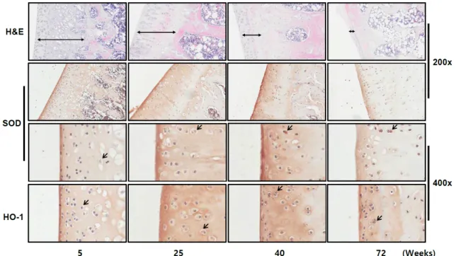
노화된 세포에서 증가하는 항산화 인자들이 쥐의 관절조직 에서도 발현이 증가하는지를 분석하고자 쥐의 나이대별 관절 연골을 분리하여 총 glutathione, SOD, HO-1의 양을 조사하였 다. 주령이 5, 25, 40, 72인 female Sprague-Dawley rats에서 무릎관절의 연골을 분리한 후 연골조직에 존재하는 gluta- thione (GSSG/GSH)의 정량은 100 mg 조직을 사용하여 균질 화한 후 total glutathione assay kit를 이용하였고, 항산화 효소 SOD와 HO-1의 발현은 관절연골의 고정과 decalcification 과 정을 거친 후 immunohistochemistry staining 방법으로 조사 하였다. 그 결과, 총 glutathione의 양은 25주령 및 40주령의 쥐의 관절연골에서 크게 증가하였고 72주령의 관절연골에서 5주령의 총 glutathione의 양보다는 크게 증가하였지만 25주 령에 비해 다소 감소하였다(Fig. 5). Fig. 6에서 보는 바와 같이, 주령이 증가할수록 관절연골 부위가 얇아지고 chondrocytes
Fig. 6. Detection of SOD expression during aging in rat articular cartilages. The results of hematoxylin and eosin (H&E) staining and immunohistochemical staining of anti-SOD antibody are shown. Magnification is 200x or 400x.
의 수가 크게 감소하였으나, 항산화 인자 SOD의 발현은 나이 대별로 증가함을 알 수 있다. 비슷하게, 다른 항산화 인자 HO-1의 발현도 SOD처럼 증가하는 양상을 보였다. 하지만 항 산화 인자들의 redox-sensitive transcriptional factor로 알려 진 nuclear factor κB (NFκB)와 NF-E2-related factor 2 (Nrf2) 의 발현의 변화는 관찰할 수 없었다.
고 찰
ROS는 생리적 조건에서 세포와 조직의 성장, 활성화, 분열 및 방어 등의 생물학적 역할을 조절하는 신호전달물질로서 관여하지만, 과도하게 증가한 양은 세포와 조직에 손상을 일 으키게 된다[7, 29]. 또한, 이 산화적 스트레스는 항산화 인자의 결여에 의한 ROS의 증가, 즉 oxidants와 세포내 redox (reduction-oxidation) 균형을 유지하는 antioxidants 간의 불 균형에 의해서도 유도된다. ROS는 다양한 생리적 그리고 병 리적 조건에서 만들어지며 normoxic 조건하의 O2 tension (21%)이 크게 기여하는 것으로 여겨진다[19]. 하지만, 관절연 골은 synovial fluid로부터 영양소를 공급받는 무혈관성 조직 으로 synovial fluid와 더불어 O2 tension이 7% 정도로 알려져 있다[11, 27]. 그 결과, chondrocytes는 낮은 O2 tension 환경에 적응되어져 있으며 다른 세포들이 가지는 미토콘드리아의 에 너지 대사 과정과 상이함을 보인다[31].
비록 관절연골이 hypoxic 조건하에 있지만, chondrocytes 는 대사와 기능을 위한 신호전달물질로 ROS를 생산한다. 또 한 chondrocytes는 골관절염 외에도 류마티스관절염, gout
(통풍) 등 병리적 조건에서 주위 면역세포들에 의해 생산되는 과도한 양의 ROS에 직면하게 되기도 한다[23, 26, 33]. 또한, 연구자들은 염증성 질환과 관련된 병리적 조건에서 ROS의 과도한 증가뿐 아니라 항산화 인자의 결여가 관찰된다는 것을 보고하였다[21]. 이는 다양한 염증성 물질의 분비를 촉진하고 세포 외 기질을 파괴하는 MMPs의 발현도 증가시켜 관절연골 의 손상을 일으키게 한다. 게다가 관절연골에 존재하는 chon- drocytes의 노화와 세포사망(apoptosis)을 촉진하여 관절연골 의 항상성을 깨고 궁극적으로 관절의 손상을 일으키게 된다.
한편, chondrocytes는 이러한 ROS의 독성에 맞서기 위해 SOD, catalase, glutathione peroxidase (GPX) 뿐 아니라 HO-1 등을 포함하는 정교하게 조직화된 항산화 효소계를 가지고 있다[2, 5, 14].
SOD, catalase, GPX에 반해 HO-1은 산화적 스트레스에 반 응하여 발현되는 유도성 효소로서 chondrocytes와 연골을 보 호하는 측면에서 실로 중요한 역할을 담당하고 있으나, 그 상 세한 기전은 아직 완전히 밝혀져 있지 않는 실정이다. 연구자 들은 HO-1이 연골의 파괴를 일으키는 MMPs와 다양한 염증 성 물질의 발현을 억제하여 관절연골을 보호한다고 보고하고 있다[4, 12]. 또한, HO-1은 연골의 퇴행(degeneration)에도 중 요한 역할을 할 것이라고 보고되고 있다[15]. 본 연구자들은 선행 연구에서, HO-1이 chondrocytes의 노화를 억제하며 HO-1의 발현을 위해 protein kinase casein kinase 2 (CK2)가 관여함을 증명하였다[18]. 이같은 연구결과에도 불구하고 chondrocytes의 활성, 노화, 세포사망 및 관절연골의 기능과 항상성에 HO-1을 비롯한 SOD, catalase, GPX 등의 항산화
효소들에 대한 정확한 역할이 규명되지 않은 실정이다.
그러므로, 관절연골의 퇴화와 관절염의 발달 과정에 대한 정확한 기전을 이해하기 위하여 chondrocytes의 세포노화와 항산화 인자의 상호 관련성에 대한 연구가 필수적이다. 본 연 구에서 chondrocytes의 세포노화 과정 및 쥐의 나이대별 관절 연골로부터 chondrocytes의 세포내 항산화 인자의 발현을 분 석하였다. 노화 유도한 chondrocytes는 세포내 총 glutathione 양과 항산화 효소 SOD 및 HO-1의 발현이 증가하였다. 나이대 별 쥐로부터 분리한 관절연골에서 항산화 인자 glutathione의 양은 40주령에서 발현이 가장 높게 관찰되었으며 72주령에 다소 감소하였고, SOD와 HO-1의 발현은 나이대별로 증가하 는 경향을 보였다. 이는 증가된 항산화 인자가 산화적 스트레 스로부터 세포와 조직을 보호하는 역할을 한다는 것을 의미한 다. 하지만, 72주령의 관절연골은 퇴화가 너무 진행되어 있음 을 관찰할 수 있고, 아울러 총 glutathione 양도 유의하게 감소 하는 경향을 보였다. 향후 실험에서, 5주령과 40주령의 사이의 나이대별 관절연골을 좀 더 세분화해서 세밀하게 조사할 필요 가 있다고 여겨진다. 이런 연구결과들의 축적은 세포 외 기질 의 분비와 관절연골 항상성 유지를 위한 chondrocytes의 활성 조절을 가능하게 하고, 이를 이용한 염증성 및 퇴행성 관절질 환의 치료적 접근을 위한 기초적 정보를 제공할 것으로 생각 한다.
감사의 글
이 논문은 부산대학교 기본연구지원사업(2년)에 의하여 연 구되었음.
References
1. Bau, B., Gebhard, P. M., Haag, J., Knorr, T., Bartnik, E. and Aigner, T. 2002. Relative messenger RNA expression profil- ing of collagenases and aggrecanases in human articular chondrocytes in vivo and vitro. Arthritis. Rheum. 46, 2648- 2657.
2. Borsiczky, B., Szabó, Z., Jaberansari, M. T., Mack, P. P. and Röth, E. 2003. Activated PMNs lead to oxidative stress on chondrocytes: a study of swine knees. Acta. Orthop. Scand.
74, 190-195.
3. Cawston, T., Billington, C., Cleaver, C., Elliott, S., Hui, W., Koshy, P., Shingleton, B. and Rowan, A. 1999. The regu- lation of MMPs and TIMPs in cartilage turnover. Ann. NY Acad. Sci. 878, 120-129.
4. Clérigues, V., Guillén, M. I., Gomar, F. and Alcaraz, M. J.
2012. Haem oxygenase-1 counteracts the effects of inter- leukin-1β on inflammatory and senescence markers in carti- lage-subchondral bone explants from osteoarthritic patients.
Clin. Sci. (Lond) 122, 239-250.
5. Deahl, S. T. 2nd., Oberley, L. W., Oberley, T. D. and Elwell, J. H. 1992. Immunohistochemical identification of super-
oxide dismutases, catalase, and glutathione-S-transferases in rat femora. J. Bone Miner. Res. 7, 187-198.
6. DeLise, A. M., Fischer, L. and Tuan, R. S. 2000. Cellular in- teractions and signaling in cartilage development. Osteoar- thritis Cartilage 8, 309-334.
7. Dickinson, B. C. and Chang, C. J. 2011. Chemistry and biol- ogy of reactive oxygen species in signaling or stress re- sponses. Nat. Chem. Biol. 7, 504-511.
8. Falchuk, K. H., Goetzl, E. J. and Kulka, J. P. 1970. Respiratory gases of synovial fluids. An approach to synovial tissue cir- culatory-metabolic imbalance in rheumatoid arthritis. Am.
J. Med. 49, 223-231.
9. Fernández, P., Guillén, M. I., Gomar, F. and Alcaraz, M.
J. 2003. Expression of heme oxygenase-1 and regulation by cytokines in human osteoarthritic chondrocytes. Biochem.
Pharmacol. 66, 2049-2052.
10. Goldring, M. B. and Goldring, S. R. 2007. Osteoarthritis. J.
Cell. Physiol. 213, 626-634.
11. Grimshaw, M. J. and Mason, R. M. 2000. Bovine articular chondrocyte function in vitro depends upon oxygen tension.
Osteoarthritis Cartilage 8, 386-392.
12. Guillén, M., Megías, J., Gomar, F. and Alcaraz, M. 2008.
Haem oxygenase-1 regulates catabolic and anabolic proc- esses in osteoarthritic chondrocytes. J. Pathol. 214, 515-522.
13. Henrotin, Y., Kurz, B. and Aigner, T. 2005. Oxygen and re- active oxygen species in cartilage degradation: friends or foes? Osteoarthritis Cartilage 13, 643-654.
14. Henrotin, Y. and Kurz, B. 2007. Antioxidant to treat osteo- arthritis: dream or reality? Curr. Drug Targets 8, 347-357.
15. Ishitobi, H., Sanada, Y., Kato, Y., Ikuta, Y., Shibata, S., Yama- saki, S., Lotz, M. K., Matsubara, K., Miyaki, S. and Adachi, N. 2018. Carnosic acid attenuates cartilage degeneration through induction of heme oxygenase-1 in human articular chondrocytes. Eur. J. Pharmacol. 830, 1-8.
16. Jallali, N., Ridha, H., Thrasivoulou, C., Underwood, C., Butler, P. E. and Cowen, T. 2005. Vulnerability to ROS-in- duced cell death in ageing articular cartilage: the role of antioxidant enzyme activity. Osteoarthritis Cartilage 13, 614- 622.
17. Kim, K. M., Kim, J. M., Yoo, Y. H., Kim, J. I. and Park, Y. C. 2012. Cilostazol induces cellular senescence and con- fers resistance to etoposide-induced apoptosis in articular chondrocytes. Int. J. Mol. Med. 29, 619-624.
18. Kim, K. M., Song, J. D., Chung, H. T. and Park, Y. C. 2012.
Protein kinase CK2 mediates peroxynitrite-induced heme oxygenase-1 expression in articular chondrocytes. Int. J. Mol.
Med. 29, 1039-1044.
19. Lane, J. M., Brighton, C. T. and Menkowitz, B. J. 1977.
Anaerobic and aerobic metabolism in articular cartilage. J.
Rheumatol. 4, 334-342.
20. Loeser, R. F. 2009. Aging and osteoarthritis: the role of chon- drocyte senescence and aging changes in the cartilage ma- trix. Osteoarthritis Cartilage 17, 971-979.
21. Mates, J. M., Perez-Gomez, C. and Nunez de Castro, I. 1999.
Antioxidant enzymes and human diseases. Clin. Biochem. 32, 595-603.
초록:연골세포 및 관절연골의 노화 과정에서 세포내 항산화 인자들의 변화
김강미1․김윤재2․김종민2․손동현1․박영철1*
(1부산대학교 의과대학 미생물학 및 면역학교실, 2동아대학교 의과대학 해부학 및 세포생물학교실)
류마티스관절염(rheumatoid arthritis)과 골관절염(osteoarthritis) 같은 관절질환은 연골세포(chondrocytes) 감 소와 관절연골(articular cartilage)의 분해를 수반한다. 최근, 연골세포의 활성과 연골 항상성(cartilage homeo- stasis)에 염증성 ROS (reactive oxygen species) burst 및 나이와 관련된 산화적 스트레스(oxidative stress)의 증가 와 관련된 연구가 활발히 진행되고 있다. 본 연구는 관절연골로부터 분리한 연골세포의 노화 과정과 나이대별 관절연골에서 항산화 인자들(antioxidants)의 변화를 조사함으로써, 연골세포와 관절연골의 노화 과정 동안 산화 적 스트레스로부터 조직을 보호하는 항산화 인자들의 역할을 규명하는데 목적이 있다. 쥐의 관절연골로부터 분리 한 연골세포의 연속 계대배양을 통한 노화 과정에서 산화적 스트레스가 증가함을 관찰하였다. 그리고, 노화 유도 한 연골세포는 세포내 총 glutathione (GSSG/GSH) 양과 항산화 효소 superoxide dismutase (SOD)와 heme oxy- genase-1 (HO-1)의 발현이 증가하였다. 다음으로, 나이대별 쥐로부터 분리한 관절연골에서 항산화 인자의 발현을 분석하였다. 항산화 인자 glutathione의 양은 40주령에서 발현이 가장 높게 관찰되었으며 72주령에 다소 감소하였 고, SOD와 HO-1의 발현은 나이대별로 현저히 증가되는 경향을 보였다. 이를 종합해 볼 때, 세포내 항산화 인자들 은 과도한 양의 ROS에 반응하여 연골세포의 노화와 나이와 관련된 관절연골의 퇴화 과정에서 중요한 역할을 하 는 것으로 보인다.
22. Martin, J. A. and Buckwalter, J. A. 2002. Aging, articular cartilage chondrocyte senescence and osteoarthritis. Bioger- ontology 3, 257-264.
23. Martin, W. J., Herst, P. M., Chia, E. W. and Harper, J. L.
2009. Sesquiterpene dialdehydes inhibit MSU crystal-in- duced superoxide production by infiltrating neutrophils in an in vivo model of gouty inflammation. Free Radic. Biol.
Med. 47, 616-621.
24. McCulloch, K., Litherland, G. J. and Rai, T. S. 2017. Cellular senescence in osteoarthritis pathology. Aging Cell 16, 210- 218.
25. Muir, H. 1995. The chondrocyte, architect of cartilage.
Biomechanics, structure, function and molecular biology of cartilage matrix macromolecules. BioEssays 17, 1039-1048.
26. Phillips, D. C., Dias, H. K., Kitas, G. D. and Griffiths, H.
R. 2010. Aberrant reactive oxygen and nitrogen species gen- eration in rheumatoid arthritis (RA): causes and con- sequences for immune function, cell survival, and ther- apeutic intervention. Antioxid. Redox Signal. 12, 743-785.
27. Rajpurohit, R., Koch, C. J., Tao, Z., Teixeira, C. M. and Shapiro, M. 1996. Adaptation of chondrocytes to low oxygen tension: relationship between hypoxia and cellular metabo- lism. J. Cell. Physiol. 168, 424-432.
28. Sandell, L. J., Xing, X., Franz, C., Davies, S., Chang, L. W.
and Patra, D. 2008. Exuberant expression of chemokine genes by adult human articular chondrocytes in response to IL-1beta. Osteoarthritis Cartilage 16, 1560-1571.
29. Sasaki, M., Kajiya, H., Ozeki, S., Okabe, K. and Ikebe, T.
2014. Reactive oxygen species promotes cellular senescence in normal human epidermal keratinocytes through epi- genetic regulation of p16(INK4a.). Biochem. Biophys. Res.
Commun. 452, 622-628.
30. Serrano, M., Lin, A. W., McCurrach, M. E., Beach, D. and Lowe, S. W. 1997. Oncogenic ras provokes premature cell senescence associated with accumulation of p53 and p16INK4a. Cell 88, 593-602.
31. Stockwell, R. A. 1991. Morphometry of cytoplasmic compo- nents of mammalian articular chondrocytes and corneal ker- atocytes: species and zonal variations of mitochondria in re- lation to nutrition. J. Anat. 175, 251-261.
32. Villalvilla, A., Gómez, R., Largo, R. and Herrero-Beaumont, G. 2013. Lipid transport and metabolism in healthy and os- teoarthritic cartilage. Int. J. Mol. Sci. 14, 20793-20808.
33. Yudoh, K., Nguyen, vT., Nakamura, H., Hongo-Masuko, K., Kato, T. and Nishioka, K. 2005. Potential involvement of oxidative stress in cartilage senescence and development of osteoarthritis: oxidative stress induces chondrocyte telomere instability and downregulation of chondrocyte function.
Arthritis Res. Ther. 7, R380-391.
34. Zwerina, J., Tzima, S., Hayer, S., Redlich, K., Hoffmann, O., Hanslik-Schnabel, B., Smolen, J. S., Kollias, G. and Schett, G. 2005. Heme oxygenase 1 (HO-1) regulates osteoclasto- genesis and bone resorption. FASEB J. 19, 2011-2013.