중대뇌동맥 뇌경색 이후 발생된 뇌출혈에 동반된
브로카실어증(Broca aphasia) 환자에 대한 한의치료 증례보고 1례
제유란, 김윤정, 황원덕 동의대학교 부속한방병원 한방내과학교실
A Case Report of Traditional Korean Medicine Treatment for Broca's Aphasia Associated with Cerebral Hemorrhage after Middle Cerebral Artery Infarction
Yu-ran Je, Yoon-jung Kim, Won-deok Hwang
Dept. of Oriental Medicine, College of Oriental Medicine, Dong-Eui University
ABSTRACT
Objective:The aim of this case report was to present the effects of traditional Korean medicine on a patient with Broca's aphasia associated with cerebral hemorrhage after middle cerebral artery infarction.
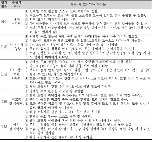
Methods: The Korean version of the Western Aphasia Battery (K-WAB) and the Evaluation of Articulator Performance were used to evaluate the language functions of the patient. Herbal medication and acupuncture were administered to improve the patient's symptoms.
Results:The inpatient and outpatient treatments improved the patient’s symptoms. The aphasia quotient (AQ) score of the K-WAB test before treatment was 6.4, but it increased to 21.4 after treatment. The Evaluation of Articulator Performance score improved by 0.0 points before treatment, by 1.0 points after 9 days of treatment, and by 1.5 points after 42 days of treatment.
Conclusions:This case report suggest that Korean medical therapy can be effective in improving the language functions of patients with Broca's aphasia.
Key words:Broca's aphasia, cerebral hemorrhage, traditional Korean medicine treatment, case report
Ⅰ. 서 론
실어증(aphasia)은 자신의 생각을 의미 있는 단 어나 문장으로 표현하지 못하거나 문장을 듣거나 읽고 이해하는 능력이 상실된 상태를 말한다. 구음 장애(dysarthria)나 무언증(mutism)과는 달리 언 어의 이해, 단어 찾기나 선택, 철자법, 문법 등의
형식적 측면에서 장애가 발생한다
1. 실어증을 일으 키는 원인으로는 뇌혈관장애, 종양, 퇴행성질환, 외 상감염 등으로 다양하나 뇌혈관장애로 인한 경우 가 가장 흔하다
2.
실어증은 뇌손상 부위 및 임상 증상의 양상에 따 라 분류 되는데 크게 브로카 실어증(Broca aphasia) 과 베르니케 실어증(Wernike aphasia) 또한 운동 성, 감각성 증상이 모두 나타나는 전실어증(global aphasia)으로 분류할 수 있다. 브로카 실어증은 뇌 의 하전두엽에 있는 브로카 영역(Broca's area)에 손상이 오는 경우로 다른 사람의 말을 정상적으로
․투고일: 2019.08.29, 심사일: 2019.11.11, 게재확정일: 2019.11.11
․교신저자: 제유란 부산광역시 부산진구 양정로 62 동의대학교부속한방병원 7층 한방의국 TEL: 051-867-5101 FAX: 051-867-5162 E-mail: [email protected]