• 검색 결과가 없습니다.

Clinical Features and Prognostic Factors in Elderly Koreans with Advanced Non-Small-Cell Lung Cancer in a Tertiary Referral Hospital

N/A
N/A
Protected

Academic year: 2021

Share "Clinical Features and Prognostic Factors in Elderly Koreans with Advanced Non-Small-Cell Lung Cancer in a Tertiary Referral Hospital"

Copied!
7
0
0

로드 중.... (전체 텍스트 보기)

전체 글

(1)

Clinical Features and Prognostic Factors in Elderly Koreans with Advanced Non-Small- Cell Lung Cancer in a Tertiary Referral

Hospital

Background: More than half of cases for advanced non-small-cell lung cancer (NSCLC) occur in elderly patients with a median age at diagnosis of 70 years. The aim of our study was to examine the clinical features and prognostic factors contributing to mortality in elderly patients with advanced NSCLC.

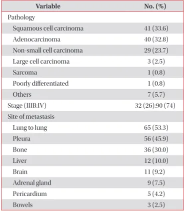
Methods: Following a retrospective review of clinical data, 122 patients aged 70 years and over with a histopathological diagnosis of locally advanced (stage IIIB, n=32) and metastatic (stage IV, n=90) NSCLC between 2005 and 2011 were enrolled.

Results: The median age was 76 years (interquartile range, [IQR], 72−80 years), and 85 (70%) patients were male. Fifty- seven (46%) patients had never smoked, and 17 (19%) were in a malnourished state with a body mass index (BMI) of <18.5 kg/m
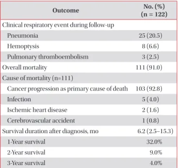
2

. The initial treatments included chemotherapy (40%) and radiotherapy (7%), but 57% of the patients received supportive care only. The 1-year survival rate was 32%, and the 3-year survival rate was 4%, with a median survival duration of 6.2 months (IQR, 2.5−15.3 months). Male gender (hazard ratio [HR], 2.2; 95% confidence interval [CI], 1.3−3.9; p=0.005), low BMI (HR, 2.3; 95% CI, 1.3−3.9; p=0.004), and supportive care only (HR, 1.9; 95% CI, 1.2−2.9; p=0.007) were independent predictors of shorter survival based on a Cox proportional hazards model.

Conclusion: Elderly patients with advanced NSCLC had a poor prognosis, particularly male patients, those with a low BMI, and those who received supportive care only.

Keywords: Carcinoma, Non-Small-Cell Lung; Aged; Prognosis; Mortality

Copyright © 2013 The Korean Academy of Tuberculosis and Respiratory Diseases. All rights reserved.

Seo Woo Kim, M.D.

1

, Mi Yeon Kim, M.D.

1

, Yoon Pyo Lee, M.D.

1

, Yon Ju Ryu, M.D., Ph.D.

1

, Seok Jeong Lee, M.D.

1

, Jin Hwa Lee, M.D., Ph.D.

1

, Jung Hyun Chang, M.D., Ph.D.

1

and Sung Shine Shim, M.D., Ph.D.

2

1

Division of Pulmonary and Critical Care Medicine, Department of Internal Medicine,

2

Department of Radiology, Ewha Medical Center and Ewha Medical Research Institute, Ewha Womans University School of Medicine, Seoul, Korea

Address for correspondence: Yon Ju Ryu, M.D., Ph.D.

Division of Pulmonary and Critical Care Medicine, Department of Internal Medicine, Ewha Medical Center and Ewha Medical Research Institute, Ewha Womans University Mokdong Hospital, Ewha Womans University School of Medicine, 1071 Anyangcheon-ro, Yangcheon-gu, Seoul 158-710, Korea Phone: 82-2-2650-2840, Fax: 82-2-2650-2559, E-mail: [email protected]

Received: Apr. 8, 2013 Revised: May 10, 2013 Accepted: May 27, 2013

cc

It is identical to the Creative Commons Attribution Non-Commercial License (http://creativecommons.org/licenses/by-nc/3.0/).

(2)

Introduction

Compared with other cancers, the incidence of lung cancer has decreased since 2000. However, it remains the leading cause of cancer mortality according to Korean national sta- tistics

1

. In addition, it has been reported that compared with other cancers, the incidence of lung cancer in individuals aged 70 years and older has increased rapidly

1

. According to a survey conducted by the Society of Korean Lung Cancer, the average age of patients initially diagnosed with lung can- cer in 2005 was 64.7 years, and ~14% were over the age of 75 years

2

. Of the patients reviewed from a therapeutic perspec- tive, 73.4% among all subjects underwent antitumor therapy, whereas only 47.1% in elderly subgroup more than 75 years of age received antitumor therapy

2

. The incidence of antitumor therapy in elderly subgroup was significantly less than those in the all subjects group.

More than 60% of patients diagnosed with non-small-cell lung cancer (NSCLC) have stage IIIB disease or higher

3

, and surgical treatment is not possible at the time of diagnosis.

Although elderly patients required initial chemotherapy and/

or radiotherapy, the treatment may be delayed secondary to family values in Korea; decisions may be made by members of the family and not from a medical standpoint

4

. In addition, physicians often do not perform aggressive treatment in el- derly patients because of concerns regarding aging, organ dys- function, and associated multiple comorbid conditions. How- ever, an increased interest in appropriate antitumor therapy for elderly patients with lung cancer could improve survival

5,6

; thus, recognition of this issue is needed.

In a previous study, prognostic factors of NSCLC included age, gender, performance status (PS), histological type, stage, and treatment

2

. Advanced age was associated with a poor clinical outcome in patients with NSCLC. Much research has focused on the clinical outcomes and prognostic factors in pa- tients with advanced NSCLC. Few studies have attempted to identify the clinical features and outcomes, and the predictive factors associated with mortality in elderly patients with ad- vanced NSCLC are not well defined. This retrospective study was undertaken to identify the clinical features and prognostic factors contributing to mortality in elderly Korean patients with advanced NSCLC.

Materials and Methods

1. Patients

Patients with a diagnosis of primary lung cancer confirmed by cytologic or histologic evaluation, who were over the age of 70 years, and who were admitted to the Ewha Womans University Hospital between January 2005 and January 2011 were enrolled. Following a review of the medical records, data

on age at the time of diagnosis, gender, smoking history, body mass index (BMI, kg/m

2

), comorbid conditions, pulmonary function, PS, histological type, treatment, and final clinical outcome were obtained. PS was based on the Eastern Coop- erative Oncology Group (ECOG) classification. Histologically, separated NSCLC and small cell lung cancer (SCLC), SCLC were excluded from the study. Histological analysis of the tumor was based on the World Health Organization classifi- cation

7

. Using the clinical TNM classification, the study was limited to patients with stage IIIB or higher disease

3

. The pa- tients who were transferred to other hospital for treatment or not followed up after diagnosis were excluded from this study subjects. Finally, 122 patients ware included. They were classi- fied into groups according to treatment modality.

2. Data collection

All patients underwent chest X-ray, chest computed tomog- raphy (CT), and evaluation of percutaneous needle aspiration samples, sputum cytology, and/or bronchoscopic biopsies.

For diagnostic confirmation of metastasis to other sites, pa- tients underwent bone scans, positron emission tomography, and brain magnetic resonance imaging or CT. Survival and causes of death were identified from the medical records, by interviews with the patients’ families/doctors, or by accessing the national death registry data. The retrospective review of medical records and radiographic findings was approved by the Institutional Review Board of Ewha Medical Center (IRB No. 13-09-02).

3. Statistical analysis

For statistical analysis, the SPSS version 18.0 (SPSS Inc., Chi- cago, IL, USA) was used throughout the study, and p<0.05 was taken to indicate statistical significance. Continuous variables were summarized by calculating the median and interquartile range (IQR). Cumulative survival probabilities were estimated using the Kaplan-Meier method, and the log-rank test was applied to compare survival curves between patient charac- teristics. Cox proportional hazard multivariate analysis was performed to identify the independent factors closely related to death. Factors associated with mortality in the univariate analysis were entered into the multivariate model, and non- significant factors were removed by means of a backward- selection procedure. Hazard ratios (HR) with 95% confidence intervals (CI) were used to report the results.

Results

1. Clinical characteristics

The clinical characteristics of the 122 patients are listed in

(3)

Table 1. The median age was 76 years (range, 72−80 years) with 85 (70%) male patients. There were 47 patients (39%) with an ECOG PS of ≥3. Nineteen percent of patients had a BMI of <18.5 kg/m

2

. Sixty-five patients (54%) had a history of smoking; 31 were current smokers and 34 were ex-smokers.

Hypertension (34%) was the most common comorbidity, followed by chronic obstructive lung disease (24%), diabetes mellitus (16%), and pulmonary tuberculosis (13%). Pulmo- nary function test results were available for 82 patients (67%) at diagnosis and showed a median predicted forced vital ca- pacity (FVC) of 77%, a predicted forced expiratory volume in 1 second (FEV

1

) of 77%, and an FEV

1

/FVC ratio of 69%. The pa-

tients were followed for a median of 4.2 months (IQR, 0.9−12.6 months).

2. Histology and metastasis

Among all 122 patients, 90 (74%) were diagnosed at stage IV. In terms of the histologic distribution, squamous cell car-

Table 1. Clinical characteristics

Variable No. (%) (n=122)

Median age, yr 76 (72−80)

Male gender 85 (70)

ECOG 2 (1−3)

0−2 75 (61)

3−4 47 (39)

BMI, kg/m

2

(n=92) 21.9 (19.0−23.9)

<18.5 kg/m

2

17 (19)

Smoking history (%)

Never smoked 57 (46)

Smoker 65 (54)

Ex-smoker 34 (28)

Current smoker 31 (26)

Amount of smoking (pack-years) 20 (0−45) Baseline pulmonary function test (n=82)

FVC, % predicted 77 (60−89)

FEV

1

, % predicted 77 (58−93)

FEV

1

/FVC ratio 69 (62−76)

Comorbidity, %

Chronic obstructive pulmonary disease 29 (24)

Mild to moderate 20 (16)

Severe to very severe 9 (7)

Pulmonary tuberculosis 16 (13)

Hypertension 41 (34)

Diabetes mellitus 19 (16)

Cerebrovascular accident 10 (8)

Ischemic heart disease 7 (6)

Prior malignancy or double primary cancer 6 (5) Values are presented as median (interquartile range) or number (%).

ECOG: Eastern Cooperative Oncology Group; BMI: body mass index; FVC: forced vital capacity; FEV

1

: forced expiratory volume in 1 second.

Table 2. Pathology and staging

Variable No. (%)

Pathology

Squamous cell carcinoma 41 (33.6)

Adenocarcinoma 40 (32.8)

Non-small cell carcinoma 29 (23.7)

Large cell carcinoma 3 (2.5)

Sarcoma 1 (0.8)

Poorly differentiated 1 (0.8)

Others 7 (5.7)

Stage (IIIB:IV) 32 (26):90 (74)

Site of metastasis

Lung to lung 65 (53.3)

Pleura 56 (45.9)

Bone 36 (30.0)

Liver 12 (10.0)

Brain 11 (9.2)

Adrenal gland 9 (7.5)

Pericardium 5 (4.2)

Bowels 3 (2.5)

Values are presented as number (%).

Table 3. Treatments

Variable No. (%)

Supportive care alone 69 (56.6)

Chemotherapy

First-line chemotherapy 49 (40.2)

Platinum-based combination 31 (25.6) Single-agent therapy 18 (14.2) Second-line chemotherapy 15 (12.3) ≥Third-line chemotherapy 3 (2.4) Radiotherapy

Radiation of lung fields 9 (7.4)

Palliative therapy 5 (4.1)

Surgery after chemotherapy 2 (1.6)

Values are presented as number (%).

(4)

cinoma (33.6%) was the most frequent histologic type, fol- lowed by adenocarcinoma (32.8%), non-small cell carcinoma (23.7%), and others. Stage IV was divided into M1a and M1b according to metastasis sites. Sixty-five patients (53%) were M1a, and 49 patients (40%) were M1b. The most common site of distant metastasis was bone (Table 2).

3. Clinical course and outcome

Sixty-nine (56.6%) patients were managed with symptom- atic supportive care without antitumor therapy. Regarding the initial antitumor therapy, 49 patients received chemotherapy, and nine received radiotherapy (Table 3). The first-line che- motherapy in most patients comprised platinum-based com- bination therapy (n=31).

The overall clinical outcomes are summarized in Table 4.

Among the 122 patients, 111 had died as of January 2011. The median survival time was 6.2 months (IQR, 2.5−15.3 months) for patients with advanced NSCLC. According to the stage, the median survival of patients with stage IIIB disease was 7.0 months (IQR, 3.3−14.5 months) and that of patients with stage IV disease was 6.0 months (IQR, 2.3−15.5 months). The 1-year survival rate was 32%, and the 3-year survival rate was 4%. The Table 4. Clinical outcomes and prognosis

Outcome No. (%)

(n = 122) Clinical respiratory event during follow-up

Pneumonia 25 (20.5)

Hemoptysis 8 (6.6)

Pulmonary thromboembolism 3 (2.5)

Overall mortality 111 (91.0)

Cause of mortality (n=111)

Cancer progression as primary cause of death 103 (92.8)

Infection 5 (4.0)

Ischemic heart disease 2 (1.6)

Cerebrovascular accident 1 (0.8)

Survival duration after diagnosis, mo 6.2 (2.5–15.3)

1-Year survival 32.0%

2-Year survival 9.0%

3-Year survival 4.0%

Values are presented as median (interquartile range) or number (%).

Figure 1. Kaplan-Meier Survival curves according to significant prognostic factors. (A) The cumulative survival rate as depicted graphically by the Kaplan-Meier method; p=0.005, ECOG ≥3 vs.

ECOG <3. (B) The cumulative survival rate as depicted graphically by the Kaplan-Meier method; p=0.003, BMI <18.5 kg/m

2

vs. BMI ≥ 18.5 kmg/m

2

. (C) The cumulative survival rate as depicted graphi- cally by the Kaplan-Meier method; p=0.002, antitumor therapy vs.

no antitumor therapy.

(5)

most common cause of death was lung cancer progression (Table 4). During the follow-up period, adverse clinical respi- ratory events occurred in 36 patients (29.5%); pneumonia (n=25) was the leading cause requiring a visit to the hospital, followed by hemoptysis (n=8) and pulmonary thromboembo- lism (n=3).

4. Prognostic factors contributing to mortality

Three factors were associated with mortality based on the Kaplan-Meier survival curves and log-rank tests (p<0.05): an ECOG PS of ≥3 (p=0.005), a BMI of <18.5 kg/m

2

(p=0.003), and symptomatic supportive care without antitumor therapy (p=0.002) (Figure 1). Patients with a poor ECOG PS of ≥3 showed a shorter median survival than those with an ECOG PS of ≤2 (123 days [95% CI, 7−238 days] vs. 221 days [95%

CI, 150−292 days]; p=0.005). Patients with a BMI of <18.5 kg/

m

2

had a median survival of 103 days (95% CI, 35−171 days), while those with a BMI of ≥18.5 kg/m

2

had a median survival of 252 days (95% CI, 148−356 days; p=0.003). The median sur- vival for patients with any one of the antitumor therapies was 262 days (95% CI, 206−318 days), while that for patients who had received symptomatic supportive care without antitumor therapy was 143 days (95% CI, 84−202 days; p=0.002). How- ever, no significant difference was found in median survival with respect to smoking history (p=0.293), stage (p=0.700), and histology type (p=0.116). After considering age, gender, smoking history, stage and histology, these three factors were introduced into the Cox regression hazard model using a backward-elevation method, which revealed that male gender (HR, 2.22; 95% CI, 1.28−3.86; p=0.005), low BMI (HR, 2.25; 95%

CI, 1.29−3.93; p=0.004), and receiving supportive care only (HR, 1.87; 95% CI, 1.19−2.93; p=0.007) were independent pre- dictive factors for a shorter survival duration (Table 5).

Discussion

The results of the present study show that elderly patients with advanced NSCLC have a poor prognosis, particularly

male patients, those with a low BMI, and those who receive symptomatic supportive care without antitumor therapy. Our results suggest that female patients, those with a good nutri- tional status, and those who have received antitumor therapy could be expected to have a better prognosis even in aged.

Since 2000, lung cancer in Korea as well as that worldwide has remained a leading cause of cancer death disease, and its incidence is expected to increase

1,8

. Previous studies have often been based on those over than 65 years of age, as a study including elderly population epidemiologically, but most re- cent studies of chemotherapy in elderly patients have been based on those over 70 years of age; thus, our study popula- tion was ≥70 years old. According to a Korean report, the most common histologic type of NSCLC is adenocarcinoma.

However, Kim et al.

9

surveyed one region in 2008 and found a higher incidence of squamous cell carcinoma than adenocari- noma; Jung et al.

4

reported the same results in 2012. Likewise, our data suggest a higher incidence of squamous cell carci- noma (33.6%) than of adenocarcinoma (32.8%). These results were thought to be associated with age

10,11

and gender

2,11

. The proportion of histologic types of cancer other than ad- enocarcinoma, such as squamous cell carcinoma, increased with age. Moreover, because this study included mostly male patients (70%), our results were expected to be similar. How- ever, in this study the third most common histologic type was non-small-cell carcinoma. In an analysis of non-small cell car- cinoma by year, no difference was found from 2005 to 2008;

however, since 2009, the frequency of this histologic result has decreased substantially. This may be because of the increas- ing sophistication of histological classification due to develop- ments in technology and immunology. Histological classifica- tion may have been underestimated in our study; additional research is needed to further clarify this issue.

Berghmans et al. reviewed conventional prognostic factors in stage III NSCLC by performing a meta-analysis

12

. According to this review, age, gender, PS, histology, and hemoglobin level were the most commonly reported prognostic factors in non- operable stages of NSCLC

12

. Female patients with lung cancer have a longer survival period than do males

13

, as reported in a survey of patients in Korea

2,11

, female patients showed a better prognosis than those of male in our study. Similarly, the most common histologic type in female patients in the present study was adenocarcinoma (60%), and most female patients were nonsmokers (76%). Other factors, such as cancer stage and initial treatment, showed no differences between the gen- ders. But Jang et al.

11

suggested that gender differences have an influence until 70 years of age, but not beyond. So further studies involving elderly female patients with lung cancer are warranted.

Some studies have shown that the ECOG PS score is in- creased in elderly patients

13

and is a prognostic factor for advanced NSCLC

14

. However, other studies have suggested no relationship between age and PS

15

. In this study, 39% of Table 5. Multivariate analysis of prognostic factors con-

tributing to mortality

Variable Hazard ratio (95%

confidence interval) p-value

Male gender 2.220 (1.278–3.858)* 0.005

Body mass index<18.5 kg/m

2

2.251 (1.289–3.929) 0.004 Supportive care alone 1.866 (1.188–2.930) 0.007

*Cox proportional hazards model was used with the backward-

elevation method with forced inclusion of variables significant in

the univariate analysis.

(6)

patients had a poor PS (ECOG PS score ≥3), and our findings suggest that such patients have a poorer prognosis, although this factor may not have been significantly associated with prognosis because of the small sample size. Our findings show that patients with a lower BMI have a poorer prognosis. BMI is known to reflect the general nutritional status, which is an important factor affecting the response to treatment and pro- gression. The influence of BMI in several types of cancers has been investigated

16

. However, of lung cancer, BMI rarely has been studied. Yang et al.

17

suggested that variations of patient’s weight could be related with survival in lung cancer, and Luo et al.

18

suggested low BMI to be a predictor of survival in pa- tients with NSCLC.

More than half of the patients in the present study did not receive antitumor therapy and were managed with symptom- atic supportive care. This result is similar to another survey of elderly patients in Korea

2

, in which antitumor therapy was an independent prognostic factor for lung cancer. Our findings also suggest that this factor affected survival, despite enroll- ment of only elderly patients. However, among 75 patients with a good PS (ECOG PS of ≤2), 40 patients did receive active antitumor therapy in this study. The authors therefore consid- er that the efforts of physicians would be required to change an awareness of antitumor therapy in elderly patients to both patients and caregivers. Undergoing an antitumor therapy in- creased the medial survival period, and thus affected survival.

Given this evidence that treatment can prolong survival, phy- sicians should advise elderly patients with advanced NSCLC accordingly. Because our study was enrolled patients before epidermal growth factor receptor tyrosine kinase inhibitor (EGFR-TKI) being used as initial antitumor therapy in Korea, only a small number of patients were treated with EGFR-TKI.

However, EGFR-TKI was known to be effective to particularly elderly and poor PS patients, for patients with either never- smoker status or adenocarcimona; histologic type

19

. NSCLC Patients should be evaluated to provide them with the most appropriate antitumor therapy based on tumor biology and be considered oral agents for initial antitumor therapy based on general status.

Our study has several drawbacks related to its retrospec- tive nature and inclusion of a relatively small number of cases from only one tertiary hospital. Our inclusion of only patients with advanced NSCLC means that the subjects were not rep- resentative of the clinical characteristics of the entire popula- tion of elderly patients with NSCLC. More large-scale studies of elderly patients with NSCLC are warranted.

In conclusion, male gender, low BMI, and symptomatic sup- portive care without antitumor therapy were poor prognostic factors for survival. According to the current results and previ- ous studies, elderly with advanced NSCLC including poor PS patients, benefit from antitumor therapy, although they tend to remain undertreated. In future, because lung cancer in el- derly patients will increase and the medical environment will

be developed at the same time, all advanced NSCLC patients should be referred for therapeutic evaluation, physicians should actively recommend to receive antitumor therapy by providing appropriate information about the lung cancer and understanding about the effects of treatment to patients and their families. And additional molecular evaluation may offer to match patients with available treatment. Therefore, al- though elderly advanced NSCLC patients, antitumor therapy is positively considered because those affected survival.

Acknowledgements

We thank Kyung Ae Kong, M.D., Ph.D. (Ewha Medical Cen- ter and Ewha Medical Research Institute, Ewha Womans Uni- versity School of Medicine, Seoul, South Korea) for assistance with the statistical analysis.

References

1. Jung KW, Park S, Kong HJ, Won YJ, Lee JY, Park EC, et al.

Cancer statistics in Korea: incidence, mortality, survival, and prevalence in 2008. Cancer Res Treat 2011;43:1-11.

2. Kim YC, Kwon YS, Oh IJ, Kim KS, Kim SY, Ryu JS, et al. Na- tional survey of lung cancer in Korea, 2005. J Lung Cancer 2007;6:67-73.

3. Goldstraw P, Crowley J, Chansky K, Giroux DJ, Groome PA, Rami-Porta R, et al. The IASLC Lung Cancer Staging Project:

proposals for the revision of the TNM stage groupings in the forthcoming (seventh) edition of the TNM Classification of malignant tumours. J Thorac Oncol 2007;2:706-14.

4. Jung SY, Park DI, Park MR, Jung SS, Kim JO, Kim SY, et al. Clin- ical characteristics of patients older than 76 with lung cancer.

Korean J Med 2012;82:562-8.

5. Gridelli C, Perrone F, Gallo C, Cigolari S, Rossi A, Piantedosi F, et al. Chemotherapy for elderly patients with advanced non- small-cell lung cancer: the Multicenter Italian Lung Cancer in the Elderly Study (MILES) phase III randomized trial. J Natl Cancer Inst 2003;95:362-72.

6. Schild SE, Stella PJ, Geyer SM, Bonner JA, McGinnis WL, Mail- liard JA, et al. The outcome of combined-modality therapy for stage III non-small-cell lung cancer in the elderly. J Clin Oncol 2003;21:3201-6.

7. The World Health Organization histological typing of lung tumours. Second edition. Am J Clin Pathol 1982;77:123-36.

8. Siegel R, Naishadham D, Jemal A. Cancer statistics, 2012. CA Cancer J Clin 2012;62:10-29.

9. Kim HS, Hyun DS, Kim KC, Lee SC, Jung TH, Park JY, et al.

The clinical characteristics and prognosis of elderly patient with lung cancer diagnosed in Daegu and Gyeongsangbukdo.

Tuberc Respir Dis 2008;65:15-22.

10. Hsu CL, Chen KY, Shih JY, Ho CC, Yang CH, Yu CJ, et al. Ad-

(7)

vanced non-small cell lung cancer in patients aged 45 years or younger: outcomes and prognostic factors. BMC Cancer 2012;12:241.

11. Jang TW, Kim YC, Kwon YS, Oh IJ, Kim KS, Kim SY, et al. Fe- male lung cancer: re-analysis of national survey of lung can- cer in Korea, 2005. J Lung Cancer 2010;9:57-63.

12. Berghmans T, Paesmans M, Sculier JP. Prognostic factors in stage III non-small cell lung cancer: a review of conventional, metabolic and new biological variables. Ther Adv Med Oncol 2011;3:127-38.

13. Rivera MP, Stover DE. Gender and lung cancer. Clin Chest Med 2004;25:391-400.

14. Brown JS, Eraut D, Trask C, Davison AG. Age and the treat- ment of lung cancer. Thorax 1996;51:564-8.

15. Montella M, Gridelli C, Crispo A, Scognamiglio F, Ruffolo P, Gatani T, et al. Has lung cancer in the elderly different charac-

teristics at presentation? Oncol Rep 2002;9:1093-6.

16. Van Cutsem E, Arends J. The causes and consequences of cancer-associated malnutrition. Eur J Oncol Nurs 2005;9 Suppl 2:S51-63.

17. Yang R, Cheung MC, Pedroso FE, Byrne MM, Koniaris LG, Zimmers TA. Obesity and weight loss at presentation of lung cancer are associated with opposite effects on survival. J Surg Res 2011;170:e75-83.

18. Luo J, Chen YJ, Narsavage GL, Ducatman A. Predictors of sur- vival in patients with non-small cell lung cancer. Oncol Nurs Forum 2012;39:609-16.

19. Langer CJ. Clinical evidence on the undertreatment of older

and poor performance patients who have advanced non-

small-cell lung cancer: is there a role for targeted therapy in

these cohorts? Clin Lung Cancer 2011;12:272-9.

수치

Table 1. The median age was 76 years (range, 72−80 years)  with 85 (70%) male patients
Figure 1. Kaplan-Meier Survival curves according to significant  prognostic factors. (A) The cumulative survival rate as depicted  graphically by the Kaplan-Meier method; p=0.005, ECOG ≥3 vs

참조

관련 문서