J. of Korean Bone & Joint Tumor Soc.
Volume 12, Number 1, June, 2006
※통신저자: 김 재 윤
서울특별시 노원구 하계 1동 281-1 을지병원 정형외과학교실
Tel: 02) 970-8736, Fax: 02) 973-3024, E-mail: [email protected]
동시성 단지성 관절 근접 다발성 연골 육종 -증례 보고-
서울대학교 의과대학 정형외과학교실, 을지의대 정형외과학교실*, 분당 제생병원�
오주한∙김재윤*∙공현식∙김우성�∙김태윤
연골 육종은 가장 흔한 원발성 골육종 중 하나로, 간엽성 연골육종을 제외하면 대체로 저 악성도 병변이며, 다발성 발현이나 원격 전이가 드문 것으로 알려져 있다. 다발성 내연골종 (Ollier’s disease)과 Maffucci‘s 증후군에서 다발성 연골육종이 발생한 례가 드물게 보고되 었으며, 아직까지 한 관절을 사이에 두고 그 근위부와 원위부에서 연골육종이 동시에 발생한 례는 보고된 적이 없었다. 저자들은 30세 남자 환자에서 폐나 내장의 전이를 동반하지 않고, 한쪽 견관절을 사이에 두고 견갑골의 견봉과 상완골에서 동시에 발생한 다발성 연골육종을 경 험하였으며, 이를“동시성 다발성 관절 근접 연골육종”이라 명하였다. 견봉에 위치한 병변은 소파술과 함께 동종 골이식과 시멘트 충전술을 시행하였으며, 근위 상완골 병변은 설상 절제 술과 시멘트 충전술을 시행하였다. 수술 후 18개월까지 재발의 증거가 관찰되지 않았으며, 통 증 없이 전 범위 운동이 가능하였다.
색인 단어: 연골 육종, 다발성, 관절 근접
서 론
연골 육종은 가장 흔한 원발성 골육종 중 하나이
며1,2,5), 기왕에 존재하던 이형성 연골로부터 발생한
다고 알려져 있다4). 간엽성 연골육종 외에는 다발성 발현이 드물고, 다발성 내연골종(Ollier’s disease) 이나 Maffucci’s 증후군과 연관되어, 혹은 전구 병 변없이 다발성으로 발생한 증례들이 드물게 보고된 경우가 있지만7,8), 아직까지 한 사지에 국한되어 (monomelic) 한 관절을 사이에 두고 그 근위부와 원위부에서 동시에 발생한 다발성 연골육종은 보고
된 바가 없다. 저자들은 전구 병변 없이 한쪽 견관절 을 사이에 두고 견갑골의 견봉과 근위 상완골에서 동시에 발생한 다발성 연골육종을 경험하였으며, 이 를 문헌 고찰과 함께 보고하는 바이다.
증 례
30세 남자 환자가 10년 동안 지속된 우측 견관절 의 간헐적 통증이, 최근 특별한 외상력 없이 악화되 어 내원하였다. 환자의 견관절 운동 범위는 정상이 었으며, 이학적 검사에서 견봉의 압통 외에 특이소
견은 없었다. 단순 방사선 사진에서 우측 견봉과 상 완골의 대결절 부위에 골용해성 병변이 관찰되었으 며(Fig. 1), 두 병변 모두 자기공명영상 T1 강조
영상에서 저신호 강도로 나타났으며, T2 강조 조영 증강 영상에서 조영 증강되었다(Fig. 2). 전신 골 주사 검사에서 우측 견봉과 상완골에 음영증가 소견
Fig. 2. (A) Coronal T1-weighted image showed intra-osseous lesions of low signal intensity in the humoral greater tuberosity and the acromion. (B) Sagittal T2-weighted fat suppressed gadolinium enhancement image demon- strate a well enhanced lesion within the acromion.
A B
Fig. 1. Plain radiographs demonstrated a vague osteolytic lesion in the humoral greater tuberosity, and multi-loculat- ed osteolytic lesions in the acromion.
이 보였으며(Fig. 3), 흉부와 복부 및 골반 전산화 단층 촬영 검사에서 원격 전이의 증거는 없었다.
환자의 증상이 오래되었고, 방사선 검사에서 주위 연부 조직의 침범 없이 한 사지에만 국한되어 병변 이 존재하였으므로, 다발성 섬유성 골 이형성증을 가장 먼저 의심하였다. 이러한 의심하에, 견봉 부위 에만 통증을 호소하였기 때문에 이 부위만 소파술을 시행하고, 대결절 부위는 관찰하기로 치료방침을 정 하였다. 완전 소파술을 시행한 후 얇아진 피질골 부 위를 동종골 이식으로 보강한 후 시멘트 충전술을 시행하였다(Fig. 4-A). 수술 중 시행한 동결 절편 조직 검사에서 세포 밀도가 높은 연골 종양이 확인 되었으며, 최종 병리 검사에서 grade 2의 연골육종 으로 확진되었다(Fig. 5-A). 이 후, 상완골 근위부 병변에서 시행한 세침 흡입 검사에서도 같은 병리 진단을 확인할 수 있었다(Fig. 5-B).
Fig. 3. Bone scan showed increased uptake in the right proximal humerus and acromion.
Fig. 4. Intraoperative photographs showed that (A) Curettage and cement filling with allograft pack- ing into the thinned cortex were performed in the acromion, and (B) Wedge resection was conduct- ed (C) The bony defect was filled with cement.
A
C
B
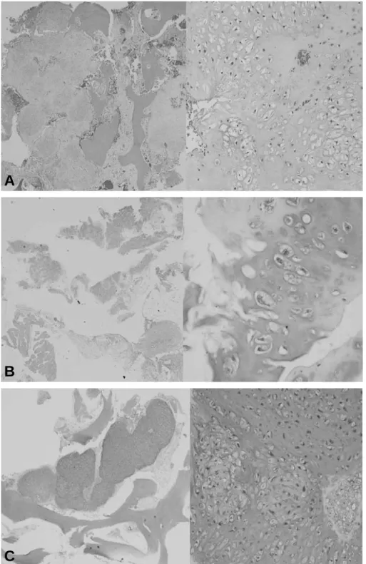
Fig. 5. Hypercellularity, plump nuclei, anisotropic cells, multinucleated cells, mitosis, and multiple chondrosarcoma cells in one lacunae with abundant cartilage matrix are noted in all 3 specimens. (A) Permanent biopsy of acromion (Left :H&E stain 40, Right :H&E stain 100). (B) Needle aspiration biopsy of humerus (Left :H&E stain 40, Right :H&E stain 200) (C) Permanent biopsy of humerus (Left :H&E stain 40, Right :H&E stain 100).
A
C
B
섬유성 골 이형성증이었다면, 증상이 없었던 근위 상완골은 단순 관찰만 하려고 하였으나, 상완골의 병변도 악성인 연골육종으로 판명되었으므로, 상완 골에 대하여도 수술적 치료를 시행하기로 결정하였 다. 상완골의 근위부 병변에 대하여 광범위 설상 절 제술을 시행한 후, 골 결손 부위에 대하여 시멘트 충 전술을 시행하였다(Fig. 4-B, C). 이전에 병소내 절제술만을 시행하였던 견봉에 위치한 병변은 재수 술하지 않고, 적극적인 추시 관찰만 하기로 하고, 재 발 소견이 의심이 되면 광범위 절제술을 시행하기로 하였다. 근위 상완골 병변에서도 grade 2의 연골육 종이 확진되었다(Fig. 5-C).
수술 후 1년에 시행한 자기공명영상에서 견봉과 근위 상완골에 위치한 시멘트는 잘 유지되었고, 재 발의 증거는 없었으며(Fig. 6), 수술 후 18개월까지 환자는 통증 없이 전 범위 견관절 운동이 가능하였 다. 향후 지속적인 추시를 통해 재발의 증거가 보이 는 즉시 광범위 절제술을 시행할 예정이다.
고 찰
연골육종은 다발성 골수종과 골육종 다음으로 흔 한 원발성 악성 골 종양이며1,2,5), 아시아에서는 두 번째로 흔하다. 대부분 저 악성도의 병변이며 드물 게 주위 림프절과 폐로 전이한다10). 다발성 연골육종 은 연골육종의 근골격계 전이와 구별하기 쉽지 않으 며, 주로 다발성 내연골종(Ollier’s disease)이나 Maffucci’s 증후군에서 발생한 것이 드물게 보고 되어 왔다7,8). 연골육종이 근골격계에 전이된 경우도 주로 질병 말기에 폐전이가 동반되면서 축성골로 전
이된다2,6,9). 본 증례에서는 오랜 병력과 함께 폐 전
이가 없고, 환측 사지의 인접 연부 조직 침범이나 축 성골 침범 소견 없이 골 내부에만 다발성 골 용해 소 견을 보여, 다발성 섬유성 이형성증으로 오진하였다.
다발성 연골육종의 분류는 Darmon 등에 의해 제 시되었는데3), 첫 번째 기준은 이환된 사지의 수이
Fig. 6. (A) Plain radiograph taken at one year postoperatively demonstrating packed cement in the acromion and the humoral greater tuberosity, and (B) No evidence of recurrence is evident in this F/U MRI taken at 1-year post- operatively.
A
B
다. 동일한 사지에 이환된 경우(monomelic)와 둘 이상의 사지 혹은 사지 이외의 부위에 이환된 경우 (non-monomelic)를 구별하였다. 단지성은 다시 동 시성(synchronous)과 전이성(metachronous)으 로, 다지성은 다발성 골연골종(Ollier’s disease)에 서 기인한 것과 그렇지 않은 것으로 나누었다. 단지 성 연골육종의 경우 각각의 병변을 독립적인 원발성 병변으로 간주하여 치료했을 경우 성공적인 치료 결 과를 보였으며, 다지성 연골육종 중 다발성 골연골 종에서 기인하지 않은 것은 예후가 불량하고, 다발 성 골연골종에서 기인한 다지성 연골 육종은 예후가 양호하다고 보고하였다3).
본 증례에서 견봉의 병변에 대하여, 전동 burr를 이용하여 철저하게 소파술을 시행한 후, 동종골 이 식과 시멘트 충전술을 시행하였으나, 이러한 치료는 grade 2 연골육종에서는 적절치 못한 것이었으므 로, 추시 관찰을 하다가 재발의 증거가 보이면 즉시 광범위 절제술을 시행하기로 하였다. 상완골 근위부 병변에 대해서는 견봉의 병변과는 별개의 병변으로 간주하였고, 광범위 설상 절제술을 시행한 후, 시멘 트 충전술을 시행하였다. 수술 후 18개월까지 재발 의 증거는 관찰되지 않았으며, 수술 전에 호소하였 던 증상도 사라졌다.
저자들이 시행한 문헌 고찰에 따르면, 관절을 사 이에 두고 관절에 근접하여 그 근위부와 원위부에 발생한 연골 육종은 지금까지 보고된 바가 없었으 며, 저자들은 이를“동시성 단지성 관절 근접 다발성 연골육종(synchronous monomelic juxta-articu- lar multicentric chondrosarcoma)”이라 명명하 였다. 또한, 각각의 병변을 독립된 것으로 간주하여 치료함으로써 아직까지는 성공적인 치료 결과를 보 였다. 본 증례에서, 견봉의 병변이 악성임을 확인함 과 동시에 견봉과 근위 상완골에 대하여 광범위 절 제술을 시행해야 하였으나, 고찰했던 바와 같이 소 파술후 골이식과 시멘트 충전술을 시행하였으며, 이
는 적절한 치료가 되지 못하였다. 이에, 지속적인 추 시 관찰을 하여, 재발의 증거가 보이면 광범위 절제 술을 시행할 예정이다.
REFERENCES
11) Dahlin DC and Henderson ED: Chondrosarcoma, a surgical and pathological problem; review of 212 Cases.Bone Joint Surg, 38-A: 1025-p38, 1956.
12) Dahlin DC and Unni KK: General aspects and data on 8542 cases. Bone tumor, 4th Ed. Thomas Springfield,: 227, 1986.
13) Damron TA, Sim FH and Unni KK: Multicentric chondrosarcoma. Clin Orthop, 328: 211-19, 1996.
14) Disler DG, Rosenberg AE, Springfield D, O’Connell JX, Rosenthal DI and Kattapuram SV: Extensive skeletal metastases from chondrosar- coma without pulmonary involvement. Skeletal Radiol, 22: 595-599, 1993.
15) Ennecking WF: Musculoskeletal Tumor Surgery.
Vol 2. New York, Churchill Livingstone: 875-997, 1983.
16) Evans HL, Ayala AG and Romsdahl MM : Prognostic factors in chondrosarcoma of bone: a clinicopathologic analysis with emphasis on histo- logic grading. Cancer, 40: 818-831, 1977.
17) Johnson JL, Webster Jr. JR and Sippy HI:
Maffucci’s syndrome (dyschondroplasia with hemangiomas). Am J Med, 28: 864-866, 1960.
18) Liu J, Hudkins PG, Swee RG and Unni KK:
Bone sarcomas associated with Ollier’s disease.
Cancer, 59: 1376-1385, 1987.
19) Marcove RC, Mike V, Hutter RV et al : Chondrosarcoma of the pelvis and upper end of the femur. An analysis of factors influencing survival time in one hundred and thirteen cases. J Bone Joint Surg., 54-A: 561-572, 1972.
10) Nakashima Y, Unni KK, Shives TC, Swee RG and Dahlin DC: Mesenchymal chondrosarcoma of bone and soft tissue. A review of 111 cases.
Cancer, 57: 2444-2453, 1986.
Synchronous Monomelic Juxta-articular Multicentric Chondrosarcoma -A Case Report-
Joo Han Oh, M.D., Jae Yoon Kim, M.D.*, Hyun Sik Gong, M.D., Woo Sung Kim, M.D. , Tae Yune Kim, M.D.
Department of Orthopaedic Surgery, Seoul National University College of Medicine, Eulji University College of Medicine*, Daejin Medical Center , Seoul, Korea
Chondrosarcoma is one of the most common types of primary bone sarcoma. With the excep- tion of the mesenchymal subtype, chondrosarcomas are usually low-grade lesions and rarely show multicentricity or distant metastasis. Only rare cases of multicentric chondrosarcomas have been reported in association with Ollier’s disease and Maffucci’s syndrome. To our knowl- edge, no report has been issued of a synchronous multicentric chondrosarcoma occurrence across a joint. We experienced a 30-year-old man with a synchronous monomelic juxta-articular multicentric chondrosarcoma across a shoulder joint in the absence of pulmonary and visceral metastases. He was treated by curettage and cement filling with allograft in the acromion and wedge resection with cement filling in the proximal humerus. At the 18-month follow-up, there was no evidence of recurrence, and the patient had full range of motion without pain.
Key Words: Chondrosarcoma, Multicentric, Monomelic, Juxta-articular
Address reprint requests to Jae Yoon Kim, M.D.
Department of Orthopaedic Surgery, Eulji University College of Medicine 281-1 Hagye 1-dong, Nowon-gu, Seoul
TEL: 82-2-970-8736, Fax: 82-2-973-3024, E-mail: [email protected]
Abstract