ISSN 2234-3806 • eISSN 2234-3814
https://doi.org/10.3343/alm.2021.41.3.318
Serum Ferritin as a Diagnostic Biomarker for Kawasaki Disease
Sung Hoon Kim , M.D.1,*, Eun Song Song , M.D.2,*, Somy Yoon , Ph.D.3, Gwang Hyeon Eom , Ph.D.3, Gaeun Kang , M.D.4, and Young Kuk Cho , M.D.5
1Department of Pediatrics, Samsung Changwon Hospital, Sungkyunkwan University School of Medicine, Changwon, Korea; 2Department of Pediatrics, Chonnam National University Hospital, Chonnam National University Medical School, Gwangju, Korea; 3Department of Pharmacology and Medical Research Center for Gene Regulation, Chonnam National University Medical School, Hwasun-gun, Jeollanam-do, Korea; 4Division of Clinical Pharmacology, Chonnam National University Hospital, Gwangju, Korea; 5Department of Pediatrics, College of Medicine Chosun University, Gwangju, Korea
Diagnosis of Kawasaki disease (KD) is occasionally delayed because it is solely based on clinical symptoms. Previous studies have attempted to identify diagnostic biomarkers for KD. Recently, patients with KD were reported to have elevated serum ferritin levels. We in- vestigated the usefulness of the serum ferritin level as a diagnostic biomarker for distin- guishing KD from other acute febrile illnesses. Blood samples were obtained from pediat- ric patients with KD (N=77) and those with other acute febrile illnesses (N=32) between December 2007 and June 2011 for measuring various laboratory parameters, including serum ferritin levels. In patients with KD, laboratory tests were performed at diagnosis and repeated at 2, 14, and 56 days after intravenous immunoglobulin treatment. At the time of diagnosis, serum ferritin levels in patients with KD (188.8 µg/L) were significantly higher than those in patients with other acute febrile illnesses (106.8 µg/L, P =0.003). The serum ferritin cut-off value of 120.8 µg/L effectively distinguished patients with KD from those with other acute febrile illnesses, with a sensitivity and specificity of 74.5% and 83.3%, respectively. Serum ferritin may be a useful biomarker to distinguish KD from other acute febrile illnesses.
Key Words: Kawasaki disease, Diagnosis, Ferritin, Biomarker
Received: March 28, 2020 Revision received: June 5, 2020 Accepted: November 28, 2020 Corresponding author:
Young Kuk Cho, M.D.
Department of Pediatrics, Chosun University Hospital, 365 Pilmun-daero, Dong-gu, Gwangju 61453, Korea Tel: +82-62-220-3040
Fax: +82-62-227-2904 E-mail: [email protected]
* These authors contributed equally to this study.
© Korean Society for Laboratory Medicine This is an Open Access article distributed under the terms of the Creative Commons Attribution Non-Commercial License (https://creativecom- mons.org/licenses/by-nc/4.0) which permits unrestricted non-commercial use, distribution, and reproduction in any medium, provided the original work is properly cited.
Kawasaki disease (KD) is an acute febrile systemic vasculitis of unknown etiology [1, 2]. Among all patients diagnosed as hav- ing KD, 15%–20% showed incomplete presentation [2]. KD di- agnosis depends on the use of adjuvant diagnostic markers, such as echocardiographic findings and laboratory biomarkers, including C-reactive protein (CRP), erythrocyte sedimentation rate (ESR), albumin, alanine aminotransferase (ALT), aspartate aminotransferase (AST), white blood cell count (WBC), hemo- globin, platelet count, and pyuria [2]. Although the laboratory findings observed in KD are not specific enough to conclusively establish a diagnosis, they can be useful, particularly in patients with a high suspicion for KD [2]. Tumor necrosis factor alpha
(TNF-α), which is released from activated macrophages under certain inflammatory conditions, induces ferritin synthesis. A se- rum ferritin level ≥200 µg/L has been associated with an incre- ased risk of myocardial infarction and, reportedly, plays an im- portant role in inflammation [3-5]. Like in patients with systemic juvenile idiopathic arthritis, elevated serum ferritin levels have been observed in patients with KD [6-8]. Serum ferritin level is being considered a useful predictor of non-responsiveness to initial intravenous immunoglobulin (IVIG) therapy in KD and a useful biomarker to distinguish between KD and systemic juve- nile idiopathic arthritis [7, 8]. However, the utility of serum ferri- tin levels as a diagnostic biomarker to distinguish between KD
2017-03-16 https://crossmark-cdn.crossref.org/widget/v2.0/logos/CROSSMARK_Color_square.svg
and other acute febrile illnesses has not been investigated. We investigated this utility by measuring serum ferritin levels in pe- diatric patients with acute- and/or convalescent-phase KD.
All procedures involving human participants were carried out in accordance with the ethical standards of the Institutional and/
or National research committee and with the 1964 Helsinki dec- laration and its later amendments or comparable ethical stan- dards. The Institutional Review Board of Chonnam National Uni- versity Hospital, Gwangju, Korea, approved this study (protocol number: I-2009-09-103).
We enrolled 77 pediatric patients (age range, 3 months–6.9 years) who were admitted to Chonnam National University Hos- pital between December 2007 and June 2011 with a diagnosis of KD. Informed written consent was obtained from the parents of all patients. All patients with KD were administered IVIG at a dose of 2 g/kg, concomitantly with medium-dose aspirin (30–50 mg/kg), during the acute phase with fever. Among the 77 pa- tients with KD who received the initial IVIG infusion, 11 (14.3%) received a second dose. At the time of admission, among the 77 patients with KD, 73 (94.8%), 69 (89.6%), 70 (90.9%), 55 (71.4%), and 45 (58.4%) showed bilateral bulbar conjunctival injection, polymorphous rash, oral mucosal changes, cervical adenopathy with lymph nodes measuring >1.5 cm in diameter, and changes in their extremities, respectively.
In addition, we enrolled 32 patients with other acute febrile diseases presenting with fever lasting more than three days. Ex- clusion criteria were the presence of hemophagocytic lympho- histiocytosis, systemic juvenile idiopathic arthritis, and viral in- fections, such as hepatitis C, avian influenza A (H5N1), dengue fever, and Mycoplasma pneumoniae pneumonia with increased serum ferritin levels [9-13].
We measured the complete blood cell count, ESR, CRP, total protein, albumin, electrolytes, blood urea nitrogen, creatinine, AST, ALT, creatine kinase (CK), CK-cardiac isoenzyme (CK-MB), myoglobin, troponin-I, N-terminal pro-brain natriuretic peptide (NT-proBNP), and ferritin in patients with KD at the time of di- agnosis and in those with other acute febrile illnesses. Blood samples (5-8 mL) was analyzed within two hours after collection [14]. In addition, we measured serum ESR, CRP, NT-proBNP, and ferritin in these patients at 2, 14, and 56 days after IVIG treatment. A microparticle enzyme immunoassay (MEIA; Ax- SYM System, Abbot Diagnostics, IL, USA) was used to quantita- tively assess serum ferritin levels.
Chi-square test was applied to assess the statistical significance of differences between independent variables and Student’s t-
of the collected data, as indicated by the Shapiro–Wilk test, to assess differences between the patients with KD and those with other acute febrile illnesses. Only white blood cell count, hemo- globin, ESR, total protein, potassium, and chloride were normally distributed. The serial changes in CRP, ESR, NT-proBNP, and ferritin, at the time of diagnosis and at 2, 14, and 56 days after IVIG treatment were analyzed by Kruskal–Wallis test, followed by Bonferroni correction. We used receiver operating characteristic (ROC) curves to determine a cut-off value to distinguish between patients with KD and patients with other acute febrile illnesses.
Continuous variables with normal distribution are expressed as the mean±standard deviation, while non-normal variables are expressed as median and range. P <0.05 was considered sta- tistically significant. The SPSS software (version 20.0, IBM SPSS Statistics for Windows, IBM Corp., Armonk, NY, USA) was used for all data analyses.
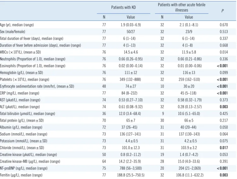
Table 1 shows the demographic features and the laboratory findings of the patients with KD and those with other acute fe- brile illnesses at the time of diagnosis. The clinical diagnoses for patients with other acute febrile illnesses (N=32) were as follows:
viral pneumonia (N=18), acute tonsillitis (N=3), acute bronchi- olitis (N=2), acute obstructive laryngitis (N=2), bacteremia (N=2), cervical lymphadenitis (N=1), acute gastroenteritis (N=1), bac- terial keratoconjunctivitis (N=1), acute otitis media (N=1), and acute gastroenteritis (N=1) (Table 1). There were no significant sex differences.
Table 2 shows changes in CRP, ESR, NT-proBNP, and ferritin, at the time of diagnosis and at 2, 14, and 56 days after IVIG treat- ment. In patients with KD, the increased ferritin levels showed a gradual drop after IVIG treatment (Table 2). There was no signif- icant sex-based difference in ferritin levels in patients with KD (male: 196.0±120.7 µg/L, vs. female: 197.0±88.1 µg/L, P =0.601).
Table 3 shows the diagnostic serum ferritin cut-off value in patients with KD and in those with other acute febrile illnesses.
We also evaluated the platelet count, ESR, CRP, and NT-proBNP cut off values, which have been identified as potential diagnos- tic markers in previous reports (Table 3) [15-17]. The serum ferritin cut-off value of 120.8 µg/L effectively distinguished pa- tients with KD from those with other acute febrile illnesses, with a sensitivity and specificity of 74.5% and 83.3%, respectively (AUC=0.830, 95% confidence interval: 0.704-0.955) (Table 3).
In our study, serum ferritin levels were significantly higher in patients with KD than in those with other acute febrile illnesses, indicating that the serum ferritin level can serve as a useful di- agnostic biomarker for KD. We determined the optimal serum
Table 1. Demographic features and laboratory findings in patients with KD and patients with other acute febrile illnesses at the time of diag- nosis
Patients with KD Patients with other acute febrile
illnesses P
N Value N Value
Age (yr), median (range) 77 1.9 (0.03–6.9) 32 2.1 (0.1–8.1) 0.670
Sex (male/female) 77 50/27 32 23/9 0.513
Total duration of fever (days), median (range) 77 6 (1–14) 32 6 (1–14) 0.337
Duration of fever before admission (days), median (range) 77 4 (1–13) 32 4 (1–8) 0.668
WBCs (×109/L), (mean±SD) 76 14.5±4.6 32 11.9±5.8 0.014
Neutrophils (Proportion of 1.0), median (range) 76 0.66 (0.26–0.95) 32 0.66 (0.21–0.86) 0.336 Eosinophils (Proportion of 1.0), median (range) 76 0.02 (0.00–0.14) 32 0.01 (0.00–0.06) <0.001
Hemoglobin (g/L), (mean±SD) 76 111±12 32 116±13 0.099
Platelets (×109/L), median (range) 76 349 (132–888) 32 259 (162–510) <0.001
Erythrocyte sedimentation rate (mm/hr), (mean±SD) 48 74±27 10 30±20 <0.001
CRP (mg/L), median (range) 77 84 (8–232) 32 45 (5–118) <0.001
AST (µkat/L), median (range) 74 0.53 (0.27–7.10) 32 0.58 (0.32–1.79) 0.373
ALT (µkat/L), median (range) 74 0.61 (0.08–9.32) 32 0.28 (0.13–2.57) 0.003
Total bilirubin (µmol/L), median (range) 36 12.0 (3.4–68.4) 9 10.6 (5.1–65.0) 0.425
Total protein (g/L), (mean±SD) 70 65±7 30 66±5 0.217
Albumin (g/L), median (range) 72 37 (26–45) 31 40 (20–44) 0.050
Sodium (mmol/L), median (range) 73 136 (127–141) 31 137 (130–143) 0.064
Potassium (mmol/L), (mean±SD) 73 4.4±0.5 31 4.2±0.5 0.075
Chloride (mmol/L), (mean±SD) 73 101.0±12.3 31 103.9±3.2 0.017
Creatine kinase (µkat/L), median (range) 50 0.8 (0.2–11.2) 19 1.4 (0.7–4.2) 0.053
Creatine kinase-MB (µg/L), median (range) 64 14.2 (2.2–35.9) 28 15.0 (4.0–33.6) 0.391
NT-proBNP (ng/L), median (range) 75 788 (56–3,500) 20 204 (21–2,069) <0.001
Ferritin (µg/L), median (range) 77 188.8 (25.5–750.5) 32 106.8 (11.1–632.2) 0.003
The statistical significance of differences between males and females was assessed by the Chi-square test. White blood cell count, hemoglobin, ESR, total protein, potassium, and chloride were analyzed using Student’s t-test. Results of the other laboratory tests were analyzed using the Mann–Whitney U-test.
Abbreviations: KD, Kawasaki disease; WBC, white blood cell count; CRP, C-reactive protein; AST, aspartate aminotransferase; ALT, alanine aminotransferase;
creatine kinase-MB, cardiac isoenzyme of creatine kinase; NT-proBNP, N-terminal pro-brain natriuretic peptide; SD, standard deviation.
Table 2. Comparison of laboratory findings between patients with KD at the time of diagnosis and at 2, 14, and 56 days after IVIG treatment and patients with other acute febrile illnesses
Laboratory Findings
Patients with KD
Patients with other acute febrile illnesses At the time of
diagnosis 2 days after
treatment 14 days after
treatment 56 days after treatment
N Value N Value N Value N Value N Value
CRP (mg/L), median (range) 77 84 (8–232) 77 22 (2–164) 74 2 (0–31) 65 0 (0–1) 32 45 (5–118)
ESR (mm/hr), (mean±SD) 48 74±27 61 78±30 74 44±27 65 11±12 10 30±20
NT-proBNP (ng/L), median (range) 75 788 (56–3,500) 76 569 (14–2,3367) 72 91 (16–648) 7 133 (22–348) 20 204 (21–2,069) Ferritin (µg/L), median (range) 77 188.8 (25.5–750.5 71 159.5 (4.8–483.0) 68 78.2 (12.4–271.1) 14 26.1 (11.0–133.5) 32 106.8 (11.1–632.2) Abbreviations: KD, Kawasaki disease; IVIG, intravenous immunoglobulin; CRP, C-reactive protein, ESR, erythrocyte sedimentation rate; NT-proBNP, N-termi- nal pro-brain natriuretic peptide; SD, standard deviation.
febrile illnesses to be 120.8 µg/L.
Ferritin is an acute-phase reactant that is utilized in clinical practice as a serum biomarker [3]. Ferritin synthesis is mark- edly induced by TNF-α and IL-1α; thus, serum ferritin levels are elevated in patients with certain inflammatory conditions [7]. El- evated serum ferritin levels have also been implicated in the de- velopment of diabetic microvascular disease through interaction with vascular endothelial growth factor (VEGF), which is involved in the pathogenesis of vasculitis [7, 18]. VEGF is also associated with the formation of coronary artery lesions (CALs) in patients with KD; however, an association between VEGF and the serum ferritin level in KD has not been established [7, 19].
While the serum ferritin level is elevated in patients with KD, markedly elevated serum ferritin level is not a common feature in KD, although it has been observed in KD patients with con- comitant hemophagocytic lymphohistiocytosis and in adult pa- tients with KD [3, 9, 20]. Nasir, et al. [9] reported three neonates who presented with clinical features suggestive of KD, with high serum ferritin levels. In their series, the patients demonstrated prompt and complete resolution of symptoms within 48 hours of IVIG treatment, without recurrence or CALs at the six-month follow-up [9]. They suggested that children and neonates with KD might have high serum ferritin levels and that serum ferritin could be a useful diagnostic biomarker for KD [9]. Mizuta, et al.
[8] reported higher mean ferritin levels in patients with systemic juvenile idiopathic arthritis (1,189 µg/L [range, 63–68,310]) than in patients with KD (147.5 µg/L [range, 14–2,376]).
In our study, WBC, CRP, and NT-proBNP levels were also sig- nificantly higher in patients with KD than in patients with other
15-17].
There were some limitations in our study. (i) Some data per- taining to patient follow-up were missing, perhaps because par- ents refused to permit blood draws as their children began to recover and appear healthy. (ii) The patients with acute febrile illnesses other than KD had heterogeneous febrile illnesses, which may have affected the measurement of serum ferritin levels ow- ing to differences in inflammation severity. (iii) The number of patients with acute febrile illnesses other than KD was signifi- cantly smaller than that of patients with KD. Therefore, large prospective investigations are required for obtaining a more ac- curate cutoff for ferritin as a diagnostic biomarker.
In conclusion, this was the first study to compare serial serum ferritin levels between patients with KD and those with other acute febrile illnesses. The serum ferritin level may be a useful biomarker to distinguish KD from other acute febrile illnesses.
ACKNOWLEDGEMENTS
None.
AUTHOR CONTRIBUTIONS
Kim SH, Song ES, Kang G, and Cho YK made substantial contri- butions to study planning and design and writing of the manu- script; Yoon S and Eom GH performed data collection, statistical analysis, and provided valuable feedback. Kim SH and Song ES contributed equally to all aspects of this paper. All authors ap- proved the final manuscript submitted and agreed to be account- able for all aspects of the work.
CONFLICTS OF INTEREST
The authors declare that they have no conflict of interest.
RESEARCH FUNDING
This study was supported by a grant (NRF-2015R1D1A1A01059017) provided by the Basic Science Research Program through the National Research Foundation of Korea (NRF) funded by the Ministry of Education, Korea.
ORCID
Sung Hoon Kim https://orcid.org/0000-0002-2128-2874 Table 3. Comparison of diagnostic serum ferritin, platelet count,
ESR, CRP, and NT-proBNP cut-off values between patients with KD and those with other acute febrile illnesses
Diagnostic markerCut-off value
Area under curve
95%
Confidence Interval
Sensi- tivity (%)
Speci- ficity
(%) P
Ferritin (µg/L) 120.8 0.830 0.704–0.955 74.5 83.3 0.009 Platelets (×109/L) 279.5 0.750 0.526–0.974 76.6 83.3 0.048 ESR (mm/hr) 37 0.897 0.741–1.000 93.6 83.3 0.002 CRP (mg/L) 44 0.812 0.013–0.988 78.7 83.3 0.013 NT-proBNP (ng/L) 571.3 0.848 0.711–0.984 66.0 83.3 0.006 Abbreviations: ESR, erythrocyte sedimentation rate; CRP, C-reactive protein;
NT-proBNP, N-terminal pro-brain natriuretic peptide; KD, Kawasaki disease.
Receiver operating characteristic (ROC) curves were used to determine a cut-off value to distinguish between patients with KD and patients with other acute febrile illnesses.
Somy Yoon https://orcid.org/0000-0001-7393-5040 Gwang Hyeon Eom https://orcid.org/0000-0002-7904-8503 Gaeun Kang https://orcid.org/0000-0001-9841-1139 Young Kuk Cho https://orcid.org/0000-0001-6811-2883
REFERENCES
1. Kawasaki T, Kosaki F, Okawa S, Shigematsu I, Yanagawa H. A new in- fantile acute febrile mucocutaneous lymph node syndrome (MLNS) pre- vailing in Japan. Pediatrics 1974;54:271-6.
2. McCrindle BW, Rowley AH, Newburger JW, Burns JC, Bolger AF, Gewitz M, et al. Diagnosis, treatment, and long-term management of Kawasaki disease: a scientific statement for health professionals from the Ameri- can Heart Association. Circulation 2017;135:e927-99.
3. Knovich MA, Storey JA, Coffman LG, Torti SV, Torti FM. Ferritin for the clinician. Blood Rev 2009;23:95-104.
4. de Godoy MF, Takakura IT, Machado RD, Grassi LV, Nogueira PR. Se- rum ferritin and obstructive coronary artery disease: angiographic cor- relation. Arq Bras Cardiol 2007;88:430-3.
5. Salonen JT, Nyyssönen K, Korpela H, Tuomilehto J, Seppänen R, Sa- lonen R. High stored iron levels are associated with excess risk of myo- cardial infarction in eastern Finnish men. Circulation 1992;86:803-11.
6. Takahara T, Shimizu M, Nakagishi Y, Kinjo N, Yachie A. Serum IL-18 as a potential specific marker for differentiating systemic juvenile idiopathic arthritis from incomplete Kawasaki disease. Rheumatol Int 2015;35:81-4.
7. Yamamoto N, Sato K, Hoshina T, Kojiro M, Kusuhara K. Utility of ferritin as a predictor of the patients with Kawasaki disease refractory to intra- venous immunoglobulin therapy. Mod Rheumatol 2015;25:898-902.
8. Mizuta M, Shimizu M, Inoue N, Kasai K, Nakagishi Y, Takahara T, et al.
Serum ferritin levels as a useful diagnostic marker for the distinction of systemic juvenile idiopathic arthritis and Kawasaki disease. Mod Rheu- matol 2016;26:929-32.
9. Nasir A, Al Tatari H, Hamdan MA. Very high serum ferritin levels in three newborns with Kawasaki-like illness. Paediatr Child Health 2012;17:201-4.
10. Ackerman Z, Pappo O, Ben-Dov IZ. The prognostic value of changes in serum ferritin levels during therapy for hepatitis C virus infection. J Med Virol 2011;83:1262-8.
11. Soepandi PZ, Burhan E, Mangunnegoro H, Nawas A, Aditama TY, Par- takusuma L, et al. Clinical course of avian influenza A (H5N1) in patients at the Persahabatan Hospital, Jakarta, Indonesia, 2005-2008. Chest 2010;138:665-73.
12. Chaiyaratana W, Chuansumrit A, Atamasirikul K, Tangnararatchakit K.
Serum ferritin levels in children with dengue infection. Southeast Asian J Trop Med Public Health 2008;39:832-6.
13. Kawamata R, Yokoyama K, Sato M, Goto M, Nozaki Y, Takagi T, et al.
Utility of serum ferritin and lactate dehydrogenase as surrogate markers for steroid therapy for Mycoplasma pneumoniae pneumonia. J Infect Chemother 2015;21:783-9.
14. Shin S, Oh J, Park H. Comparison of three blood collection tubes for 35 biochemical analytes: the Becton Dickinson Barricor Tube, serum sepa- rating tube, and plasma separating tube. Ann Lab Med 2021;41:114-9.
15. Dahdah N, Siles A, Fournier A, Cousineau J, Delvin E, Saint-Cyr C, et al.
Natriuretic peptide as an adjunctive diagnostic test in the acute phase of Kawasaki disease. Pediatr Cardiol 2009;30:810-17.
16. Lee H, Kim H, Kim HS, Sohn S. NT-pro BNP: a new diagnostic screen- ing tool for Kawasaki disease. Korean J Pediatr 2006;49:539-44.
17. Lee DW, Kim YH, Hyun MC, Kwon TC, Lee SB. NT-proBNP as a useful tool in diagnosing incomplete Kawasaki disease. Korean J Pediatr 2010;
53:519-24.
18. Guo L, Jiang F, Tang YT, Si MY, Jiao XY. The association of serum vascu- lar endothelial growth factor and ferritin in diabetic microvascular dis- ease. Diabetes Technol Ther 2014;16:224-34.
19. Ohno T, Yuge T, Kariyazono H, Igarashi H, Joh-o K, Kinugawa N, et al.
Serum hepatocyte growth factor combined with vascular endothelial growth factor as a predictive indicator for the occurrence of coronary artery lesions in Kawasaki disease. Eur J Pediatr 2002;161:105-11.
20. Cunha BA, Pherez FM, Alexiadis V, Gagos M, Strollo S. Adult Kawasa- ki’s disease with myocarditis, splenomegaly, and highly elevated serum ferritin levels. Heart Lung 2010;39:164-72.