ISSN 2234-3806 • eISSN 2234-3814
68 www.annlabmed.org https://doi.org/10.3343/alm.2020.40.1.68 Ann Lab Med 2020;40:68-71
https://doi.org/10.3343/alm.2020.40.1.68
Brief Communication
Clinical Microbiology
Evaluating Diagnostic Tests for Helicobacter pylori
Infection Without a Reference Standard: Use of Latent Class Analysis
Dong Wook Jekarl , M.D.1,2, Hyunyu Choi , B.S.3, Ji Yeon Kim , M.T.3, Seungok Lee , M.D.2,3, Tae Geun Gweon , M.D.4, Hae Kyung Lee , M.D.2,5, and Yonggoo Kim , M.D.1,2
1Department of Laboratory Medicine, Seoul St. Mary’s Hospital, College of Medicine, The Catholic University of Korea, Seoul, Korea; 2Laboratory for Development and Evaluation Center, The Catholic University of Korea, Seoul, Korea; 3Department of Laboratory Medicine, Incheon St. Mary’s Hospital, College of Medicine, The Catholic University of Korea, Seoul, Korea; 4Department of Internal Medicine, Incheon St. Mary’s Hospital, College of Medicine, The Catholic University of Korea, Seoul, Korea; 5Department of Laboratory Medicine, Uijeongbu St. Mary’s Hospital, College of Medicine, The Catholic University of Korea, Seoul, Korea
Evaluation of diagnostic tests requires reference standards, which are often unavailable.
Latent class analysis (LCA) can be used to evaluate diagnostic tests without reference standards, using a combination of observed and estimated results. Conditionally indepen- dent diagnostic tests for Helicobacter pylori infection are required. We used LCA to con- struct a reference standard and evaluate the capability of non-invasive tests (stool antigen test and serum antibody test) to diagnose H. pylori infection compared with the conven- tional method, where histology is the reference standard. A total of 96 healthy subjects with endoscopy histology results were enrolled from January to July 2016. Sensitivity and specificity were determined for the LCA approach (i.e., using a combination of three tests as the reference standard) and the conventional method. When LCA was used, sensitivity and specificity were 83.8% and 99.4% for histology, 80.0% and 81.9% for the stool anti- gen test, and 63.6% and 89.3% for the serum antibody test, respectively. When the con- ventional method was used, sensitivity and specificity were 75.8% and 71.1% for the stool antigen test and 77.7% and 60.7% for the serum antibody test, respectively. LCA can be applied to evaluate diagnostic tests that lack a reference standard.
Key Words: Helicobacter pylori, Latent class analysis, Stool antigen test, Reference stan- dard, Serum antibody test, Diagnosis
Received: April 28, 2019 Revision received: June 10, 2019 Accepted: July 30, 2019
Corresponding author: Hae Kyung Lee, M.D.
Department of Laboratory Medicine, College of Medicine, The Catholic University of Korea, Uijongbu St. Mary’s Hospital, 271 Cheonbo-ro, Uijeongbu 11765, Korea Tel: +82-31-820-3959
Fax: +82-31-847-6226 E-mail: [email protected]
© Korean Society for Laboratory Medicine This is an Open Access article distributed under the terms of the Creative Commons Attribution Non-Commercial License (http://creativecom- mons.org/licenses/by-nc/4.0) which permits unrestricted non-commercial use, distribution, and reproduction in any medium, provided the original work is properly cited.
Helicobacter pylori is a microaerophilic gram-negative flagellate associated with gastritis, peptic ulcer disease, mucosa-associ- ated lymphoid tissue lymphoma, and gastric cancer [1, 2]. Over half of the world’s population, including Korean population, has been infected by H. pylori [3, 4]. Although endoscopic interven- tion and eradication of H. pylori has decreased the seropreva- lence rate of H. pylori infection from 66.9% (1998) to 51.0%
(2015), the Korean national prevalence is similar to the average worldwide prevalence [4]. H. pylori infection is diagnosed using
both invasive methods, such as culturing, histology, and rapid urease tests, and non-invasive methods, including the urea breath test, serological tests, and stool antigen test [5, 6]. Non-invasive diagnostic methods are considered adequate to reflect the global infection state as they cover ≥0.5 m2 of the gastric mucosa [7].
Histology is regarded as the reference standard for diagnosing H. pylori infection; however, its accuracy is affected by biopsy site, size, number of biopsy specimens, staining method, and drug history [5, 8]. We therefore believe that a reference stan-
1 / 1 CROSSMARK_logo_3_Test
2017-03-16 https://crossmark-cdn.crossref.org/widget/v2.0/logos/CROSSMARK_Color_square.svg
Jekarl DW, et al.
LCA for H. pylori infection diagnostic tests
https://doi.org/10.3343/alm.2020.40.1.68 www.annlabmed.org 69
dard created using statistical methods would be more accurate than the current reference standard.
Latent class analysis (LCA) can be used to evaluate diagnostic tests without reference standards, by creating a reference stan- dard using a combination of observed and estimated results [9].
LCA reveals hidden groups or disease states in multivariate di- chotomous or categorical data [10, 11]. One limitation of LCA is the underlying assumption that the tests are independent of each other, raising the possibility that there are more than two latent classes in the data.
We examined whether LCA can be used to construct a refer- ence standard to diagnose H. pylori infection through a combi- nation of results from histology, a stool antigen test using an im- munochromatographic method (Ag-ICA, BioTracer H. pylori Ag Rapid Card; NanoEntek, Seoul, Korea), and a serum antibody test using an immunochromatographic method (Ab-ICA, Bio- Tracer H. pylori Rapid Card; NanoEntek). We compared the per- formance of these tests under LCA and under the conventional method, in which histology is the reference standard.
This retrospective study was approved by the Institutional Re- view Board of Incheon St. Mary’s Hospital, Incheon, Korea (XC- 15DDME0103U). Informed consent was waived as the study posed only minimal risk to the subjects. A cohort of 96 healthy subjects (median age: 63 years [range: 51–83 years]; 50 men and 46 women) undergoing a routine health check-up were en- rolled from January to July 2016 at Incheon St. Mary’s Hospital.
Stool and remnant serum specimens after blood tests with a volume of more than 1 mL were collected in the same day that the subjects underwent endoscopy and biopsy and were stored at -20°C. The serum was isolated in the same day of blood col- lection. The stool was thawed for the H. pylori Ag-ICA and was then re-stored at -20°C, in case of future re-testing. H. pylori
IgG, IgM, and IgA antibody tests were performed. Endoscopic and histology findings were reviewed from electronic medical records.
The H. pylori Ag-ICA was performed according to the manu- facturer’s instructions. The test utilizes a monoclonal antibody against H. pylori antigens. A swab was dipped into the stool spec- imen and then mixed with the 100 mM Tris buffer in the con- tainer. Three to five drops (120–150 µL) of the buffer-diluted stool specimen mixture were passed through a filtered tip and then placed in the specimen port of the test cassette. The ap- pearance of a red line in the interpretation window after 10 min- utes at room temperature (18–25°C) indicated a positive control band; an additional red band appeared if the specimen con- tained H. pylori. To determine seroprevalence, we examined anti-H. pylori IgG, IgA, and IgM antibodies, using the Ab-ICA, wherein three drops (120 µL) of serum specimen were applied to the port of the test cassette and interpreted after 10 minutes.
The positive or negative test result of biopsy, Ag-ICA, and Ab- ICA was entered in the model. Unknown disease state, that is, the subclass with and without H. pylori infection, could be a hidden or latent class. LCA assumes that tests are conditionally independent, and the data fit the model. A two-class model with two latent variables, H. pylori infection and H. pylori non-infec- tion, and a three-class model with infection, non-infection, and intermediate state (indeterminate state), were considered. Model fit was evaluated using the Bayesian information criterion (BIC), Akaike information criterion (AIC), and Pearson goodness of fit and likelihood ratios. The BIC and AIC values are unitless, and lower values are considered for model selection. For the Pear- son goodness of fit and likelihood ratios, a higher P is consid- ered for model selection. This analysis was performed using the R package, poLCA. The sensitivity, specificity, positive predictive
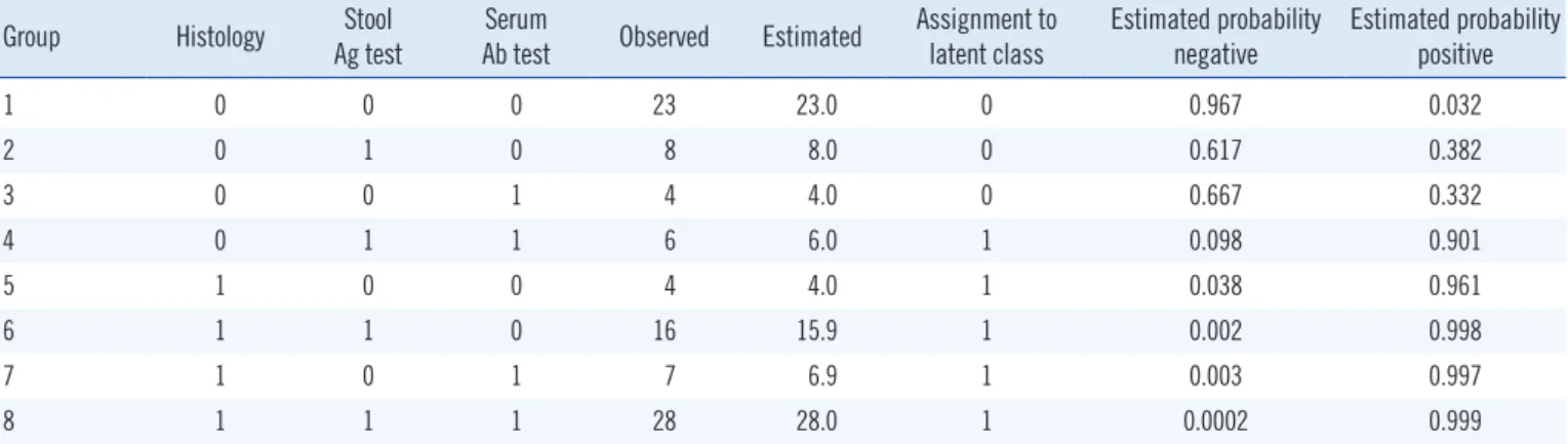
Table 1.Tested data, expected values, assignment to latent class, and probability of the assignment class based on a two-class model Group Histology Stool
Ag test
Serum
Ab test Observed Estimated Assignment to latent class
Estimated probability negative
Estimated probability positive
1 0 0 0 23 23.0 0 0.967 0.032
2 0 1 0 8 8.0 0 0.617 0.382
3 0 0 1 4 4.0 0 0.667 0.332
4 0 1 1 6 6.0 1 0.098 0.901
5 1 0 0 4 4.0 1 0.038 0.961
6 1 1 0 16 15.9 1 0.002 0.998
7 1 0 1 7 6.9 1 0.003 0.997
8 1 1 1 28 28.0 1 0.0002 0.999
Abbreviations: 0, negative result; 1, positive result; Ab, antibody; Ag, antigen.
Jekarl DW, et al.
LCA for H. pylori infection diagnostic tests
70 www.annlabmed.org https://doi.org/10.3343/alm.2020.40.1.68 values, and negative predictive values and accuracies with 95%
confidence interval (CI) were calculated using the randomLCA package [12-14].
The overall prevalence of H. pylori infection based on biopsy was 57.3% (55/96): 62.0% (31/50) in men and 52.1% (24/46) in women. It did not differ significantly between subjects <60 years and those ≥60 years (51.3% [19/37] vs 61.0% [36/59], P =0.868). The seroprevalence of H. pylori infection was 46.8%
(45/96).
The two-class model had better values (BIC, 384.5; AIC, 366.6;
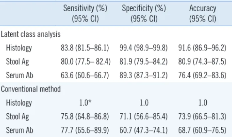
P from Pearson goodness of fit, 0.000019; P from likelihood ra- tio, 0.000019) than the three-class model (BIC, 402.8; AIC, 374.6; P from Pearson goodness of fit, 1.377 ×10⁻¹⁰; P from likelihood ratio, 1.379×10⁻¹⁰). Accordingly, we selected the two- class model. The observed and estimated distributions based on results of three tests are listed in Table 1. When LCA was used, histology had the highest sensitivity, followed by the Ag- ICA and Ab-ICA. Histology exhibited the highest specificity, fol- lowed by the serum antibody and stool antigen tests (Table 2).
Unexpectedly, sensitivity and specificity were higher for all tests under LCA than the conventional method, except for sensitivity of the serum antibody test.
Our results showed that diagnostic capability was 5–10% higher for the LCA two-class model than for the conventional method.
Thus, using LCA could support diagnosis in the absence of a reference standard. The results of our two-class LCA model are in line with a study showing that the sensitivity and specificity of histology are 85–95% and almost 100%, respectively [5]. The sensitivity and specificity of Ag-ICA vary widely: 48.9–92.2%
and 88.9–94.4%, respectively [5, 15]. Sensitivity and specificity of Ab-ICAs are 55.6–97.8% and 60.3–96.8%, respectively [5].
The low specificity of Ag-ICA (71.1%) in our study was due to the use of histology as the reference standard; most previous studies used a composite reference standard [5, 6]. Despite its low specificity, Ag-ICA can be used in combination with the Ab- ICA in LCA. If the serum antibody test had concordant results with the stool antigen test, the LCA might show higher specificity than the conventional method, whereas discordant results might result in similar or lower specificity.
In previous studies, histology results were combined with those of other tests to construct a composite reference standard [6]. A combination of tests has been used to classify definite infection or probable infection based on the number of positive test re- sults [6, 16]. LCA and the conventional method provided similar results, as shown in Table 2. However, the 5–10% increase in sensitivity and specificity indicates that LCA has improved ability to evaluate H. pylori infection diagnostic tests. Thus, LCA might provide a reliable reference standard in the absence of invasive methods for diagnosing H. pylori infection.
A limitation of this study was that the sample size was relatively small. Further, information on the number and site of the biopsy specimens was not available, which might affect the positive rate of H. pylori infection.
In conclusion, LCA could be applied to evaluate diagnostic tests that lack a reference standard. Sensitivity and specificity increased using the LCA, except for the sensitivity of serum an- tigen tests.
Acknowledgements
We thank Sunmi Han and Jiyeon Sung for technical support. All reagents were provided by NanoEntek.
Author Contributions
DWJK analyzed the data and wrote the manuscript. HC and JYK collected the samples and performed tests. GTG and SL analyzed the data and reviewed medical records. YK and HKL analyzed the data, reviewedthe manuscript, and supervised this study.
Conflicts of Interest
None declared.
Table 2. Performance of diagnostic tests for Helicobacter pylori in- fection according to latent class analysis based on a two-class mod- el (performed without a reference standard) and the conventional method
Sensitivity (%)
(95% CI) Specificity (%)
(95% CI) Accuracy (95% CI) Latent class analysis
Histology 83.8 (81.5–86.1) 99.4 (98.9–99.8) 91.6 (86.9–96.2) Stool Ag 80.0 (77.5– 82.4) 81.9 (79.5–84.2) 80.9 (74.3–87.5) Serum Ab 63.6 (60.6–66.7) 89.3 (87.3–91.2) 76.4 (69.2–83.6) Conventional method
Histology 1.0* 1.0 1.0
Stool Ag 75.8 (64.8–86.8) 71.1 (56.6–85.4) 73.9 (66.5–81.3) Serum Ab 77.7 (65.6–89.9) 60.7 (47.3–74.1) 68.7 (60.9–76.5)
*Histology results were used as the reference standard for the conventional method.
Abbreviations: CI, confidence interval; Ag, antigen; Ab, antibody.
Jekarl DW, et al.
LCA for H. pylori infection diagnostic tests
https://doi.org/10.3343/alm.2020.40.1.68 www.annlabmed.org 71
Research Funding
This study was partially supported by Incheon St. Mary’s Hospi- tal Fund for basic research.
ORCID
Dong Wook Jekarl https://orcid.org/0000-0002-6269-5501 Hyunyu Choi https://orcid.org/0000-0003-1318-0250 Ji Yeon Kim https://orcid.org/0000-0001-9465-8484 Seungok Lee https://orcid.org/0000-0003-4538-8427 Tae Geun Gweon https://orcid.org/0000-0002-0884-7228 Hae Kyung Lee https://orcid.org/0000-0001-8545-9272 Yonggoo Kim https://orcid.org/0000-0003-2808-3795
REFERENCES
1. Kabir S. The role of interleukin-17 in the Helicobacter pylori induced in- fection and immunity. Helicobacter 2011;16:1-8.
2. Bjorkman DJ and Steenblik M. Best practice recommendations for diag- nosis and management of Helicobacter pylori - synthesizing the guide- lines. Curr Treat Options Gastroenterol 2017;15:648-59.
3. Hooi JKY, Lai WY, Ng WK, Suen MMY, Underwood FE, Tanyingoh D, et al. Global prevalence of Helicobacter pylori infection: systematic review and meta-analysis. Gastroenterol 2017;153:420-9.
4. Lee JH, Choi KD, Jung HY, Baik GH, Park JK, Kim SS, et al. Seropreva- lence of Helicobacter pylori in Korea: a multicenter, nationwide study conducted in 2015 and 2016. Helicobacter 2018;23:e12463.
5. Wang YK, Kuo FC, Liu CJ, Wu MC, Shih HY, Wang SS, et al. Diagnosis of Helicobacter pylori infection: current options and developments. World J Gastroenterol 2015;21:11221-35.
6. Jekarl DW, An YJ, Lee S, Lee J, Kim Y, Park YJ, et al. Evaluation of a new- ly developed rapid stool antigen test using an immunochromatographic assay to detect Helicobacter pylori. Jpn J Infect Dis 2013;66:60-4.
7. Hirschl AM and Makristathis A. Non-invasive Helicobacter pylori diag- nosis: stool or breath tests? Dig Liver Dis 2005;37:732-4.
8. Malfertheiner P, Megraud F, O’Morain CA, Gisbert JP, Kuipers EJ, Axon AT, et al. Management of Helicobacter pylori infection–the Maastricht V/
Florence consensus report. Gut 2017;66:6-30.
9. Rutjes AWS, Reitsma JB, Coomarasamy A, Khan KS, Bossyut PMM.
Evaluation of diagnostic test when there is no gold standard. A review of methods. Health Technol Assess 2007;11:1-86.
10. Rindskopf D and Rindskopf W. The value of latent class analysis in medi- cal diagnosis. Stat Med 1986;5:21-7.
11. Wiegand RE, Cooley G, Goodhew B, Banniettis N, Kohlhoff S, Gwyn S, et al. Latent class modeling to compare testing platforms for detection of antibodies against the Chlamydia trachomatis antigen Pgp3. Sci Rep 2018;8:4232.
12. R Core Team. R: a language and environment for statistical computing.
R Foundation for Statistical Computing, Vienna, Austria. http://www.r- project.org (Updated on Jun 2017).
13. Linzer DA and Lewis JB. poLCA: an R package for polytomous variable latent class analysis. J Stat Softw 2011;42:1-29.
14. Beath KJ and Heller GZ. Latent trajectory modelling of multivariate bina- ry data. Stat Model 2009;9:199-213.
15. Kalali B, Formichella L, Gerhard M. Diagnosis of Helicobacter pylori:
changes towards the future. Diseases 2015;3:122-35.
16. Thijs JC, van Zwet AA, Thijs WJ, Oey HB, Karrenbeld A, Stellaard F, et al. Diagnostic tests for Helicobacter pylori: a prospective evaluation of their accuracy, without selecting a single test as the gold standard. Am J Gastroenterol 1996;91:2125-9.