Copyright © 2018 Korean Neurological Association 259
JCN
Open AccessIs Corticospinal Tract Degeneration Caused by Sjögren Syndrome?
Dear Editor,
A 36-year-old woman with a history of dry eyes and recurrent oral ulcers presented with an 8-month history of progressive motor weakness of both legs. She was documented as ex- periencing mild weakness (4+/5) of both legs with no sensory loss 2 months after onset.
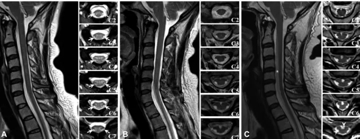
Cervical MRI revealed a T2 hyperintense lesion from C2 to C7 without cord swelling or con- trast enhancement (Fig. 1A). The rest of the spinal cord and the brain MRI were normal. A cerebrospinal fluid (CSF) examination was unrevealing. She was treated with intravenous and oral corticosteroids based on the presumptive diagnosis of transverse myelitis (TM).
Her motor weakness continued to worsen. On admission examination, she showed spas- tic paraplegia with 3/5 strength with no sensory or bladder/bowel signs or symptoms. Her deep tendon reflexes were all brisk, and the Babinski sign was positive bilaterally. Follow-up cervical MRI revealed focal areas of atrophy along the bilateral lateral columns, with spar- ing of the posterior columns (Fig. 1B). Workup to exclude metabolic or infectious causes of myelitis were unremarkable. Electromyography findings excluded amyotrophic lateral sclero- sis, and no pathologic variants related to hereditary spastic paraplegia were found on whole- exome sequencing. Serologic studies revealed positive anti-Ro/SSA (102.3 U/mL), IgG β2- glycoprotein 1 antibody, and anticardiolipin antibody, and negative for the AQP4 antibody.
The ocular staining scores were 4/3 in both eyes, and salivary scintigraphy showed delayed ex- cretion in the parotid and submandibular glands. A salivary gland biopsy confirmed chronic lymphocytic sialadenitis, consistent with primary Sjögren syndrome (pSS). The patient was ultimately diagnosed with progressive cervical myelopathy due to pSS.
Her paraplegia gradually worsened (1/5) despite continued immunotherapy including corticosteroids and cyclophosphamide. Cervical MRI performed 1 year later revealed prom- inent atrophy in the regions of the lateral and anterior corticospinal tracts (CSTs) bilaterally (Fig. 1C). Her symptoms were confined to pure motor paraplegia during the 5-year follow- up after the treatment.
This case fulfilled the diagnostic criteria for definite pSS,1 with selective atrophy of CSTs following chronic progressive cervical myelopathy, which has not been reported previously. In previous reports of myelitis due to pSS, patients presented with various courses including chronic progressive myelitis, acute or subacute TM, and multiple sclerosis-like diseases.2 There is one previous report of pSS presenting as selective atrophy of dorsal columns within the cervi- cal cord.3 The mechanism underlying spinal cord atrophy in the present case might have been Wallerian degeneration or delayed axonal loss following demyelination from chronic pro- gressive cervical myelitis.3,4 Notably, the MRI and CSF examinations revealed no evidence of inflammation, and her disease course appeared to be unresponsive to corticosteroids, which suggests noninflammatory mechanisms as work.
There are numerous accounts of neurodegeneration in pSS and other systemic rheuma- tologic diseases without evidence of inflammation,5 suggesting that pSS could alone have Tai-Seung Nama,b
Michael Levyc Sang-Hoon Kimb Kyung-Wook Kangb Byoung Joon Kimd Seung-Han Leea,b
a Department of Neurology, Chonnam National University Medical School, Gwangju, Korea
b Department of Neurology,
Chonnam National University Hospital, Gwangju, Korea
c Department of Neurology, Johns Hopkins University, Baltimore, MD, USA
d Department of Neurology, Samsung Medical Center, Sungkyunkwan University School of Medicine, Seoul, Korea
pISSN 1738-6586 / eISSN 2005-5013 / J Clin Neurol 2018;14(2):259-260 / https://doi.org/10.3988/jcn.2018.14.2.259
Received December 5, 2017 Revised January 24, 2018 Accepted January 25, 2018 Correspondence Michael Levy, MD, PhD Department of Neurology, Johns Hopkins University, Pathology 509, 600 N. Wolfe Street, Baltimore, MD 21287, USA Tel +1-443-287-4612 Fax +1-888-523-4168 E-mail mlevy@jhmi.edu Tai-Seung Nam, MD, PhD Department of Neurology, Chonnam National University Medical School, 42 Jebong-ro, Dong-gu, Gwangju 61469, Korea Tel +82-62-220-6171 Fax +82-62-236-0839 E-mail nts0022@hanmail.net
cc This is an Open Access article distributed under the terms of the Creative Commons Attribution Non-Com- mercial License (http://creativecommons.org/licenses/by-nc/4.0) which permits unrestricted non-commercial use, distribution, and reproduction in any medium, provided the original work is properly cited.
LETTER TO THE EDITOR
260 J Clin Neurol 2018;14(2):259-260
Corticospinal Tract Degeneration and Sjögren Syndrome
JCN
been responsible for the selective noninflammatory CST de- generation in this case.5 Additional possibilities include vascu- lar insufficiency or primary lateral sclerosis (PLS). pSS-associ- ated vascular myelopathy is more compatible with an etiology of pure motor paraparesis following selective degeneration of CSTs. CSTs in the spinal cord have greater spinal cord blood flow and metabolic activity, which make them more vulnera- ble to ischemic injury.6 This case also technically fulfills the diagnostic criteria for PLS proposed by Pringle et al.7 Howev- er, this case seems less likely to be PLS considering the young age at onset, rapid worsening of paraplegia and CSTs atrophy, lack of progression to other body regions, and the presence of another disease (pSS) that was more like to underlie the my- elopathy.8
The study protocol was approved by the Institutional Re- view Board at Chonnam National University Hospital, and the subject consented to the publication of her case.
Conflicts of Interest
The authors have no financial conflicts of interest.
Acknowledgements
This study was supported by a grant (CRI 13902-24) from Chonnam Na- tional University Hospital Biomedical Research Institute.
REFERENCES
1. Shiboski SC, Shiboski CH, Criswell L, Baer A, Challacombe S, Lan- franchi H, et al. American College of Rheumatology classification criteria for Sjögren’s syndrome: a data-driven, expert consensus ap- proach in the Sjögren’s International Collaborative Clinical Alliance cohort. Arthritis Care Res 2012;64:475-487.
2. Massara A, Bonazza S, Castellino G, Caniatti L, Trotta F, Borrelli M, et al. Central nervous system involvement in Sjögren’s syndrome: un- usual, but not unremarkable--clinical, serological characteristics and outcomes in a large cohort of Italian patients. Rheumatology 2010;49:
1540-1549.
3. Lin CC, Chiu MJ. Teaching neuroimage: cervical cord atrophy with dorsal root ganglionopathy in Sjögren syndrome. Neurology 2008;70:
4. McGavern DB, Murray PD, Rivera-Quiñones C, Schmelzer JD, Low e27.
PA, Rodriguez M. Axonal loss results in spinal cord atrophy, electro- physiological abnormalities and neurological deficits following demye- lination in a chronic inflammatory model of multiple sclerosis. Brain 2000;123:519-531.
5. Kim MJ, Lee MC, Lee JH, Chung SJ. Cerebellar degeneration associ- ated with Sjögren’s syndrome. J Clin Neurol 2012;8:155-159.
6. Blisard KS, Follis F, Wong R, Miller KB, Wernly JA, Scremin OU. De- generation of axons in the corticospinal tract secondary to spinal cord ischemia in rats. Paraplegia 1995;33:136-140.
7. Pringle CE, Hudson AJ, Munoz DG, Kiernan JA, Brown WF, Ebers GC. Primary lateral sclerosis. clinical features, neuropathology and di- agnostic criteria. Brain 1992;115:495-520.
8. Floeter MK, Mills R. Progression in primary lateral sclerosis: a pro- spective analysis. Amyotroph Lateral Scler 2009;10:339-346.
Fig. 1. Cervical MRI scans of the patient. A: At 2 months after symptom onset, a T2-weighted sagittal image shows no significant abnormality, but T2- weighted axial images show suspicious focal hyperintensities (arrows) in lateral motor tracts bilaterally in the cervical spinal cord. B: MRI images ob- tained 8 months after the onset show spinal cord atrophy primarily of the lateral motor tracts, with preservation of the posterior columns. C: At 20 months after the onset, a T2-weighted sagittal image shows a linear hyperintensity (asterisk) extending to the upper thoracic levels of the vertebral column (from C2 to T1), and T2-weighted axial images show more-prominent atrophy in both the lateral and ventral columns (arrow heads), which correspond to the area of lateral and anterior corticospinal tracts.
A B C