Received:April 24, 2018, Revised:June 26, 2018, Accepted:June 28, 2018 Corresponding to:Kwang Nam Kim http://orcid.org/0000-0003-4024-5128
Department of Pediatrics, Hallym University Sacred Heart Hospital, 22 Gwanpyeong-ro 170beon-gil, Dongan-gu, Anyang 14068, Korea. E-mail:[email protected]
Copyright ⓒ 2018 by The Korean College of Rheumatology. All rights reserved.
This is a Open Access article, which permits unrestricted non-commerical use, distribution, and reproduction in any medium, provided the original work is properly cited.
Prevalence of Lower Bone Mineral Density and Its Associated Factors in Korean Children and Adolescents with Juvenile Idiopathic Arthritis
JinShik Shin, Min Jae Kang, Kwang Nam Kim
Department of Pediatrics, Hallym University Sacred Heart Hospital, Anyang, Korea
Objective. This study examined the prevalence of low-bone mineral density (BMD) and its associated factors in Korean children and adolescents with juvenile idiopathic arthritis (JIA). Methods. Thirty-nine patients with JIA were included in this cross-sec- tional study. The patients were examined for their bone age (BA) and bone mineral density (BMD). The BMD was measured using dual-energy X-ray absorptiometry on the lumbar spine. Each BMD value was converted to a Z-score by comparing the reference values of the healthy control group in terms of the age and sex of each patient, which was measured using the same device. A Z-score of less than −2.0 was defined as a low BMD. Laboratory tests were performed to detect the serum calcium, phosphorus, alkaline phosphatase, and 25-hydroxyvitamin D levels. Results. The mean age at the time of the examination was 12.2±3.6 years, and the study comprised a total of 39 patients (16 males, 23 females). Patients with systemic JIA had a lower BMD, which was calculated based on the CA, BA, and HA, than those with non-systemic JIA (P=0.020, P=0.049, and P=0.024, respectively); the corticosteroid user group also showed a lower BMD, which had been calculated based on the CA, BA, and HA, than the corticosteroid non-user group (p=0.002, p=0.022, and p=0.188, respectively). Conclusion. This study suggests that JIA patients have a lower BMD than control subjects. Therefore, treatment, and education are warranted while treating patients with JIA, particularly those requiring oral corticosteroids or those with systemic JIA and appropriate laboratory tests. (J Rheum Dis 2018;25:248-254)
Key Words. Juvenile idiopathic arthritis, Bone mineral density
INTRODUCTION
Juvenile idiopathic arthritis (JIA) is one of the most common chronic inflammatory diseases that occur in one or more joints, with arthritis lasting at least 6 weeks in children and adolescents under the age of 16 years [1].
There are several extra-articular manifestations and se- quelae related to JIA, one of which has a negative impact on bone health. JIA is a representative disease that ad- versely affects bone mineral density (BMD) during child- hood and adolescence, when peak bone mass (PBM) is at- tained [2,3]. Decreased BMD is commonly observed in patients with JIA, which can be seen from childhood
through adulthood [3-6]. Several studies have demon- strated that there is a decrease in bone mineral content and BMD in JIA patients [3,7]. Decreased BMD is asso- ciated with an increased risk of osteopenia and osteopo- rosis, which increases the risk of fracture [7-9]. Many fac- tors, including the disease itself, are associated with de- creased BMD in JIA patients; decreased BMD, also, may be associated with low physical activity, reduced joint mo- tility, and corticosteroid therapy [10-12].
To the best of our knowledge, to date, there have been no studies that evaluate BMD and its associated factors in Korean children and adolescents with JIA. Acquiring knowledge of factors influencing bone mass is an im-
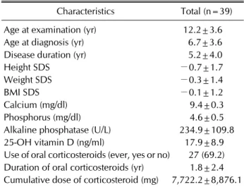
Table 1. Clinical characteristics of patients with juvenile idiopathic arthritis
Characteristics Total (n=39) Age at examination (yr) 12.2±3.6 Age at diagnosis (yr) 6.7±3.6 Disease duration (yr) 5.2±4.0
Height SDS −0.7±1.7
Weight SDS −0.3±1.4
BMI SDS −0.1±1.2
Calcium (mg/dl) 9.4±0.3
Phosphorus (mg/dl) 4.6±0.5
Alkaline phosphatase (U/L) 234.9±109.8 25-OH vitamin D (ng/ml) 17.9±8.9 Use of oral corticosteroids (ever, yes or no) 27 (69.2) Duration of oral corticosteroids (yr) 1.8±2.4 Cumulative dose of corticosteroid (mg) 7,722.2±8,876.1 Data are expressed as mean±standard deviation or number (%). SDS: standard deviation score, BMI: body mass index.
portant first step in developing strategies to optimize bone health. Therefore, the aim of this study was to eval- uate the prevalence of low BMD and factors affecting BMD in Korean children and adolescents with JIA.
MATERIALS AND METHODS
Patients
A total of 39 JIA patients aged between 7 and 19 years who were being followed up in the Department of Pediatrics, Hallym University Sacred Heart Hospital from February 2017 to May 2017 were enrolled. All patients were diagnosed by a pediatric rheumatologist before the age of 16 years and assigned the JIA subtype according to the criteria of the International League of Associations for Rheumatology (ILAR) classification [13]. Patients with other chronic diseases (endocrinal, neurological, cardiac, and renal) were excluded from this study.
Medical records were retrospectively reviewed to check for subtype of JIA; age at diagnosis; disease duration; cur- rent and previous treatment with oral corticosteroids;
and serum calcium, phosphate, alkaline phosphatase and 25-OH vitamin D levels. 25-OH vitamin D levels was clas- sified as normal (≥20 ng/ml), deficient (<20 ng/ml) [14].
The control group was consisted of 514 healthy Korean children (262 girls and 252 boys) aged 5∼20 years, and their cost of dual energy X-ray absorptiometry (DXA) ex- amination was supported by the Korean Society of Pediatric Endocrinology Research Fund [15].
The study protocol was approved by the Ethnics Committees of Hallym University Sacred Heart Hospital (IRB no. 2018-04-015-002) and informed consent was obtained from all patients and/or parents.
Anthropometric and BMD measurements
Patients' height and weight were measured and recorded using the same measuring device (DS-103; Dong Sahn Jenix Co., Ltd., Seoul, Korea) by the same observer on the day of DXA examination. Body mass index (BMI) was cal- culated as weight divided by height squared (kg/m²).
Age-related reference values of height, weight, and BMI were obtained from the 2007 Korean National Growth Charts [16].
Height, weight, and BMI were normalized for chrono- logical age (CA) by converting the values to standard de- viation score (SDS). SDS was calculated by subtracting the patient’s measured value from the mean age-related
reference value; dividing this difference with the standard deviation (SD) of the age-related reference value.
Besides CA, the age of each patient was expressed as bone age (BA) and height age (HA). BA was calculated us- ing the Greulich & Pyle atlas method by the same pedia- tric endocrinologist, after taking an X-ray scan of the left hand, including carpal bone X-ray. HA was defined as the age that corresponds to the child's height when plotted at the 50th percentile on a growth chart.
BMD values obtained for the lumbar spine (L1∼L4) us- ing DXA instrument (Lunar Prodigy Advance, Madison, WI, USA). Each BMD value was converted to a Z-score by comparing the reference values of the control group for each age and sex measured by the same device [15]. We defined a Z-score of less than –2.0 as low BMD and ana- lyzed BMD on the basis of CA, BA, and HA.
Statistical analysis
All statistical analyses were performed using a software program (PASW Statistics, Version 18.0; IBM Co., Armonk, NY, USA). Continuous variables were ex- pressed as the mean±SD, and the categorical variables were expressed as frequency (%). Differences between the patient and controls groups were assessed using the Mann–Whitney test. Spearman’s rank correlation co- efficients were used to test the relationship between the duration of the disease and the dosage of corticosteroids.
The association between different variables and BMD Z-score in the lumbar spine was investigated using multi-
Table 2. BMD of patients according to levels of 25-OH vitamin D
Level of 25-OH vitamin D Deficient (<20 ng/ml), n=25 (64%) Normal (≥20 ng/ml), n=14 (36%) p-value
BMD Z-score CA −0.94±1.53 −0.64±1.82 0.26
BA −1.18±1.94 −0.66±1.48 0.37
HA −0.38±1.20 −0.36±1.26 0.91
Data are expressed as mean±standard deviation. BMD: bone mineral density, CA: chronological age, BA: bone age, HA: height age.
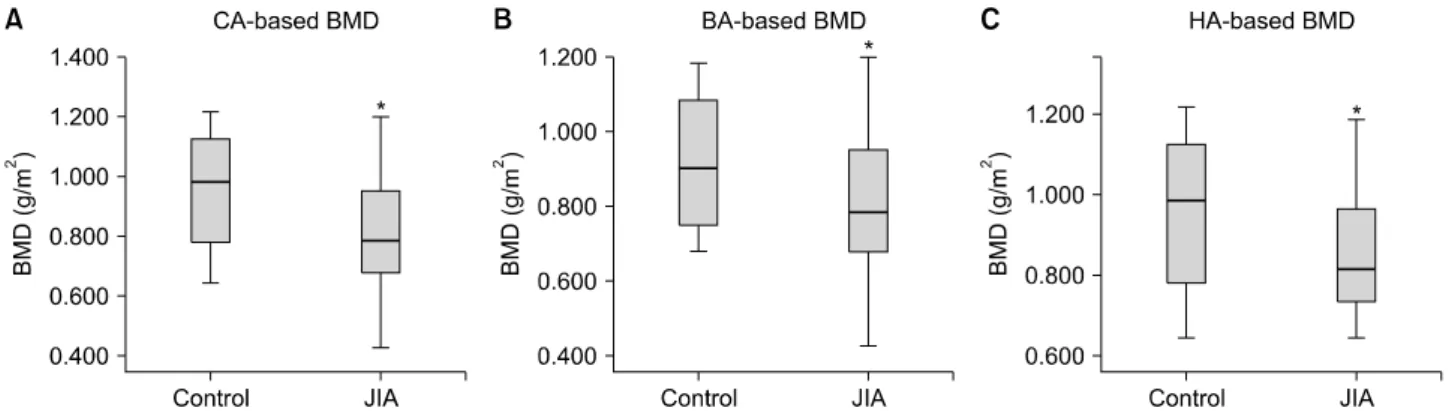
Figure 1. Comparison of BMD between JIA and control group. CA: chronological age, BA: bone age, HA: height age, BMD: bone mineral density, JIA: juvenile idiopathic arthritis. *p-value<0.05.
ple linear regression analysis. A p-value of <0.05 was considered to be statistically significant.
RESULTS
Patients
Among the 39 patients with JIA, 23 (59%) were females and 16 (41%) were males; and 9 (23%) had oligoarticular;
10 (26%), polyarticular; 16 (41%), systemic; and 4 (10%), enthesitis-related arthritis subtype. The mean age at diagnosis was 6.7±3.6 years, and the mean disease du- ration was 5.2±4.0 years. Demographic and clinical char- acteristics of patients are described in Table 1.
Age and BMD types
For JIA patients, the mean BMD was 0.81±0.18 and the mean HA-based BMD was 0.85±0.15, which were lower than the values for the control group. There was a sig- nificant difference in BMD, calculated based on CA, BA, and HA, between JIA patients and healthy controls (p=0.005, p=0.045, and p=0.035, respectively) (Figure 1). The mean CA-based, BA-based, and HA-based BMD Z-scores were –0.83±1.62, −0.99±1.79, and −0.38±
1.20, respectively. The number of patients with low CA-based, BA-based, and HA-based BMD was 8 (21%), 9 (23%) and 4 (10%), respectively.
25-OH vitamin D and BMD values
There were 25 patients (64%) with a 25-OH vitamin D (<20 ng/ml) deficiency. Of these patients, 6 (24%) had a CA-based low BMD, 6 (24%) has a BA-based low BMD, and 3 (12%) had an HA-based low BMD. The number of patients with a normal 25-OH vitamin D level (>20 ng/ml) was 14 (36%). Of these 14 patients, 2 (14%) had a CA-based low BMD, 3 (21%) had a BA-based low BMD, and 1 (7%) had a HA-based low BMD. No statistically sig- nificant difference was observed between BMD values of the lumbar spine, calculated with respect to CA, BA, and HA, and serum 25-OH vitamin D levels (p=0.26, p=0.37, and p=0.91, respectively) (Table 2).
Comparison of BMD values
This study comprised 16 patients with systemic JIA (41%), 23 patients with non-systemic JIA (59%). Compared with patients with non-systemic JIA, patients with sys- temic JIA had lower BMD values, which were calculated with respect to CA, BA, and HA; this results showed a statistically significant difference (CA-based BMD:
p=0.020, BA-based BMD: p=0.049, and HA-based BMD:
p=0.024) (Table 3).
Sixty-nine percent of patients had received oral cortico- steroids at least once. When corticosteroids user group
Table 3. Comparison of BMD between systemic JIA and non-systemic JIA patients
Systemic JIA, n=16 (41%) Non-systemic JIA, n=23 (59%) p-value
BMD Z-score CA −1.42±1.27 −0.42±1.74 0.020
BA −1.72±2.21 −0.48±1.24 0.049
HA −0.76±1.02 −0.11±1.27 0.024
Data are expressed as mean±standard deviation. BMD: bone mineral density, JIA: juvenile idiopathic arthritis, CA: chronological age, BA: bone age, HA: height age.
Table 4. Comparison of BMD between corticosteroid users and corticosteroid non-users
Corticosteroid users, n=27 (69%) Corticosteroid non-users, n=12 (31%) p-value
BMD Z-score CA −1.39±1.35 0.28±1.59 0.002
BA −1.50±1.76 0.02±1.40 0.022
HA −0.54±1.22 –0.06±1.14 0.188
Data are expressed as mean±standard deviation. BMD: bone mineral density, CA: chronological age, BA: bone age, HA: height age.
Table 5. Regression analysis of clinical variables and BMD
Variable
BMD Z-score
CA base BA base HA base
CI at 95%
β p-value
CI at 95%
β p-value
CI at 95%
β p-value Lower
limit
Upper limit
Lower limit
Upper limit
Lower limit
Upper limit Weight
SDS
0.507 1.055 0.781 0.001 0.332 1.030 0.681 0.048 0.214 0.689 0.451 0.303
Height SDS
0.334 0.835 0.585 0.008 0.252 0.848 0.550 0.021 0.526 0.739 0.632 <0.001
Disease duration
−0.135 0.132 −0.002 0.202 −0.124 0.169 0.023 0.151 −0.224 −0.048 −0.136 0.014
BMD: bone mineral density, CA: chronological age, BA: bone age, HA: height age, CI: confidential interval, SDS: standard deviation score.
and corticosteroids non-user group were compared, the user group had a lower BMD than the non-user group.
There was a statistically significant difference between the corticosteroid user and non-user groups in terms of CA- and BA-based BMD values, but not for HA-based BMD value (p=0.002, p=0.022, and p=0.188, re- spectively) (Table 4). However, there was no significant difference in CA-, BA-, and HA-based BMD values be- tween the two groups with respect to the duration of cor- ticosteroid use (p=0.231, p=0.251, and p=0.080, re- spectively).
Other clinical variables and laboratory parameters There was a significant correlation between the duration of the disease and the dosage of corticosteroids (p=
0.014).
Multiple linear regression analysis was performed for BMD in the lumbar spine against weight, height, BMI, disease duration. BMD had a positively significant corre- lation with the weight and the height of the patient at the time of DXA. However, disease duration was only asso- ciated with HA-based BMD (Table 5).
Serum levels of calcium, phosphate and alkaline phos- phatase were normal in all patients.
DISCUSSION
Our study is the first trial in Korea that evaluates BMD and its related factors in JIA children and adolescents in Korea. In most studies, BMD and its associated factors for JIA patients were evaluated based only on the chrono- logical age. However, a majority of JIA patients in their rapid growth phase are likely to be medicated with corti- costeroids and various immunosuppressive agents, and this may affect growth spurts and pubertal maturation. To solve the problem of BMD being closely related to the aforementioned growth spurts and pubertal maturation, we compared BMD not only with CA but also BA and HA.
In our study, the JIA patient group had markedly lower BMD than the control group, and the number of patients with low CA-, BA-, and HA-based BMD was 8 (21%), 9 (23%), and 4 (10%), respectively. We found a significant correlation between reduced BMD and systemic JIA, and we also found that the use of corticosteroids significantly affected the reduction of BMD. However, we did not find a significant correlation between 25-OH vitamin D levels, disease duration, and BMD.
During childhood and adolescence phases in patients with JIA, when the PBM is attained in the healthy people, an increase in bone mass is inhibited by various mecha- nisms, namely the onset of inflammatory disease or drug therapy [3]. Osteopenia or osteoporosis occurs in all of the JIA subtypes, most typically in systemic and poly- articular disease subtypes [17,18]. Our study also showed a significant association between systemic JIA subtype and reduced BMD.
Children and adolescents with JIA have to take several medications such as disease-modifying antirheumatic drugs (DMARDs) and corticosteroids that have adverse effects on bone mineralization and acquisition of an opti- mal PBM [3]. In our study, decreased BMD appeared to be associated with the use of corticosteroids. In patients with JIA, a correlation between reduced BMD and the use of corticosteroids was reported in several studies [19-21].
However, no relation was found between BMD and the duration of corticosteroid use in the present study. This is probably due to the gradual tapering of corticosteroid dosage, and many patients continue to receive a low-dose corticosteroids treatment. Tengstrand et al. [22] reported that administration of low-dose corticosteroid treatment for several years has persisting anti-inflammatory effects in rheumatoid arthritis and no further negative impact on BMD.
According to 2017 American College of Rheumatology (ACR) Guideline for the prevention and treatment of glu- cocorticoid-Induced osteoporosis, calcium (1,000 mg/day) and vitamin D intake (600 IU/day) were recommended for individuals aged 4∼17 years and who had received glucocorticoids for ≥3 months [23]. Dey et al. [19] re- ported that it was important to improve the dietary intake of calcium and vitamin D. Johnston et al. [24] also showed that calcium supplementation enhanced the rate of increase in BMD in pre-pubertal children.
Weight and height have a positive impact on BMD in the current study. Several studies suggested that weight and height all correlated positively with BMD at every skeletal site in individuals of both sexes, irrespective of their pu- bertal stage [25-27]. Accordingly, it is recommended that children and adolescents should maintain adequate weight and muscle strength through an appropriate diet and weight-bearing physical activity [2].
In this study, a deficiency of 25-OH vitamin D (<20 ng/ml) was found in 64% patients, and no relationship was found between BMD and 25-OH vitamin D level.
Serum levels of calcium, phosphorus, and alkaline phos- phatase were normal in all patients. Several other studies have reported diverse results in JIA patients with regard to the levels of calcium, phosphorus, 25-OH vitamin D.
Although in a study by Pepmueller et al. [28], vitamin D levels in JIA patients were not found to be lowered, the calcium levels were significantly lower. Lien et al. [18] re- ported that calcium, phosphorus, and vitamin D levels were all normal in JIA patients. Conversely, Hillman et al.
[29] reported that JIA patients had a low serum calcium level and normal 25-OH vitamin D and phosphorus levels.
In contrast to our expectation, the disease duration was not significantly associated with BMD in JIA patients; this result was found to be consistent with the findings of the study by Dey et al. [19]. On the other hand, Islam et al.
[30] reported that the disease duration had a positive re- lationship with lower BMD in JIA patients.
This study is limited by the small sample size within each age/sex group, and the cross-sectional characteristic of the data. In addition, there are only a few studies on the normal BMD of Korean children and adolescents accord- ing to age, sex, and pubertal development compared to the number of studies on those of other countries.
Although there are many patients who need to undergo DXA such as the children and adolescents with JIA, there is no insurance standard established by the National
Health Insurance System of Korea for performing DXA on children. Currently, there are no standard recom- mendations or guidelines to perform a DXA exam on children. However, according to the 2013 Pediatric Position Development Conference convened by The International Society for Clinical Densitometry (ISCD), it is recommended to evaluate the bone health of both chil- dren and adolescents who may benefit from interventions to decrease their elevated risk of a clinically significant fracture [31].
CONCLUSION
Our study is the first report on the BMD of JIA children and adolescents in Korea. We found that JIA patients had lower BMD than the control subjects, and osteopenia was a frequent complication of JIA. In JIA patients, low BMD is caused by various factors such as low physical activity, reduced joint motility, and corticosteroid treatment.
Therefore, while treating JIA patients, especially those re- quiring oral corticosteroids or those with systemic JIA subtype, appropriate test, treatment, and education re- garding bone health are warranted. Moreover, it is im- portant to improve the dietary intake of calcium and vita- min D; further large-scale follow-up studies are required in the future to strengthen our findings and conclusions.
CONFLICT OF INTEREST
No potential conflict of interest relevant to this article was reported.
REFERENCES
1. Petty RE, Laxer RM, Lindsley CB, Wedderburn L. Textbook of pediatric rheumatology e-book. Philadelphia (PA), Elsevier Health Sciences, 2015.
2. Naka H, Iki M, Morita A, Ikeda Y. Effects of pubertal devel- opment, height, weight, and grip strength on the bone min- eral density of the lumbar spine and hip in peripubertal Japanese children: Kyoto kids increase density in the skel- eton study (Kyoto KIDS study). J Bone Miner Metab 2005;23:463-9.
3. Brabnikova Maresova K. Secondary osteoporosis in patients with juvenile idiopathic arthritis. J Osteoporos 2011;2011:
569417.
4. McDonagh JE. Osteoporosis in juvenile idiopathic arthritis.
Curr Opin Rheumatol 2001;13:399-404.
5. Masi L, Cimaz R, Simonini G, Bindi G, Stagi S, Gozzini A, et al. Association of low bone mass with vitamin d receptor gene and calcitonin receptor gene polymorphisms in juve-
nile idiopathic arthritis. J Rheumatol 2002;29:2225-31.
6. Tortolani PJ, McCarthy EF, Sponseller PD. Bone mineral density deficiency in children. J Am Acad Orthop Surg 2002;10:57-66.
7. Burnham JM, Shults J, Weinstein R, Lewis JD, Leonard MB.
Childhood onset arthritis is associated with an increased risk of fracture: a population based study using the General Practice Research Database. Ann Rheum Dis 2006;65:
1074-9.
8. Huber AM, Ward LM. The impact of underlying disease on fracture risk and bone mineral density in children with rheumatic disorders: A review of current literature. Semin Arthritis Rheum 2016;46:49-63.
9. Nusman CM, Anink J, Otten MH, van Rossum MA, van Rijn RR, Maas M, et al. Bone health of patients with juvenile idio- pathic arthritis: a comparison between dual-energy X-ray absorptiometry and digital X-ray radiogrammetry. Eur J Radiol 2015;84:1999-2003.
10. Oh YJ, La KS, Rhie YJ, Lee KH, Park SH, Choung JT, et al.
Bone mineral density and correlation factors in normal chil- dren and adolescence. J Korean Soc Pediatr Endocrinol 2009;14:38-44.
11. Lim JS. Pediatric dual-energy X-ray absorptiometry: inter- pretation and clinical and research application. Korean J Pediatr 2010;53:286-93.
12. Stagi S, Masi L, Capannini S, Cimaz R, Tonini G, Matucci-Cerinic M, et al. Cross-sectional and longitudinal evaluation of bone mass in children and young adults with juvenile idiopathic arthritis: the role of bone mass determi- nants in a large cohort of patients. J Rheumatol 2010;
37:1935-43.
13. Petty RE, Southwood TR, Manners P, Baum J, Glass DN, Goldenberg J, et al. International League of Associations for Rheumatology classification of juvenile idiopathic arthritis:
second revision, Edmonton, 2001. J Rheumatol 2004;31:
390-2.
14. Holick MF. Vitamin D deficiency. N Engl J Med 2007;
357:266-81.
15. Lim JS, Hwang JS, Lee JA, Kim DH, Park KD, Cheon GJ, et al.
Bone mineral density according to age, bone age, and pu- bertal stages in Korean children and adolescents. J Clin Densitom 2010;13:68-76.
16. Moon JS, Lee SY, Nam CM, Choi JM, Choe BK, Seo JW, et al.
2007 Korean National Growth Charts: review of devel- opmental process and an outlook. Korean J Pediatr 2008;51:1-25.
17. Henderson CJ, Specker BL, Sierra RI, Campaigne BN, Lovell DJ. Total-body bone mineral content in non-cortico- steroid-treated postpubertal females with juvenile rheuma- toid arthritis: frequency of osteopenia and contributing factors. Arthritis Rheum 2000;43:531-40.
18. Lien G, Flatø B, Haugen M, Vinje O, Sørskaar D, Dale K, et al. Frequency of osteopenia in adolescents with early-onset juvenile idiopathic arthritis: a long-term outcome study of one hundred five patients. Arthritis Rheum 2003;48:
2214-23.
19. Dey S, Jahan A, Yadav TP, Bhagwani DK, Sachdev N.
Measurement of bone mineral density by dual energy X-ray absorptiometry in juvenile idiopathic arthritis. Indian J Pediatr 2014;81:126-32.
20. Hämäläinen H, Arkela-Kautiainen M, Kautiainen H,
Haapasaari J, Leirisalo-Repo M. Bone mineral content in young adults with active or inactive juvenile idiopathic ar- thritis and in controls. Scand J Rheumatol 2010;39:219-22.
21. Thornton J, Pye SR, O'Neill TW, Rawlings D, Francis RM, Symmons DP, et al. Bone health in adult men and women with a history of juvenile idiopathic arthritis. J Rheumatol 2011;38:1689-93.
22. Tengstrand B, Larsson E, Klareskog L, Hafström I.
Randomized withdrawal of long-term prednisolone treat- ment in rheumatoid arthritis: effects on inflammation and bone mineral density. Scand J Rheumatol 2007;36:351-8.
23. Buckley L, Guyatt G, Fink HA, Cannon M, Grossman J, Hansen KE, et al. 2017 American College of Rheumatology Guideline for the Prevention and Treatment of Glucocorti- coid-Induced Osteoporosis. Arthritis Care Res (Hoboken) 2017;69:1095-110.
24. Johnston CC Jr, Miller JZ, Slemenda CW, Reister TK, Hui S, Christian JC, et al. Calcium supplementation and increases in bone mineral density in children. N Engl J Med 1992;327:82-7.
25. Frost HM, Schönau E. The "muscle-bone unit" in children and adolescents: a 2000 overview. J Pediatr Endocrinol Metab 2000;13:571-90.
26. Kotaniemi A, Savolainen A, Kröger H, Kautiainen H,
Isomäki H. Weight-bearing physical activity, calcium in- take, systemic glucocorticoids, chronic inflammation, and body constitution as determinants of lumbar and femoral bone mineral in juvenile chronic arthritis. Scand J Rheumatol 1999;28:19-26.
27. Lien G, Selvaag AM, Flatø B, Haugen M, Vinje O, Sørskaar D, et al. A two-year prospective controlled study of bone mass and bone turnover in children with early juvenile idio- pathic arthritis. Arthritis Rheum 2005;52:833-40.
28. Pepmueller PH, Cassidy JT, Allen SH, Hillman LS. Bone mineralization and bone mineral metabolism in children with juvenile rheumatoid arthritis. Arthritis Rheum 1996;
39:746-57.
29. Hillman L, Cassidy JT, Johnson L, Lee D, Allen SH. Vitamin D metabolism and bone mineralization in children with ju- venile rheumatoid arthritis. J Pediatr 1994;124:910-6.
30. Islam MF, Islam MI, Talukdar MK, Rahman SA. Bone miner- al density in children with juvenile idiopathic arthritis: a hospital based study. Bangladesh J Child Health 2013;37:
18-21.
31. Gordon CM, Leonard MB, Zemel BS; International Society for Clinical Densitometry. 2013 Pediatric Position Development Conference: executive summary and reflections. J Clin Densitom 2014;17:219-24.