https://doi.org/10.4174/astr.2021.101.2.79 Annals of Surgical Treatment and Research
Adrenal ganglioneuroma resected for suspicious
malignancy: multicenter review of 25 cases and review of the literature
Dorit Esther Zilberman1, Tomer Drori1, Gadi Shlomai2,3, Haggi Mazeh4, Boris Fishman3, Shay Golan5, Hen Hendel5, Monica Laniado6, Zohar A. Dotan1
1Department of Urology, Chaim Sheba Medical Center, Tel-Hashomer, affiliated to the Sackler School of Medicine, Tel-Aviv University, Tel-Aviv, Israel
2Institute of Endocrinology, Chaim Sheba Medical Center, Tel-Hashomer, affiliated to the Sackler School of Medicine, Tel-Aviv University, Tel-Aviv, Israel
3Department of Internal Medicine D, Chaim Sheba Medical Center, Tel-Hashomer, affiliated to the Sackler School of Medicine, Tel- Aviv University, Tel-Aviv, Israel
4Department of Surgery, Hadassah Hebrew University Medical Center, Mount Scopus, Jerusalem, Israel
5Department of Urology, Rabin Medical Center, Petah Tikva affiliated to the Sackler School of Medicine, Tel-Aviv University, Tel- Aviv, Israel
6Department of General Surgery, Bnai-Zion Medical Center, Haifa, Israel
Received December 28, 2020, Revised April 14, 2021, Accepted June 5, 2021
Corresponding Author: Dorit E. Zilberman
Department of Urology, Chaim Sheba Medical Center, Tel-Hashomer, 52621 Israel
Tel: +972-3-5302231, Fax: +972-3-5351892 E-mail: [email protected]
ORCID: https://orcid.org/0000-0002-2092-4732
Copyright ⓒ 2021, the Korean Surgical Society
cc Annals of Surgical Treatment and Research is an Open Access Journal. All articles are distributed under the terms of the Creative Commons Attribution Non- Commercial License (http://creativecommons.org/licenses/by-nc/4.0/) which permits unrestricted non-commercial use, distribution, and reproduction in any medium, provided the original work is properly cited.
Purpose: We reviewed the experience with adrenal ganglioneuroma (AGN) pathologically confirmed following adrenalectomy in medium- to high-volume medical centers.
Methods: The medical records of all adrenalectomy cases in 4 medical centers between 2006 and 2020 were retrospectively reviewed for demographics, clinical, radiological and laboratory findings, surgical treatment, pathology results, and outcomes.
Results: Twenty-five out of 875 adrenalectomy cases (2.9%) were pathologically confirmed as AGN. Those patients’ average age was 40.5 years (range, 4–76 years), 13 (52.0%) were males, and 18 lesions (72.0%) were right-sided. One patient had a family history of neurofibromatosis, and another had a succinate dehydrogenase gene mutation. Abdominal/back pain attributed to mass effect was the most common symptom. All 25 patients underwent abdominal computerized tomography scanning in which the average maximal tumor diameter was 6.61 cm. The mean pre- and postcontrast Hounsfield units (HU) values were 35.2 and 59, respectively; and the mean late-phase HU value was 71.1. Twenty-two patients (88.0%) underwent minimally invasive surgery. The average tumor diameter recorded in the final pathology report was 7 cm. Isolated AGN was diagnosed in 21 cases (84.0%), and the additional components reported for the remaining 4 cases included pheochromocytoma (2), ganglioneuroblastoma (1), and neurofibroma (1). The average follow-up length was 16.8 months (range, 1–136 months), during which there was no recurrence or death.
Conclusion: AGN is a rare, slow-growing, large benign tumor with radiological characteristics similar to those seen in malignant tumor. Final diagnosis is established by pathology after surgical resection, preferably minimally invasive, with an overall excellent prognosis.
[Ann Surg Treat Res 2021;101(2):79-84]
Key Words: Adrenal glands, Adrenal gland neoplasms, Benign neoplasms, Ganglioneuroma
INTRODUCTION
Adrenalectomy is an integral procedure of both general and urological surgeons. The finding of a large, progressively growing adrenal mass that is enhanced on CT always arouses concern for an underlying malignant tumor and leads to the scheduling of prompt surgical resection.
In rare cases, however, malignant-appearing masses turn out to be benign, one of which is an adrenal ganglioneuroma (AGN).
Ganglioneuromas are large neoplasms originating within the neural crest and consisting of ganglion cells, Schwann cells, and nerve fibers [1]. The adrenal gland is the most involved site [2].
The incidence of AGN is reported to vary between 0.3% to 4% of all adrenal masses [1,3], or 1 per million in the general population [2]. There are many case reports of AGN in the English literature. One study found 41 AGN cases reported between 1961 and 2009 [4], another counted 230 cases between 1978 and 2008 [5], while a third study found 150 cases between 2000 and 2016 [1].
As of 2021, there are only 5 case series in the English literature reporting over 20 cases and originating in China, South Korea, and the United States [3,6-9]; another 3 originating in Italy and China and report 10–17 cases each [1,10,11] and 4 case series of 6–9 cases each from Canada [4], Italy [2], Greece [5], and Belgium [12]. The largest studies from the Far East and the
United States are single-center experiences and relate to 29–45 cases recorded between 1988 and 2019 [3,6-9].
In the present study, we sought to retrospectively review the collective experience with AGN pathologically confirmed following adrenalectomy in 4 medium- to high-volume medical centers in Israel. To the best of our knowledge, this is the largest study on this topic coming from the Middle East region.
METHODS
Four medical centers participated in the present study.
Following approval of each medical center’s institutional review board, all adrenalectomy cases registered between 2006 and 2020 were retrospectively reviewed. The recorded data included patients’ age, sex, tumor side, relevant family history, radiological and laboratory findings, associated clinical symptoms, date of surgery and surgical technique, maximal diameter of the tumor and other tumor components according to pathological findings, tumor recurrence, and follow-up duration. Data acquired from all medical centers were integrated and interpreted. The results are given in average (range) unless otherwise specified.
This study has been approved by the Institutional Review Board of Chaim Sheba Medical Center (No. SMC-18-5818) with a waiver for informed consent.
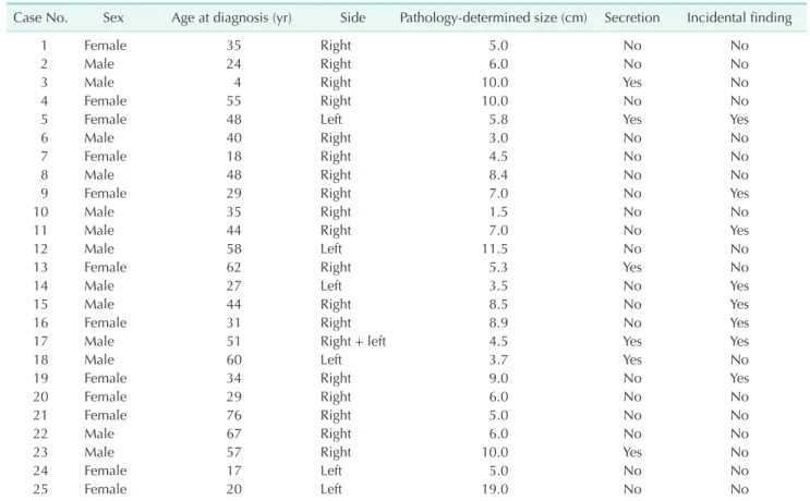
Table 1. Demographic data
Case No. Sex Age at diagnosis (yr) Side Pathology-determined size (cm) Secretion Incidental finding
1 Female 35 Right 5.0 No No
2 Male 24 Right 6.0 No No
3 Male 4 Right 10.0 Yes No
4 Female 55 Right 10.0 No No
5 Female 48 Left 5.8 Yes Yes
6 Male 40 Right 3.0 No No
7 Female 18 Right 4.5 No No
8 Male 48 Right 8.4 No No
9 Female 29 Right 7.0 No Yes
10 Male 35 Right 1.5 No No
11 Male 44 Right 7.0 No Yes
12 Male 58 Left 11.5 No No
13 Female 62 Right 5.3 Yes No
14 Male 27 Left 3.5 No Yes
15 Male 44 Right 8.5 No Yes
16 Female 31 Right 8.9 No Yes
17 Male 51 Right + left 4.5 Yes Yes
18 Male 60 Left 3.7 Yes No
19 Female 34 Right 9.0 No Yes
20 Female 29 Right 6.0 No No
21 Female 76 Right 5.0 No No
22 Male 67 Right 6.0 No No
23 Male 57 Right 10.0 Yes No
24 Female 17 Left 5.0 No No
25 Female 20 Left 19.0 No No
RESULTS
A total of 875 adrenalectomy cases were reviewed between 2006 and 2020, among which the final pathology was AGN in 25 patients (2.9%). The demographic data of those patients are detailed in Table 1. Their average age was 40.5 years (range, 4–76 years), and 13 (52.0%) were males.
The AGNs of 8 patients (32%) who were entirely asymptomatic were found incidentally. The clinical symptoms of the remaining 17 patients were as follows; abdominal pain/discomfort (11 patients, 44.0%), back pain (3 patients, 12.0%), weight loss (3 patients, 12.0%), and hot flashes and hypertension (1 patient each, 4.0%). Eighteen patients (72.0%) had right-side lesions, and 1 patient had bilateral AGNs. One patient had a family history of neurofibromatosis (NF) 1, and 1 patient had a succinate dehydrogenase (SDHD) gene mutation.
Only 6 cases (24.0%) had pathological metabolite hypersecretion detected in 24-hour urine collection, specifically, vanillylmandelic acid (1 observation) and catecholamines (CA;
epinephrine, norepinephrine, metanephrine, normetanephrine, and dopamine) (9 observations in total). The specific levels, as well as normal ranges, are detailed in Table 2.
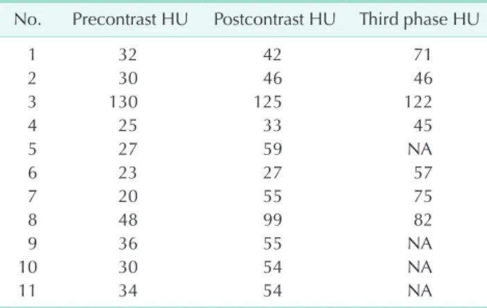
The radiological diagnosis was based on a CT scan in all 25 cases. The estimated average maximal tumor diameter on CT was 6.61 cm (range, 1.5–20 cm). Pre- and postcontrast Hounsfield
units (HU) as well as late-phase HU were available for 11 patients, and the postcontrast HU values were higher than the precontrast ones in 10 of them (Table 3). The average pre- and postcontrast HU values were 35.2 and 59.0, respectively, and the average late-phase HU value was 71.1. Tumor calcification was detected in 4 cases (16.0%).
Twenty-two patients (88.0%) underwent minimally invasive surgery for removal of the adrenal mass (21 by laparoscopy and 1 by robot-assisted surgery), while the remaining 3 had open surgery. The average tumor diameter in the final pathology report was 7 cm (range, 1.5–19.0 cm). Isolated AGN was diagnosed in 21 cases (84.0%), and the additional components reported for the remaining 4 cases included pheochromocytoma (PC) in 2, ganglioneuroblastoma (GNB) in 1, and neurofibroma in 1.
Positive surgical margins were detected in 3 cases (12.0%).
Notably, a coexisting high secretion of CA was detected in 2 of the 3 cases in which a PC component was present, and coexisting hypertension was observed in the 3rd case.
The average follow-up duration for the entire group was 16.8 months (range, 1–136 months). All 25 study patients were free from local recurrence and metastatic progression. Data acquired from the national population registry revealed that they all were alive at study closure.
Table 2. Detected metabolite levels (6 of 25 patients)
Metabolite No. of
patients Level Normal range
VMA, creatinine (µg/gr) 1 48 1–10
Metanephrine (µg/24 hr) 2 3,887 10–300 2,746
Normetanephrine (µg/24 hr) 2 2,346 90–450 439
Epinephrine (µg/24 hr) 2 126 0–20
111
Norepinephrine (µg/24 hr) 1 254 5–40
Dopamine (µg/24 hr) 2 1,050 50–300
992 VMA, vanyllylmandelic acid.
Table 3. Tumor density on computerized tomographic scans pre- and postcontrast as well as late phase (11 of 25 patients)
No. Precontrast HU Postcontrast HU Third phase HU
1 32 42 71
2 30 46 46
3 130 125 122
4 25 33 45
5 27 59 NA
6 23 27 57
7 20 55 75
8 48 99 82
9 36 55 NA
10 30 54 NA
11 34 54 NA
HU, Hounsfield unit; NA, not applicable.
A B
Fig. 1. Adrenal ganglioneuroma in a 24-year-old patient. (A) Macroscopic appearance. (B) To m o g ra p h i c a p p e a ra n c e . Written informed consent has been obtained for these images.
DISCUSSION
The international neuroblastoma (NB) pathology classification divides neuroblastic tumors into 4 subcategories based on their morphology, clinical features, and behavior; NB, nodular GNB, intermixed GNB, and ganglioneuroma [13]. Ganglioneuroma is a rare, differentiated neuroblastic tumor of the adrenal gland originating in the neural crest cells [14]. It is found within the adrenal medulla in 20%–30% of the cases [4]. They are macroscopically described as lobulated and gray-white [12], although they also appeared encapsulated and well-demarcated in this and other studies [8,11] (Fig. 1).
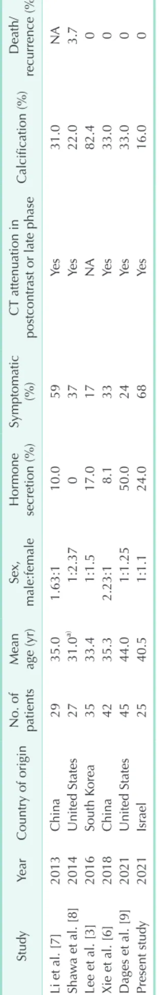
Table 4 summarizes the findings of the present study in comparison to those found in previous large case series (i.e.,
≥20 cases). As demonstrated in the present and previous case series [1,3,6-11], the peak incidence of AGN is the 4th–5th decade of life [13,14].
A male-to-female ratio of 2:1 has been reported for adrenomedullary tumors [15]; however, an equal ratio for both sexes has been found in the present study as well as in other studies [9,14]. AGN has been reported to accompany hereditary familial syndromes, such as NF1 and NF2 [1,2,14], and 1 study also reported a single AGN case with an associated multiple endocrine neoplasia 2 syndrome [8].
We had 1 case associated with NF1 and another case with SDHD gene mutation. Most AGNs are hormonally silent [3,14];
however, some of them may be associated with hormonal secretion. Glucocorticosteroid (i.e., cortisol), mineralocorticoid (i.e., aldosterone), and androgen (i.e., testosterone and its precursors) secretion have been reported in numerous cases [1,4,6,7].
Secretion of CA and their metabolites have been observed in our study as well as in others [2-7]. In the present cohort, pathological CA metabolite secretion was observed in 24% of our cases. This may be partly explained by the origination of AGN in the adrenal cortex rather than the adrenal medulla.
Another explanation could derive from the PC component known to be associated with some of the AGN originating within the medulla [4,15]. In our study, a PC component was present in 3 cases of which 2 demonstrated relatively high urinary CA secretion.
Notwithstanding, the extent of hormone secretion does not always suffice to produce clinical symptoms. In our study, hypertension was present in 1 of the 3 cases producing CA, similar to the findings of other studies [3,4,6,7].
Cortisol secretion was not associated with clinical symptoms in 1 study [1] but reported to have led to menstrual disorders in another study [6]. Rondeau et al. [4] described virilization in 1 case in which increased testosterone secretion was detected, and Li et al. [7] described central obesity due to urinary 17-hydroxysteroid hypersecretion.
Table 4. Summary of ≥20 AGN case series published in the English literature StudyYearCountry of origin No. of patients
Mean age (yr)Sex, male:femaleHormone secretion (%)Symptomatic (%)CT attenuation in postcontrast or late phaseCalcification (%)Death/ recurrence (%) Li et al. [7]2013China2935.01.63:110.059Yes31.0NA Shawa et al. [8]2014United States2731.0a) 1:2.37 037Yes22.03.7 Lee et al. [3]2016South Korea3533.41:1.517.017NA82.40 Xie et al. [6]2018China4235.32.23:1 8.133Yes33.00 Dages et al. [9]2021United States4544.01:1.2550.024Yes33.00 Present study2021Israel2540.51:1.124.068Yes16.00 NA, not applicable. a) Median.
Interestingly, many reports describe AGN as an asymptomatic, incidental finding. Asymptomatic cases reportedly vary between 71.4% and 100% [1-6,12], although in the same breath symptoms attributable to AGN, i.e., most commonly, abdominal pain, back pain, and abdominal discomfort [1-3,6,11,12]; and less commonly fatigue and malaise [7,11] have also been mentioned. In our study, similar to the findings of Shawa et al. [8], abdominal and back symptoms were relatively common (56%), with a mass effect having been suggested as a possible mechanism [3,8].
A CT scan is the most common imaging tool implemented to characterize an adrenal mass. An AGN as demonstrated on a CT scan is homogenous and of low attenuation in the precontrast phase, mild attenuation in the postcontrast phase, and sometimes either homogenous or heterogeneous in appearance in the late phase [3,11]. This pattern was repeatedly observed by us as well as by others [4-6,8,9,12]. Calcification is another indication for AGN seen on CT; however, its prevalence widely varies between studies from 11.1% [2] to as high as 82.4%
[3]. It should be kept in mind however, that calcifications, heterogeneity, and postcontrast attenuation are seen in malignant adrenal tumors as well [16].
MRI has been suggested as an alternative diagnostic tool for AGN. Low signal intensity on T1 and high signal intensity on T2-weighted images are expected [2,7], but the malignant tumor cannot be excluded, and the final diagnosis is most definitive by surgical histology.
AGN develops in childhood but it is not recognized before early adulthood, and it is rarely diagnosed after the age of 60 years [2]. These findings are in line with the single patient under 10 years of age (i.e., a 4-year-old) as well as the 3 patients who were older than 60 years of age at diagnosis in our cohort.
The tumor rarely metastasizes or recurs [14]. No tumor recurrence or death due to AGN was observed in the present study or in any other studies in the literature [1-6,8-11], although 1 mortality in association with AGN was reported due to CA hypersecretion [17]. While the overall prognosis is excellent, surgical excision should remain the standard of care due to the inability of radiology to discriminate between AGN from a malignant tumor.
The present study summarizes the experience of 4 medium- to high-volume medical centers throughout a period of 14 years.
To the best of our knowledge, this is the first case series on this topic coming from the Middle East region and is among
the largest AGN case series published in English literature.
Its retrospective nature and relatively low case numbers are its main disadvantages. Another weak point is that there was no standardization of the CT scans, or of the laboratory and histopathology results among the participating institutions, which could potentially affect the interpretation of the main findings.
In summary, AGN is a rare, slow-growing, large benign tumor with peak occurrence during the 4th–5th decade of life. There is no gender predominance. The tumor may be hormone- secreting, however not necessarily to the level that would create associated symptoms. The most common symptom is abdominal/back pain which is attributed to a mass effect. Given its radiologic characteristics, which cannot be discriminated from a malignant adrenal tumor, the final diagnosis should be made by surgical resection, preferably laparoscopic if possible.
The overall prognosis of AGN is excellent.
ACKNOWLEDGEMENTS
Conflict of Interest
No potential conflict of interest relevant to this article was reported.
ORCID iD
Dorit Esther Zilberman: https://orcid.org/0000-0002-2092-4732 Tomer Drori: https://orcid.org/0000-0001-6334-4554
Gadi Shlomai: https://orcid.org/0000-0002-8563-0858 Haggi Mazeh: https://orcid.org/0000-0002-8425-1776 Boris Fishman: https://orcid.org/0000-0003-3835-7438 Shay Golan: https://orcid.org/0000-0002-9205-5820 Hen Hendel: https://orcid.org/0000-0003-0250-3202 Monica Laniado: https://orcid.org/0000-0002-0810-0247 Zohar A. Dotan: https://orcid.org/0000-0002-5771-6793
Author Contribution
Conceptualization: GS, ZAD Formal Analysis: DEZ, TD
Investigation: TD, GS, HM, BF, SG, HH, ML Methodology: GS, ZAD
Project Administration: ZAD Writing – Original Draft: DEZ
Writing – Review and Editing: All authors
REFERENCES
1. Iacobone M, Torresan F, Citton M, Schiavone D, Viel G, Favia G. Adrenal ganglioneuroma: the Padua Endocrine
Surgery Unit experience. Int J Surg 2017;41 Suppl 1:S103-8.
2. Spinelli C, Rossi L, Barbetta A, Ugolini C, Strambi S. Incidental ganglioneuromas:
a presentation of 14 surgical cases and literature review. J Endocrinol Invest 2015;38:547-54.
3. Lee JH, Chai YJ, Kim TH, Choi JY, Lee KE, Kim HY, et al. Clinicopathological features of ganglioneuroma originating from the adrenal glands. World J Surg 2016;40:2970-5.
4. Rondeau G, Nolet S, Latour M, Braschi S, Gaboury L, Lacroix A, et al. Clinical and biochemical features of seven adult adrenal ganglioneuromas. J Clin Endocrinol Metab 2010;95:3118-25.
5. L i no s D, Ts i r l i s T, K ap r a l o u A , K iriakopoulos A, Tsak ayannis D, Papaioannou D. Adrenal ganglioneu- romas: incidentalomas with misleading clinical and imaging features. Surgery 2011;149:99-105.
6. Xie J, Dai J, Zhou WL, Sun FK. Adrenal ganglioneuroma: features and outcomes of 42 cases in a Chinese population.
World J Surg 2018;42:2469-75.
7. Li J, Yang CH, Li LM. Diagnosis and treatment of 29 cases of adrenal ganglioneuroma. Eur Rev Med Pharmacol Sci 2013;17:1110-3.
8. Shawa H, Elsayes KM, Javadi S, Morani A, Williams MD, Lee JE, et al. Adrenal ganglioneuroma: features and outcomes of 27 cases at a referral cancer centre. Clin Endocrinol (Oxf) 2014;80:342-7.
9. Dages KN, Kohlenberg JD, Young WF Jr, Murad MH, Prokop L, Rivera M, et al.
Presentation and outcomes of adrenal ganglioneuromas: a cohort study and a systematic review of literature. Clin Endocrinol (Oxf) 2021;95:47-57.
10. Li L, Shao J, Gu J, Wang X, Qu L. Adrenal ganglioneuromas: experience from a retrospective study in a Chinese population. Urol J 2014;11:1485-90.
11. Qing Y, Bin X, Jian W, Li G, Linhui W, Bing L, et al. Adrenal ganglioneuromas: a 10- year experience in a Chinese population.
Surgery 2010;147:854-60.
12. Maweja S, Materne R, Detrembleur N, de Leval L, Defechereux T, Meurisse
M, et al. Adrenal ganglioneuroma. A neoplasia to exclude in patients with adrenal incidentaloma. Acta Chir Belg 2007;107:670-4.
13. Lam AK. Update on adrenal tumours in 2017 World Health Organization (WHO) of endocrine tumours. Endocr Pathol 2017;28:213-27.
14. Mylonas KS, Schizas D, Economopoulos KP. Adrenal ganglioneuroma: what you need to know. World J Clin Cases 2017;5:373-7.
15. Lin X, Wu D, Chen C, Zheng N. Clinical characteristics of adrenal tumors in children: a retrospective review of a 15- year single-center experience. Int Urol Nephrol 2017;49:381-5.
16. Fassnacht M, Kenn W, Allolio B. Adrenal tumors: how to establish malignancy? J Endocrinol Invest 2004;27:387-99.
17. Suárez-Peñaranda JM, Gómez-Otero I, Muñoz JI, Pedreira-Pérez M. [Cardiac death associated with an adrenal ganglioneuroma]. Rev Esp Cardiol 2008;61:551-2. Spanish.