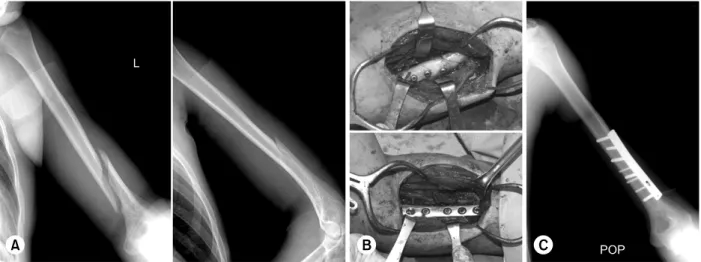
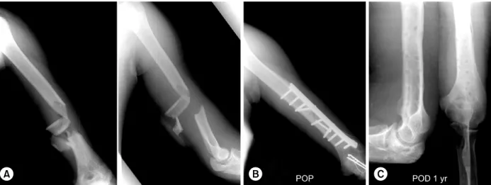
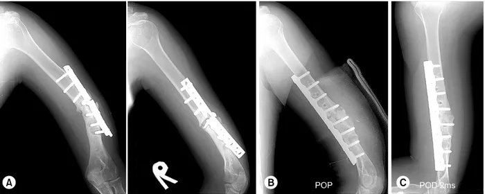
상완골 간부 골절: 금속판 고정술 (Humeral Shaft Fracture: Plating)
전체 글
수치


관련 문서
The greater tubercle is palpable on the line from the lateral epicondyle of the distal humerus in the direction of the humeral longitudianl axis and just below the acromion
limit the crack growth by increasing # of site of crazing or
이것은 이용허락규약 (Legal Code) 을 이해하기 쉽게 요약한 것입니다. 귀하는 원저작자를 표시하여야 합니다.. Operative treatment for nonunion of humerus shaft fracture ---
3.7 Fracture interface after tensile shear test with plunge depth 43 Table.. 3.13 Fracture interface after tensile shear test with dwell time
Through this result, it was confirmed that brittle fracture did not occur in high Mn steel and ductile fracture occurred in spite of cryogenic
Purpose: Calcaneal fracture is a rare fracture, which accounts for about 2% of all fractures, but is one of the most common fractures in the ankle bone.. There is
A 27-year-old man with spondyloarthropathy, oblique coronal fat-saturated T2-weighted (A) and oblique coronal postcontrast fat-saturated T1-weighted (B) images show
The clinical outcome and complication for treating proximal femoral shaft fracture were compared and analyzed through the group treated with closed