Department of Thoracic and Cardiovascular Surgery,
1Seoul National University Children’s Hospital, Seoul National University College of Medicine,
2Pusan National University Hospital, Pusan National University School of Medicine
†This article was presented with the title of ‘extracardiac Fontan operation and right ventricular exclusion procedure in case of severe dilated right ventricle and tricuspid regurgitation’ at the 254th Seoul & Gyeonggi province monthly meeting (2011. 10. 21).
Received: July 15, 2013, Revised: November 2, 2013, Accepted: November 11, 2013, Published online: June 5, 2014
Corresponding author: Woong-Han Kim, Department of Thoracic and Cardiovascular Surgery, Seoul National University Hospital, Seoul National University College of Medicine, 101 Daehak-ro, Jongno-gu, Seoul 110-744, Korea
(Tel) 82-2-2072-3637 (Fax) 82-2-3672-3637 (E-mail) [email protected]
C
The Korean Society for Thoracic and Cardiovascular Surgery. 2014. All right reserved.
CC
This is an open access article distributed under the terms of the Creative Commons Attribution Non-Commercial License (http://creative- commons.org/licenses/by-nc/3.0) which permits unrestricted non-commercial use, distribution, and reproduction in any medium, provided the original work is properly cited.
A 13-year-old girl, who had undergone the total correction of partial atrioventricular septal defect at the age of 4 years, was admitted with severe tricuspid regurgitation in echocardiography. She had received one-and-a-half ven- tricle repair during follow-up. Her right ventricle showed global akinesia, and the ejection fraction of the left ven- tricle was 25% with paradoxical interventricular septal motion. We performed right ventricular exclusion adjunct to the Fontan procedure. She is doing well two years after the operation without complications.
Key words: 1. Right ventricle exclusion 2. Congenital heart disease (CHD) 3. CHD, Fontan
4. Magnetic resonance imaging
CASE REPORT
A 13-year-old girl had a history of partial atrioventricular septal defect (pAVSD) total correction at the age of 4 years.
She underwent mitral valve repair, atrial septal defect closure with autologous pericardial patch. However, a right ven- tricular assist device was applied at the operation because of the akinetic right ventricle (RV). Two years later, based on our echocardiography results, the patient was transferred due to severe tricuspid regurgitation and RV chamber enlargement with RV dysfunction. She underwent tricuspid septal commis- suroplasty, De Vega-type tricuspid annuloplasty, right atrial reduction plasty, and isthmus ablation. Further, we performed
one-and-a-half ventricle repair. In spite of the operation, the patient’s RV function progressively decreased.
She had symptoms of dyspnea on exertion and palpitation
during follow-up. Six years after the one-and-a-half ventricle
repair, cardiac magnetic resonance imaging was performed to
evaluate her cardiac function and measure the left ventricle
(LV) volume. The patient’s RV end diastolic volume index and
RV ejection fraction were 500.4 mL/m
2, and 13.2%,
respectively. The LV stroke volume index and ejection fraction
were 38.2 mL/m
2and 28%, respectively. Echocardiographic
evaluation showed global RV akinesia and LV ejection fraction
of 25% with paradoxical interventricular septal motion. We sus-
pected that the RV enlargement affected both the RV and the
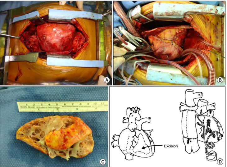
Fig. 1. Preoperative and postoperative heart appearance. (A) The markedly dilated RV is shown. (B) After RV exclusion, the heart size decreased. (C) The free wall of the RV is resected. (D) Schematic representation of the operation. RV, right ventricle.
LV function and it resulted in decreased LV contractility. Thus, we decided to exclude the enlarged RV from the systemic circulation.
She underwent extracardiac conduit Fontan operation with polytetrafluoroethylene (PTFE, Gore-Tex; WL Gore &
Associates, Flagstaff, AZ, USA) 24-mm tube graft, RV ex- clusion, atrial septectomy, and permanent pacemaker implantation. RV exclusion procedures include tricuspid valve obliteration (from the RV side; 5-0 Prolene double layer, re- inforcement suture from the RA side; 4-0 Polyester PTFE pledget-supported interrupted mattress suture) and pulmonary valve obliteration (6-0 Prolene running suture) to reduce the RV volume with no flow connection, thrombin soaked gel-foam packing to the RV, and RV free-wall wide re-
section, and it was performed under the condition of cardiac arrest (Fig. 1). Permanent pacemaker bipolar leads were im- planted at the LV apex, RV apex, left atrial roof, and RA free wall owing to a history of frequent atrial flutter and junctional rhythm. We did not perform arrhythmia surgery because the patient underwent an electrophysiology study and radiofrequency catheter ablation for supraventricular ar- rhythmia before the operation. Her palpitation symptom was relieved after radiofrequency catheter ablation. The car- diopulmonary bypass time was 308 minutes, and the aortic cross clamp time was 146 minutes.
We performed the computed tomographic angiography not
cardiac magnetic resonance imaging to evaluate the patient’s
postoperative cardiac function and chamber size, because she
Fig. 2. Postoperative echocardiography and computed tomographic angiography findings. (A) Immediate postoperative echocardiography. (B) The RV cavity is nearly collapsed in the latest echocardiography. (C) Total thrombosed RV in the computed tomographic angiography conducted 1 week after the operation. RV, right ventricle.
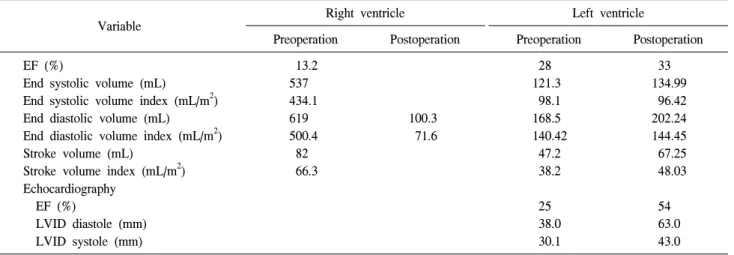
Table 1. Preoperative and postoperative cardiac function
a)Variable Right ventricle Left ventricle
Preoperation Postoperation Preoperation Postoperation EF (%)
End systolic volume (mL)
End systolic volume index (mL/m
2) End diastolic volume (mL)
End diastolic volume index (mL/m
2) Stroke volume (mL)
Stroke volume index (mL/m
2) Echocardiography
EF (%)
LVID diastole (mm) LVID systole (mm)
13.2 537 434.1 619 500.4 82 66.3
100.3 71.6
28 121.3 98.1 168.5 140.42 47.2 38.2
25 38.0 30.1
33 134.99 96.42 202.24 144.45 67.25 48.03
54 63.0 43.0 EF, ejection fraction; LVID, left ventricle internal dimension at end.
a)