• 검색 결과가 없습니다.

The posterior interosseous nerve (PIN) is a deep

N/A
N/A
Protected

Academic year: 2021

Share "The posterior interosseous nerve (PIN) is a deep "

Copied!
5
0
0

로드 중.... (전체 텍스트 보기)

전체 글

(1)

Introduction

The posterior interosseous nerve (PIN) is a deep

branch of radial nerve, liable to compression. Compres- sion can occur spontaneously (entrapment) or second- ary to mass lesions (lipoma, ganglion). 1 The common entrapment sites are as followings: fibrous band of the radial head, radial recurrent vessels at the level of radial neck, extensor carpi ulnaris brevis, arcade of Frohse and distal part of supinator muscle. 1-3 Here we report a case of PIN neuropathy due to torsion of the nerve caused by compression of Leash of Henry, a recurrent branch of radial artery. Electrodiagnostic findings were

ISSN 1229-6066 https://doi.org/10.18214/jkaem.2019.21.1.32 J Korean Assoc EMG Electrodiagn Med 21(1):32-36, 2019

J Korean Assoc

Electrodiagn Med EMG

Copyright © by Korean Association of EMG Electrodiagnostic Medicine

This is an Open Ac cess article distributed under the terms of the Creative Commons Attribution Non-Commercial License (http://creativecommons.org/licenses/by-nc/4.0) which permits unrestricted non-commercial use, distribution, and reproduction in any medium, provided the original work is properly cited.

Received March 6, 2019 Revised April 17, 2019 Accepted May 10, 2019

Corresponding Author: Hee-Kyu Kwon

Department of Physical Medicine & Rehabilitation, Korea University Anam Hospital, 73 Goryeodae-ro, Seongbuk-gu, Seoul 02841, Korea

Tel: 82-2-920-6471, Fax: 82-2-929-9951, E-mail: hkkwon@korea.ac.kr

요골동맥의 압박에 의한 후골간 신경 비틀림과 그로 인한 신경병증: 증례 보고

이해인

1

, 김 단

1

, 김동휘

2

, 박종웅

3

, 권희규

1

고려대학교 1안암병원 재활의학과, 2안산병원 재활의학과, 3안암병원 정형외과

Posterior Interosseous Neuropathy Caused by Torsion due to Leash of Henry: A Case Report

Hae In Lee 1 , Dahn Kim 1 , Dong Hwee Kim 2 , Jong Woong Park 3 , Hee-Kyu Kwon 1

Departments of

1

Physical Medicine & Rehabilitation,

3

Orthopedic Surgery, Korea University Anam Hospital, Seoul,

2

Department of Physical Medicine & Rehabilitation, Korea University Ansan Hospital, Ansan, Korea

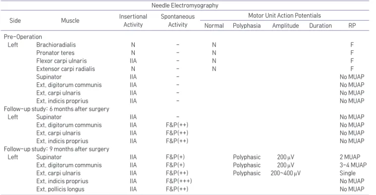
A 34-year-old man was referred for electrodiagnosis of left forearm and elbow pain with concurrent left wrist and finger extension weakness. Symptom developed abruptly, after waking up from sleep. After 11 days of onset, electromyography was performed. In nerve conduction study, the left radial motor response was unobtainable and in needle electromyography, abnormal spontaneous activities and no motor unit potential were noted in the left supinator, extensor digitorum communis, extensor carpi ulnaris and extensor indicis proprius muscles, suggesting left posterior interosseous neuropathy involving the supinator muscle and all the distal muscles to it. Subsequent ultrasonography of the left upper arm revealed multiple nerve torsions, and surgical exploration finally confirmed that the main torsion site was caused by the recurrent branch of radial artery, the Leash of Henry. We report an unusual case of spontaneous posterior interosseous nerve neuropathy due to nerve torsion caused by Leash of Henry.

Key Words: posterior interosseous nerve syndrome, electrodiagnosis, nerve compression syndromes

(2)

compatible with PIN neuropathy, ultrasonography ex- posed multiple PIN torsions and finally, operative find- ings confirmed the main torsion site caused by Leash of Henry. This case emphasizes correct diagnosis based on electrophysiologic findings, ultrasonography and operative findings with the follow up studies.

Case Report

A 34-year-old man visited department of Physical Medicine and Rehabilitation presenting left forearm and elbow pain with concurrent left wrist and finger extension weakness which started 10 days ago, abrupt- ly without any trauma history, after waking up from sleep. Left forearm pain started first, and then wrist extension and finger extension weakness progressively developed. First, the patient visited a private clinic and an orthopedic surgeon prescribed a long arm splint and the patient applied it for approximately 10 days. How- ever, the pain aggravated especially with elbow exten- sion and pronation. X-ray findings showed no sign of

fracture or any other abnormality. On physical exami- nation, Medical Research Council (MRC) grades of left extensor carpi radialis was 5/5 but extensor carpi ulna- ris was 0/5 and finger metacarpophalangeal extension 0/5. No sensory abnormalities were present. Range of motion was restricted in elbow extension.

Eletrodiagnostic examination was performed 11 days after onset. Nerve conduction study (NCS) showed no left radial motor response. Needle electromyography (EMG) showed increased insertional activities and no motor unit potential were noted in the left supina- tor, extensor digitorum communis, extensor carpi ulnaris and extensor indicis proprius muscles but the brachioradialis and extensor carpi radialis revealed normal findings (Table 1). In conclusion, the patient was diagnosed as complete left posterior interosseous neuropathy involving from supinator to all the distal muscles innervated by the PIN. Additionally, the patient underwent sonography. Sonography of the left upper arm revealed multiple torsions of PIN (Fig. 1A, B). The most obvious torsion was noticed at just proximal to

Table 1. Needle Electromyography Data

Needle Electromyography

Side Muscle Insertional

Activity

Spontaneous Activity

Motor Unit Action Potentials

Normal Polyphasia Amplitude Duration RP Pre-Operation

Left Brachioradialis N - N F

Pronator teres N - N F

Flexor carpi ulnaris IIA - N F

Extensor carpi radialis N - N F

Supinator IIA - No MUAP

Ext. digitorum communis IIA - No MUAP

Ext. carpi ulnaris IIA - No MUAP

Ext. indicis proprius IIA - No MUAP

Follow-up study: 6 months after surgery

Left Supinator IIA - No MUAP

Ext. digitorum communis IIA F&P(++) No MUAP

Ext. carpi ulnaris IIA F&P(++) No MUAP

Ext. indicis proprius IIA F&P(++) No MUAP

Follow-up study: 9 months after surgery

Left Supinator IIA F&P(+) Polyphasic 200 μV 2 MUAP

Ext. digitorum communis IIA F&P(+) Polyphasic 200 μV 3-4 MUAP

Ext. carpi ulnaris IIA F&P(++) Polyphasic 200~400 μV Single

Ext. indicis proprius IIA F&P(+++) No MUAP

Ext. pollicis longus IIA F&P(++) No MUAP

Ext: extensor, N: normal, IIA: increased insertional activities, F&P: fibrillation potentials & positive sharp wave, RP: recruitment pattern, F:

full, MUAP: motor unit action potentials, Single: single unit recruitment pattern

(3)

innervating the supinator muscle and demonstrated an hourglass-like appearance (Fig. 1C). Color Doppler im- age demonstrated increased vascularity at the main PIN torsion site (Fig. 1D).

The patient underwent surgery 26 days after symp- tom onset. Incision extending from the biceps muscle to the border of the brachioradialis was made. With further dissection, compression and torsion of PIN at

the level of and just proximal to arcade of Froshe by recurrent branches of the radial artery, the Leash of Henry, was discovered (Fig. 2A, B). The vessel’s loca- tion was in accordance with the ultrasonographic findings showing increased vascularity near the nerve torsion site. The orthopedic surgeon ligated the Leash of Henry, performed decompression and neurolysis of the PIN and finally resected the degenerated portion of

A B C

Fig. 2. (A) Surgical findings confirmed Leash of Henry (black arrow) being the culprit of causing PIN torsion. (B) Ligation of Leash of Henry re- veals nerve torsion (white arrow) more clearly. Notice the color change of the nerve. (C) The surgeon resected the degenerated portion of the PIN and performed microscopic neurorrhaphy (arrowhead).

A B

C D

Fig. 1. (A, B) Sonographic findings of

PIN showing torsion at multiple sites

at distal humerus level (arrow). (C)

Most obvious torsion (arrowhead) was

noticed just proximal to innervating

the supinator muscle. (D) Color Dop-

pler image shows increased vascular-

ity near the torsion site.

(4)

the nerve and performed direct neurorrhaphy (Fig. 2C).

Long arm cast with elbow flexed 90 degrees was ap- plied for one month.

Follow-up electrodiagnostic examination was per- formed 6 months and 9 months after surgery. The patient no longer complained of pain but no motor improvement was noticed. On physical examination, MRC grades of lefgt extensor carpi radialis was 5/5 but extensor carpi ulnaris was 0/5 and finger meta- carpophalangeal extension 0/5. Radial motor NCS and needle EMG revealed no interval change 6 months after surgery but 9 months later, needle EMG revealed rein- nervation evidence in supinator, extensor digitorum communis, and extensor carpi ulnaris muscles (Table 1).

Discussion

The radial nerve divides into two branches at the level of the lateral epicondyle: the superficial radial nerve and the PIN. The PIN supplies the radial and dorsal forearm muscles: extensor carpi radialis, supi- nator, extensor digitorum communis, extensor carpi ulnaris, extensor pollicis longus/brevis, and extensor indicis. 1-3 The PIN is predominantly a motor nerve, thus patients with posterior interosseous neuropathy will suffer metacarpophalangeal joint extension weakness, or so-called finger drop, and radial wrist deviation on extension, but no sensory symptoms. 1-4 The PIN is sus- ceptible to compression at five sites. 1,2,4 First is located at the level of the radial head, where fibrous bands confluence with brachialis, brachioradialis, extensor carpi radialis brevis and the supinator. Second is at the level of the radial neck, where radial recurrent arter- ies (Leash of Henry) cross the nerve. Third is beneath the tendon of extensor carpi radialis brevis. Fourth and the most common site for compression is the arcade of Frohse, located at the most superior part of the supi- nator muscle. Finally, the distal edge of supinator can cause compression.

Usually NCS is sufficient to diagnose nerve compres- sion, but in PIN, NCS can be inconclusive that needle

EMG is crucial to confirm the diagnosis and demarcate the lesion site in many cases. In this case, NCS could only diagnose left radial neuropathy but needle EMG could localize the lesion involving supinator to all the distal muscles innervated by PIN. Regarding the fact that the supinator is involved in this patient, we can as- sume that the nerve injury level is above the supinator.

This patient revealed restricted elbow motion due to pain, especially in elbow extension and pronation and this phenomenon suggested nerve torsion rather than compression. 5,6 Ultrasonography is known to localize the lesion so we additionally performed upper arm ul- trasound examination and multiple sites of hourglass- like appearance of the nerve were exposed. 7 With sur- gical exploration, the Leash of Henry was discovered to be the main culprit causing PIN torsion.

Spontaneous PIN torsion is uncommon. 5,6 Various the- ories exist trying to explain the mechanism of sponta- neous nerve torsion but the etiology remains unclear. 7 However, in this case, the operative findings showed leash of Henry directly above the main PIN torsion site, suggesting that leash of Henry caused PIN torsion.

Without surgical findings the etiology may have re- mained inconclusive.

In all the reported cases with PIN neuropathy, pa- tients underwent EMG, ultrasonography and eventually surgery. 8 Studies concerning prognosis of PIN neu- ropathy have confirmed timely surgical exploration is beneficial both diagnostically and therapeutically. 8,9 In this case, taking electrodiagnostic and ultrasono- graphic findings into account, we identified that the nerve torsion caused all the patient’s symptoms and immediately consulted the orthopedic surgeon. With the help of electrodiagnostic and sonographic findings, the surgeon was able to reasonably assume the lesion site and minimize surgical incision and operation time.

The surgeon untangled the twisted PIN and the patient

no longer complained pain after surgery. The prognosis

of spontaneous nerve torsion is usually good. 7 In previ-

ous cases of PIN torsion, most reported spontaneous

incomplete or complete recovery and surgical explora-

(5)

tion fastened recovery. 8,9 However, this patient took 9 months to show some sign of reinnervation, which is relatively slower than previous cases. This could be explained by the fact that this patient had multiple tor- sions along the PIN. Also, PIN degeneration and atro- phy was already noticed in the surgical field, implicat- ing slow recovery (Fig. 2).

In conclusion, we report a case of PIN neuropathy caused by nerve torsion due to anomalous compres- sion of vessels that has been successfully diagnosed with electrodiagnosis and ultrasonography and decom- pressed by surgery. Thus, when a PIN compression or torsion is suspected, prompt and precise electrodiag- nostic confirmation followed by subsequent ultraso- nography and eventually surgery is important for the patient’s prognosis.

References

1. Anania P, Fiaschi P, Ceraudo M, Balestrino A, Zaottini F, Martinoli C, et al: Posterior interosseous nerve entrapments:

review of the literature. Is the entrapment distal to the ar- cade of Frohse a really rare condition? Acta Neurochir (Wien) 2018: 160: 1857-1864

2. Bevelaqua AC, Hayter CL, Feinberg JH, Rodeo SA: Posterior interosseous neuropathy: electrodiagnostic evaluation. Hss j 2012: 8: 184-189

3. Lawley AR, Saha S, Manfredonia F: Posterior interosseous neuropathy: the diagnostic benefits of a multimodal ap- proach to investigation. Clin Case Rep 2016: 4: 437-441 4. Dang AC, Rodner CM: Unusual compression neuropathies of

the forearm, part I: radial nerve. J Hand Surg Am 2009: 34:

1906-1914

5. Endo Y, Miller TT, Carlson E, Wolfe SW: Spontaneous nerve torsion: unusual cause of radial nerve palsy. Skeletal Radiol 2015: 44: 457-461

6. Fernandez E, Di Rienzo A, Marchese E, Massimi L, Lauretti L, Pallini R: Radial nerve palsy caused by spontaneously occur- ring nerve torsion. Case report. J Neurosurg 2001: 94: 627- 629

7. Guerra WK, Schroeder HW: Peripheral nerve palsy by tor- sional nerve injury. Neurosurgery 2011: 68: 1018-1024; dis- cussion 1024

8. Kim DH, Murovic JA, Kim YY, Kline DG: Surgical treatment and outcomes in 45 cases of posterior interosseous nerve entrapments and injuries. J Neurosurg 2006: 104: 766-777 9. Ochi K, Horiuchi Y, Tazaki K, Takayama S, Nakamura T,

Ikegami H, et al: Surgical treatment of spontaneous posterior

interosseous nerve palsy: a retrospective study of 50 cases. J

Bone Joint Surg Br 2011: 93: 217-222

수치

Table 1.	Needle	Electromyography	Data

참조

관련 문서

1) 신경전도 속도검사 (Nerve conduction velocity test).. 162. Electrodiagnostic

다양한 번역 작품과 번역에 관한 책을 읽는 것은 단순히 다른 시대와 언어, 문화의 교류를 넘어 지구촌이 서로 이해하고 하나가

The index is calculated with the latest 5-year auction data of 400 selected Classic, Modern, and Contemporary Chinese painting artists from major auction houses..

Green color indicates that the output shaft of the geared motor rotates in the same direction as the output shaft of the motor..

The purpose of this study was to analyze the impaction pattern of the impacted mandibular third molar and the relationship with the inferior alveolar nerve

1. Free radical initiator abstract a hydrogen from polymer chains 2. Through chain transfer of propagating chain with polymer chain 3. Polymer mixtures are mechanically

• Junction between motor neuron and muscle fiber. – Motor

activation(electroencephalolgram: left alpha wave, right alpha wave, left sensory motor rhythm, right sensory motor rhythm, right mid-beta wave, left attention concentration