• 검색 결과가 없습니다.

Lung Cancer Associated with Sarcoidosis−

N/A
N/A
Protected

Academic year: 2021

Share "Lung Cancer Associated with Sarcoidosis−"

Copied!
3
0
0

로드 중.... (전체 텍스트 보기)

전체 글

(1)

Korean J Thorac Cardiovasc Surg 2011;44:301-303 □ Case Report □ DOI:10.5090/kjtcs.2011.44.4.301 ISSN: 2233-601X (Print) ISSN: 2093-6516 (Online)

− 301 −

*Department of Thoracic and Cardiovascular Surgery, St. Mary’s Hospital, Catholic Cancer Center, College of Medicine, The Catholic University of Korea

Received: November 18, 2010, Revised: May 19, 2011, Accepted: May 31, 2011

Corresponding author: Jae-Kil Park, Department of Thoracic and Cardiovascular Surgery, St. Mary’s Hospital, Catholic Cancer Center, College of Medicine, The Catholic University of Korea, 505, Banpo-dong, Seocho-gu, Seoul 137-040, Korea

(Tel) 82-2-2258-2858 (Fax) 82-2-594-8644 (E-mail) jaekpark@catholic.ac.kr

C

The Korean Society for Thoracic and Cardiovascular Surgery. 2011. All right reserved.

CC
This is an open access article distributed under the terms of the Creative Commons Attribution Non-Commercial License (http://creative- commons.org/licenses/by-nc/3.0) which permits unrestricted non-commercial use, distribution, and reproduction in any medium, provided the original work is properly cited.

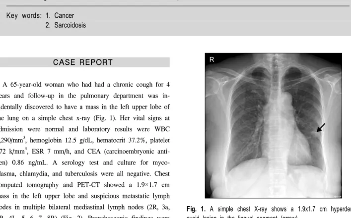
Fig. 1. A simple chest X-ray shows a 1.9x1.7 cm hyperdense, ovoid lesion in the lingual segment (arrow).

Lung Cancer Associated with Sarcoidosis

− A case report −

Jae Jun Kim, M.D.*, Jae-Kil Park, M.D.*, Young-Pil Wang, M.D.*, Soo Hwan Choi, M.D.*, Keon Hyon Jo, M.D.*

Sarcoidosis is a somewhat common pulmonary disease, but the concurrence of lung cancer and sarcoidosis in the same patient is very rare. Because sarcoidosis usually presents as mediastinal lymphadenopathies, this concurrence in a lung cancer patient detected radiologically is apt to be misunderstood to be mediastinal metastases, and it is thus considered to be an unresectable disease. We report a case of lung cancer associated with sarcoidosis that developed in a 65-year-old woman who underwent surgery. Radiological studies revealed a 1.9x1.7 cm mass in the left upper lobe with multiple enlarged bilateral mediastinal lymph nodes (2R, 3a, 4R, 4L, 5, 6, 7, 8R). Pathologic findings showed that the mass was a well-differentiated adenocarcinoma and all of the enlarged mediastinal lymph nodes were granulomas without cancer metastasis. We report this case with a review of the literature.

Key words: 1. Cancer 2. Sarcoidosis

CASE REPORT

A 65-year-old woman who had had a chronic cough for 4

years and follow-up in the pulmonary department was in-

cidentally discovered to have a mass in the left upper lobe of

the lung on a simple chest x-ray (Fig. 1). Her vital signs at

admission were normal and laboratory results were WBC

4,290/mm

3

, hemoglobin 12.5 g/dL, hematocrit 37.2%, platelet

172 k/mm

3

, ESR 7 mm/h, and CEA (carcinoembryonic anti-

gen) 0.86 ng/mL. A serology test and culture for myco-

plasma, chlamydia, and tuberculosis were all negative. Chest

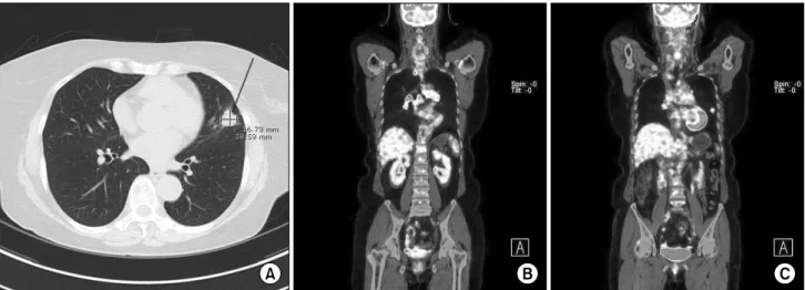
computed tomography and PET-CT showed a 1.9×1.7 cm

mass in the left upper lobe and suspicious metastatic lymph

nodes in multiple bilateral mediastinal lymph nodes (2R, 3a,

4R, 4L, 5, 6, 7, 8R) (Fig. 2). Bronchoscopic findings were

(2)

Jae Jun Kim, et al

− 302 −

Fig. 2. Chest CT (A) and PET-CT (B, C) show a suspicious malignant tumor in the lingular segment and multiple bilateral mediastinal lymph node metastases.

Fig. 3. Pathologic appearance of the resected specimen. (A) Lung tumor: a well-differentiated adenocarcinoma was found. (B) Lymph node:

a noncaseating epithelioid cell granuloma but no cancer metastasis was observed (H&E stain, ×200).

non-specific and PCNA (percutaneous needle aspiration) of the mass under chest CT revealed adenocarcinoma. At first we considered it an unresectable lung cancer with con- tralateral metastatic mediastinal lymph nodes (N3). We biop- sied the right paratracheal lymph nodes (2R, 4R) under me- diastinoscopy and the pathology reports showed that they were benign and non-caseous granuloma. We chose to oper- ate and performed a thoracotomy for a left upper lobectomy and mediastinal lymph node dissection. After the operation, the pathology report showed that the mass was adenocar- cinoma and there was no cancer metastasis in the dissected

lymph nodes with the exception of noncaseating epithelioid cell granuloma (Fig. 3) that was consistent with sarcoidosis.

The tuberculosis polymerase chain reaction (Tb-PCR) test from the specimens was negative. The stage was estimated to be IB (T2N0M0). The patient was discharged on the sixth postoperative day without any complications.

DISCUSSION

Curative resection for lung cancer is difficult when the

cancer has metastasized to the mediastinal lymph nodes. To

(3)

Lung Cancer with Sarcoidosis

− 303 − determine the optimal treatment strategy, especially in- dications for surgery, mediastinal lymph nodes must be eval- uated pathologically before the operation. There are many reasons why mediastinal lymphadenopathy may look like can- cer metastases radiologically, and one of them is sarcoidosis [1].

Sarcoidosis is a systemic granulomatous disease of un- known etiology that mainly develops between 25 and 40 years of age and primarily affects the lungs and the lym- phatic system [1,2]. The generally accepted pathogenic hy- pothesis is that certain environmental factors could promote sarcoidosis in genetically susceptible hosts [2]. The character- istic lesion of sarcoidosis is a noncaseating granuloma that consists of epithelioid cells and T lymphocytes [2]. Diverse clinical courses are observed according to the epidemiologic factors, involved organs, and disease severity [2]. Spontane- ous remissions occur in about two thirds of patients, but the clinical course is chronic or progressive in 10 to 30%.

Mortality, ranging between 1 and 5%, is mostly related to progressive respiratory failure, the central nervous system, or myocardial involvement [2,3]. The mainstay of treatment for sarcoidosis is systemic corticosteroid administration [2]. How- ever, the indication, optimal dose, and duration of cortico- steroid therapy continue to be controversial [1,2].

The association of sarcoidosis with malignancy has been published in several studies with conflicting results, due to the clinical and radiographic features of the disease, which may be similar to malignancies such as lymphoma or lung cancer, sometimes lacking histologic confirmation [4,5]. The relationship between sarcoidosis and lung cancer has been published in several studies. Brincker and Wilbek studied 2,544 patients and found that lung cancer occurred 3 times more frequently in patients with sarcoidosis [5,6], but the ex- act relationship between sarcoidosis and lung cancer is still unclear [5]. Sakula presented 3 possibilities: (1) sarcoidosis develops before the development of the lung cancer, and is more or less related and may even induce the malignant change; (2) sarcoidosis develops as a reaction to the lung

cancer; (3) sarcoidosis develops before the development of the lung cancer, the occurrence of the lung cancer and sarcoi- dosis being completely coincidental [7]. Sakula’s 1st type is represented by cancer that develops from postinflammatory scar tissue, in other words, scar cancer. The 2nd type is diffi- cult to apply because one cannot know exactly when sarcoi- dosis developed before it is clinically apparent [6]. In our case, sarcoidosis and lung cancer occurred simultaneously, there was no sarcoidosis in the lung mass and no scar lesion.

This case was the 3rd of Sakula’s types. Despite there cur- rently being conclusive evidence, sarcoidosis is suggested to have no influence on the prognosis of such patients [3,6].

Several cases with concurrent sarcoidosis and lung cancer have been reported. In most of these cases, sarcoidosis ex- isted for multiple years before the development of lung cancer. Simultaneous diagnosis of both diseases, as in our case, is rarer.

We found and treated the patient with coincidence of sar- coidosis and lung cancer successfully.

REFERENCES

1. McNeill M, Zanders TB, Morris MJ. A 49-year-old man with concurrent diagnoses of lung cancer, sarcoidosis, and multiple regions of adenopathy on positron emission tomo- graphy. Chest 2009;135:546-9.

2. Wu JJ, Schiff KR. Sarcoidosis. Am Fam Physician 2004;70:

312-22.

3. Tomimaru Y, Higashiyama M, Okami J, et al. Surgical re- sults of lung cancer with sarcoid reaction in regional lymph nodes. Jpn J Clin Oncol 2007;37:90-5.

4. Sato Y, Sasano S, Oyama K, Sakuraba M, Onuki T, Nitta S.

Lung cancer associated with sarcoidosis. Jpn J Thorac Cardiovasc Surg 2003;51:21-4.

5. Bouros D, Hatzakis K, Labrakis H, Zeibecoglou K. Associa- tion of malignancy with diseases causing interstitial pulmo- nary changes. Chest 2002;121:1278-89.

6. Yamasawa H, Ishii Y, Kitamura S. Concurrence of sarcoi- dosis and lung cancer. A report of four cases. Respiration 2000;67:90-3.

7. Sakula A. Bronchial carcinoma and sarcoidosis. Br J Cancer

1963;17:206-12.

수치

Fig. 1. A simple chest X-ray shows a 1.9x1.7 cm hyperdense,  ovoid lesion in the lingual segment (arrow).
Fig. 2. Chest CT (A) and PET-CT (B, C) show a suspicious malignant tumor in the lingular segment and multiple bilateral mediastinal  lymph node metastases

참조

관련 문서

Beichel,“Automated 3-D segmentation of lungs with lung cancer in CT data using a novel robust active shape model approach,” IEEE Trans.. 저작물의

- The strains in a beam in pure bending vary linearly with distance from the neutral surface regardless of the shape of the stress-strain curve of the material.. Normal

5 Because I know what it is like to be a new student in a strange school, I want to help him fit in. I know what it is like to be a new student in a strange school, so I

Grade 3 QTc prolongation Withhold until recovery to Grade ≤ 1, then resume at 200 mg twice daily a Grade 4 QTc prolongation Permanently discontinue. a In case

a prospective analysis of 50 cases. Preoperative staging of non-small-cell lung cancer with positron-emission tomography. Staging of non- small-cell lung cancer

Patients with breast cancer; ovarian cancer; renal cell carcinoma; pancreatic neuroendocrine cancer; colorectal cancer; head and neck cancer; non-small cell lung

According to the result of this study, to do effective nursing that improve health promoting behaviors of early-stage lung cancer patients, nurses should

(1) Differences: Ground/ Height/ Elevation/ Terrain (p.7, Li et al, 2005) - Ground: the solid surface of the earth, a solid base or foundation, a.. surface of the earth, bottom