11
Immune Network 서 론
많은 세포독성 림프구(CTL; Cytotoxic T lymphocyte, NK; Natural killer cell)는 종양에 대한 효과적인 면역인자 로 보고되어져 왔다(1,2). 적절한 조건하에서 그 세포들 은 종양세포를 인식하고 수 분내에 세포자살사(apopto-
sis)또는 괴사(Necrosis) 시킨다(3,4). CTL과 NK세포는 세 포가 중재된 반응에 관련되어 그 역할을 한다. CTL은 적 응 면역계의 일부이고, NK세포는 자연적인 면역계라고 할 수 있다. 이 두세포는 세포융해 분자인 perforin과 그 와 관련된 과립 단백질 분해효소(granzyme)이 포함되어 있는 세포질성 과립들이 관여한 세포융해성 기작을 수 행한다(5,6). 그중 NK세포는 CTL세포가 인식하지 못한 MHC class I이 결여된 표적세포를 인식하는 능력을 가지 고 있어 위와 같은 과정을 수행하는 중요한 역할을 한다 (7). NK세포는 다른 기원의 종양세포를 죽이는 세포로 처음 확인되어졌으며, 그 세포는 바이러스에 의해 감염
활성화시킨 사람의 림프구
1Biocell 면역연구소, 2연세대학교 의과대학 소화기내과
홍선민
1․이동욱
1․강진구
2․김한수
2․조성훈
1Human Activated Lymphocyte Treated with Anti-CD3, CD16, CD56 Monoclonal Antibody and IL-2
Seon-Min Hong
1, Dong-Wook Lee
1, Jin-Gu Kang
2, Han-Soo Kim
2, Sung-Hoon Cho
11Biocell Co. Immune Research Institute and 2Department of Medicine, Yonsei University College of Medicine
ABSTRACT
Background: Throughtout the last three decades, the therapy of leukemias and
lymphoma has set the stage for curative cancer therapy in systemic malignant disease.This was the result of an integrated work of basic reaserch and clinical investigators leading to more aggressive albeit tolerable protocol of chemotherapy and radiotherapy.
High dose therapy marks the most elaborated strategies in this field today. However, intensification of conventional therapeutic modalities as mentioned has to be based on new approaches and the exploration of new antineoplastic mechanisms. This insight has resulted in immune therapy of cancer. Among the cells of the immune system, natural killer (NK) cells and T cells are of major interest for the development of therapeutic strategies. Methods: Cytotoxicity to target cells was measured by LDH release method, Characterization of activated lymphocyte was measured by Flow cytometry analysis.
Anti-CD3, 16, 56 monoclonal antibody and IL-2 were used for the activation of NK and T cell. The analysis of effect of activated lymphocyte, in vivo, were used by Balb/c nude mouse. Results and Conclusion: Cytotoxicity to K562 cells was significantly higher in the mixture group of NK and T cells than that of a group of activating T cells. The survivors and the rate of reduction of size of tumor craft of nude mouse group treatment with activated lymphocyte was higher than that of the group without treatment with activated lymphocyte. Therefore, this results are suggested that the activated lymphocytes by anti-CD3, CD16 and CD56 can reduce the malignancy effect of lymphoma. (Immune Network 2005;5(1):11-15)
Key Words: Activated lymphocyte, CD3, CD16, CD56, IL-2, natural killer cell, T cell,
human, nude mouse, lymphoma, K562, raji책임저자:홍선민, Biocell 면역연구소
ꂕ 462-120, 경기도 성남시 중원구 상대원동 234-1 포스 테크노 418호
Tel: 031-737-5575, Fax: 031-737-5576 E-mail: hgansa@naver.com
된 정상세포도 죽이는 능력을 가진다. 그리고 형태적으 로는 큰 과립림프구이며, T세포의 표지인자나 표면단백 질인 Ig이 존재하지 않는다. NK세포의 종양에 대한 세포 독성은 vitro상태에서 인터루킨-2 (IL-2)와 인터페론-α와 같은 세포조절물질이 존재하면 증가하게 된다. NK세포 에서는 CD16 (FcγRIII)이 특이한 표면단백질이며 CD56 도 주된 표면 단백질이다. 그리고 T세포에서 발현되는 CD3는 결여되어있다(8).
암환자의 면역기능이 저하되면 면역결핍상태에서는 암 발생이 증가한다는 것이 알려지면서 암에 대한 면역 학적 치료법에 관하여 연구가 진행되어 왔다. 그 중 하나 가 종양숙주에 항암작용이 있는 면역세포를 투여하여 항암효과를 기대하는 양자면역요법이다(9). 1976년 인터 루킨-2의 발견으로 양자면역요법에 있어서 큰 발전을 이 루었다(10). 이 림프구 조절물질은 T세포뿐만 아니라 NK세포의 증식과 성장을 가능하게 하며 그들 세포에서 림프구 조절물질의 분비를 촉진, 증강시킨다(11).
본 연구에서는 현재 많이 연구되고 있는 세포면역치료 요법 중에서 NK세포와 NKT세포 그리고 T세포를 혼합 한 세포를 negative isolation 방법을 사용하여 분리하지 않고 공혈자로부터 채취한 혈액에서 림프구를 분리하고 anti-CD3, CD16, CD56 단일항체와 IL-2를 사용하여 활성 화시킨 림프구와 anti-CD3 단일항체와 IL-2를 사용하여 활성화시킨 림프구의 세포성상과 세포독성을 비교 분석 하여 활성화를 위해 사용된 항체에 따른 림프구의 활성 을 조사하고, Burkitt 림프종세포주인 사람의 B세포 림프 종을 유발하는 Raji세포를 누드마우스에 주입한 후 종괴
를 유발시키고, anti-CD3, CD16과 CD56 단일항체에 의 해 자극되어 활성화된 사람의 림프구를 투여하여 종괴 에 대한 영향과 마우스의 생존도를 조사함으로써 림프 종에 대한 활성화 림프구의 영향을 알아보고자 한다.
재료 및 방법
세포. 마우스를 이용한 생체실험을 위해서 종괴를 형 성시키는 세포로는 사람의 B세포 림프종을 유발하는 Burkitt 림프종 세포주인 Raji세포(ATCC: CCL-86)를 사 용한다. 그리고 세포독성은 사람의 leukemia세포주인 K562세포(연세대 소화기내과에서 배양중인 세포)를 사 용한다. 이 세포주들은 RPMI1640에서 10% 소태아 혈청 과 1%의 gentamycin을 첨가하고, 5% CO2 하의 습윤된 공기, 37oC에서 배양하였다.
실험동물. 6주령의 BALB/c 누드마우스를 구입하여, 청 정동물사역구역에서 동일 먹이와 동일 수분의 조건에서 사육하였다.
암세포의 이식. Raji 세포 1×10⁷개를 누드마우스의 오 른쪽 옆구리에 있는 피하조직에 주입하였다. 종양덩어 리는 늑골 융선을 덮으면서, 측면대부분을 차지하게 된 다. 이식 후 동일한 조건에서 사육하였다.
활성화림프구의 준비. 정상인 공혈자의 말초혈액을 헤 파린 처리된 10 ml 진공채혈관(BD VacutainerTM)에 채혈 하고 히스토파크(Histopaque, Sigma #1077-1, endotoxin tested, 밀도 1.077 g/ml) 밀도구배 원심법에 의해 림프구 를 분리한다. 분리된 림프구의 잔존 적혈구를 제거하기 위해서 0.83% 염화암모늄(NH4Cl)용액을 처리하고, 원심
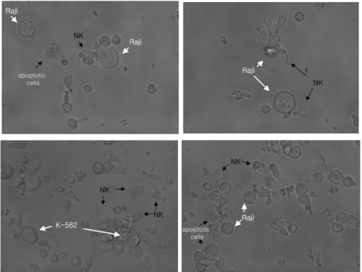
Figure 1. Activated lymphocytes attack- ing Raji, K562 cancer cell lines. Micro- scopic sections of cancer cells and acti- vated lymphocytes incubated together at the rate of 1:10 for I hour (×400, inverted microscope).
NK Raji
Raji
apoptotic cells
Raji
NK
K-562 NK
NK Raji
NK
apoptotic cells
분리로 침전된 림프구 1×10⁷의 세포를 CD3 (OKT3)로 자극시키고, IL-2 (1000 IU/ml)와 anti-CD16 또는 CD56 단일항체를 처리하여 RPMI1640배양액에 공혈자의 혈장 을 10%넣어주고, 5% CO2하의 습윤된 공기, 37oC에서 4 일간 75 cm2의 플라스크에서 배양한 후, 활성화된 상태 가 확인되면 225 cm2의 플라스크로 옮기고 CD16과 CD56을 처리한 후 3일간 배양한다. 다시 그 배양된 세포 를 1000 ml용 CO2 투과백(Nipro Co.)에서 다시 IL-2 (175 IU/ml)를 처리한후 7일간 배양한다.
활성화림프구의 투여. 종양세포 주입 후 4일째에 마우 스는 종양세포의 graft가 발달하였다. 상기 마우스를 무 작위적으로 4개의 투여그룹중의 하나로 할당하였다: (a) 대조군(5마리) (b) PBS투여 처리군(5마리) (c) PBMC 투 여 처리군(5마리) (d) 활성화림프구처리군. 종양이 가로 5 mm, 세로 5 mm 이상 되는 날(대략 7일째)에 각 실험군 의 마우스에게 26게이지 바늘을 사용하여 꼬리의 미세 정맥을 통해서 1×10⁸개의 사람의 활성화된 림프구를 투 여하였다.
Flow Cytometry Analysis. 배양된 세포를 수확한 후 FAC
Scan용 시험관 CD3+CD19, CD3+CD56항체를 10μl씩 넣고, 수확한 세포를 5×10⁵씩 넣는다. 대조군으로 γ1/γ1 (simultest control)항체를 준비하고 동일한 수의 세포 를 넣는다. PBS 200μl씩을 각각에 첨가하고, 냉장고에서
염색한 후 원심분리하여 침전된 염색된 세포에 1% phara- formaldehyde을 첨가한다. 세포는 FACSCalibur (BD Bio- sciences, San Jose, CA)로 분석한다.
Cytotoxic assay. 세포독성을 측정하기 위해서 Cytoto-
xicity Detection Kit (LDH)(Roche Applied sciences)를 사용 하고 Elisa reader를 사용하여 측정하였다. 96well 조직배 양용 플레이트에 각 well에 희석률(1:5. 1:10)을 달리 하여 표적세포(K562, Raji세포)와 활성화림프구를 혼합 하여 6시간 배양한 후 측정하였다.결 과
Fig. 1은 Raji와 K562세포와 배양된 활성화된 림프구 를 혼합하여 배양하였을 때 그 암세포주를 공격하는 그 림이다. 각 NK세포는 부정형 또는 다량의 세포질을 포 함한 큰 세포로서 암세포를 공격하여 apoptosis를 유도하 는 현상을 관찰할 수 있다.
Table I은 활성화세포의 성상에 따른 K562세포에 대한 세포독성효과를 나타낸 표로써 T세포를 활성화시킨 세 포의 경우보다 본 회사의 활성화 기법에 따라 배양된 세 포의 세포독성이 유의하게 증가되었음을 보여주는 결과 로써 NK세포의 비율이 높을수록 세포독성이 높음을 나 타낸다. anti-CD3 단일항체와 IL-2를 사용하여 림프구를 활성화시켜 배양하였을 경우(1)에 활성화된 림프구 세포 의 성상은 T세포가 74±5.2%이고, K562세포에 대한 세포 독성은 2.2±0.8%이고, (2)는 anti-CD3와 CD16 및 CD56 Table I. Cytotoxic effects according to characterization of
activated lymphocytes. Cytotoxicity was analyzed by Elisa reader, using the method of LDH release. K562 cells and activated lymphocyte were incubated together at the rate of 1:10. (1), lymphocytes were activated by using anti-CD3 monoclonal antibody and IL-2. (2), lymphocytes were activated by using anti-CD3, CD16 and CD56 monoclonal antibody and IL-2. (3), lymphocytes were activated by using anti-CD3 and CD16 monoclonal antibody and IL-2.
0 20 40 60 80 100 120
1 2 3
Cytotoxicity (%)
ꠧꠧꠧꠧꠧꠧꠧꠧꠧꠧꠧꠧꠧꠧꠧꠧꠧꠧꠧꠧꠧꠧꠧꠧꠧꠧꠧꠧꠧꠧꠧꠧꠧꠧꠧꠧꠧꠧꠧꠧꠧꠧꠧꠧꠧꠧꠧꠧꠧꠧꠧ
(%) 1 2 3
ꠏꠏꠏꠏꠏꠏꠏꠏꠏꠏꠏꠏꠏꠏꠏꠏꠏꠏꠏꠏꠏꠏꠏꠏꠏꠏꠏꠏꠏꠏꠏꠏꠏꠏꠏꠏꠏꠏꠏꠏꠏꠏꠏꠏꠏꠏꠏꠏꠏꠏꠏ
NK 0.3±0.1 23.53±3.2 68.71±2.9
T 74±5.2 42.44±3.1 9.83±1.1
ꠏꠏꠏꠏꠏꠏꠏꠏꠏꠏꠏꠏꠏꠏꠏꠏꠏꠏꠏꠏꠏꠏꠏꠏꠏꠏꠏꠏꠏꠏꠏꠏꠏꠏꠏꠏꠏꠏꠏꠏꠏꠏꠏꠏꠏꠏꠏꠏꠏꠏꠏ
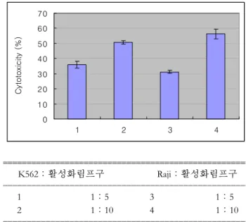
Table II. Cytotoxic effects to the target cells of Raji and K562 cells. Cytotoxicity was analyzed by Elisa reader, using the method of LDH release, Raji, K562 cells and activated lymphocyte were incubated together at the rate of 1:5 and 1:10, respectively.
(1 and 2), the rate of K562 cells and activated lymphocytes are 1:5 and 1:10. (3 and 4), the rate of Raji cellsa nd activated lymphocytes are 1:5 and 1:10, respectively.
0 10 20 30 40 50 60 70
1 2 3 4
Cytotoxicity (%)
ꠧꠧꠧꠧꠧꠧꠧꠧꠧꠧꠧꠧꠧꠧꠧꠧꠧꠧꠧꠧꠧꠧꠧꠧꠧꠧꠧꠧꠧꠧꠧꠧꠧꠧꠧꠧꠧꠧꠧꠧꠧꠧꠧꠧꠧꠧꠧꠧꠧꠧꠧ
K562:활성화림프구 Raji:활성화림프구
ꠏꠏꠏꠏꠏꠏꠏꠏꠏꠏꠏꠏꠏꠏꠏꠏꠏꠏꠏꠏꠏꠏꠏꠏꠏꠏꠏꠏꠏꠏꠏꠏꠏꠏꠏꠏꠏꠏꠏꠏꠏꠏꠏꠏꠏꠏꠏꠏꠏꠏꠏ
1 1:5 3 1:5
2 1:10 4 1:10
ꠏꠏꠏꠏꠏꠏꠏꠏꠏꠏꠏꠏꠏꠏꠏꠏꠏꠏꠏꠏꠏꠏꠏꠏꠏꠏꠏꠏꠏꠏꠏꠏꠏꠏꠏꠏꠏꠏꠏꠏꠏꠏꠏꠏꠏꠏꠏꠏꠏꠏꠏ
단일항체와 IL-2를 사용하여 배양한 결과이고, (3)은 anti-CD3과 CD16 단일항체를 사용하여 배양한 결과로 써, 세포의 성상은 각각 NK세포가 23.53±3.2, 68.71±
2.9%이고, T세포는 42.44±3.1, 9.83±1.1%이었다. 각각 의 K562세포에 대한 세포독성은 98.3±1.2, 52.3±1.7%
이었다.
다음의 결과는 Balb/c 누드마우스에 1×10⁷개의 Raji세 포를 각각의 실험군에 주입하여 종괴를 형성시킨 후 형 성된 종괴 (A), 꼬리의 미세정맥으로 PBS를 주입한 경우 (B), PBMC (C)를 주입한 경우 그리고 활성화된 세포를 주입한 경우(D)에 대한 실험결과로써 Table II는 K562세 포와 Raji세포를 표적세포로하여 활성화된 세포를 1:5, 1:10의 비율로 혼합시킨 후 LDH release방법으로 Elisa reader를 사용하여 측정한 결과로써 각각의 표적세포에 대한 세포독성률은 각각 35.97±2.4, 50.63±1.1% 그리고 31.2±0.9, 56.3±3.2%를 나타내고, 누드마우스에 주입한 활성화림프구의 성상은 NK세포가 39.63±2.8%이고, T
세포가 45.48±4.1%이었다. 세포독성이 유의한 결과를 보이는 세포를 위의 누드마우스에 투여한 결과 누드마 우스의 종괴의 변화와 누드마우스의 생존에 미치는 영 향을 관찰하여 나타낸 결과를 Table III과 Fig. 2에 표시하 였다. 각각의 실험군에서 활성화된 세포를 투여한 누드 마우스의 종괴의 크기의 증가는 A, B, C의 실험군에 비 해 현저히 증가추세가 둔화됨을 보이고 있고, 또한 누드 마우스의 생존에도 영향을 미친다. Fig. 2는 활성화된 세 포를 투여한 후 종괴의 크기가 줄어든 결과를 보여준다.
고 찰
세포독성 T 림프구와 자연살해세포(NK)세포는 림프 구와 표적세포사이에서 형성된 시냅스와 유사한 접합부 위로 perforin과 granzyme이 내포된 특별한 세포외분비성 분비 과립에 의해 표적세포를 융해시킨다(12,13). 여러 가지 인간의 질병들에는 세포외 과립분비 과정을 통해 서 림프구에 의해 조정되는 세포독성효과가 결여되어 있는 것으로 보고되었다(14). NK세포는 vitro 상태에서 인간의 종양에 대해서 그리고 vivo 상태의 설치류에서 항종양 활성을 보이는 것으로 보고되어졌다(15). vitro 상 태에서 배양된 표적세포인 종양세포나 vivo 상태에서 종 양을 융해시키는 NK세포의 능력에 대한 조사는 이후 NK세포를 암의 치료에 적용시킬 수 있는 것으로 예상되 어 흥미를 유발한다. 그러나 몇몇의 보고에 의하면 실질 적으로 단지 IL-2에 의해서 유도된 NK세포인 LAK세포 는 melanoma의 증례에서 치료효과가 보고되었을 뿐 그 다지 효과적인 결과를 보이지 않았다. NK세포는 그 단 독으로서 종양세포를 융해시킬 수 있는 능력이 있으나 T세포나 수지상세포와 같이 처리하였을 때 비 홉친스 림프종에서 더 나은 항 종양 효과를 보이는 것으로 보고 되어졌다(16).
본 연구의 결과에서 Fig. 1은 암세포주인 Raji와 K562 세포를 표적세포로 본 회사의 배양법으로 림프구를 활 성화시켜 1:10의 비율로 혼합하였을 때 NK세포가 각 암세포를 공격하는 그림으로 NK세포는 각기 두가지 형 태로 나타나는데 한가지의 형태는 세포질이 많은 큰 형 태로 그 세포안에는 과립들이 관찰되며, 또 다른 형태는 Table III. Tumor size (mm3) and survival of mice observed at 3 days intervals in five nude mices after 7 days of tumor injection and subsequently injection of PBS, PBMC and activated lymphocytes, respectively
ꠧꠧꠧꠧꠧꠧꠧꠧꠧꠧꠧꠧꠧꠧꠧꠧꠧꠧꠧꠧꠧꠧꠧꠧꠧꠧꠧꠧꠧꠧꠧꠧꠧꠧꠧꠧꠧꠧꠧꠧꠧꠧꠧꠧꠧꠧꠧꠧꠧꠧꠧꠧꠧꠧꠧꠧꠧꠧꠧꠧꠧꠧꠧꠧꠧꠧꠧꠧꠧꠧꠧꠧꠧꠧꠧꠧꠧꠧꠧꠧꠧꠧꠧꠧꠧꠧꠧꠧꠧꠧꠧꠧꠧꠧꠧꠧꠧꠧꠧꠧꠧꠧꠧꠧꠧꠧꠧꠧ
처리군 day0 day3 day6 day9 day13
ꠏꠏꠏꠏꠏꠏꠏꠏꠏꠏꠏꠏꠏꠏꠏꠏꠏꠏꠏꠏꠏꠏꠏꠏꠏꠏꠏꠏꠏꠏꠏꠏꠏꠏꠏꠏꠏꠏꠏꠏꠏꠏꠏꠏꠏꠏꠏꠏꠏꠏꠏꠏꠏꠏꠏꠏꠏꠏꠏꠏꠏꠏꠏꠏꠏꠏꠏꠏꠏꠏꠏꠏꠏꠏꠏꠏꠏꠏꠏꠏꠏꠏꠏꠏꠏꠏꠏꠏꠏꠏꠏꠏꠏꠏꠏꠏꠏꠏꠏꠏꠏꠏꠏꠏꠏꠏꠏꠏ
1 PBS 121 473.1 836.7 (6;2) 1306 (8;1) day 10, All dead
2 PBMC 130.9 367.6 913.9 (5;1) 1200.8 (8;2) day 10, All dead
3 NKM1 106.8 195.2 557.7 (8;1) 869.5 (10;2) day 13, All dead
4 NKM2 112.3 244.7 523.8 895.6 (11;2) day 13, All dead
ꠏꠏꠏꠏꠏꠏꠏꠏꠏꠏꠏꠏꠏꠏꠏꠏꠏꠏꠏꠏꠏꠏꠏꠏꠏꠏꠏꠏꠏꠏꠏꠏꠏꠏꠏꠏꠏꠏꠏꠏꠏꠏꠏꠏꠏꠏꠏꠏꠏꠏꠏꠏꠏꠏꠏꠏꠏꠏꠏꠏꠏꠏꠏꠏꠏꠏꠏꠏꠏꠏꠏꠏꠏꠏꠏꠏꠏꠏꠏꠏꠏꠏꠏꠏꠏꠏꠏꠏꠏꠏꠏꠏꠏꠏꠏꠏꠏꠏꠏꠏꠏꠏꠏꠏꠏꠏꠏꠏ ( ), dead day and number of mice: (6;2), dead day was 6, and the number is 2 mice: NKM, NK cell and T cell.
Figure 2. The photograph of tumor at 6 days in nude mice were injected PBS, PBMC and activated lymphocytes after 7 days of tumor injection. (A), Control, treatment with nothing after tumor injection, (B)(C)(D), a mouse treated with PBS, PBMC and activated lymphocytes, respectively.
A B
D
C
부정형으로 각각은 암세포의 주변에 결합하여 암세포를 apoptosis를 유발하는 것으로 관찰된다. Table I은 림프구 를 활성화시키는 배양방법에 따라 활성화된 림프구는 각기 다른 성상을 나타내는데, 각 세포의 성상에 따라 T세포가 많이 배양시킨 세포군과 NK세포를 많은 비율 로 배양시킨 세포군보다 NK세포와 NKT세포 그리고 T 세포를 고루 활성화시켜 배양시킨 세포군의 경우 K562 세포에 대한 세포독성이 더욱 증가함을 보여준다. 그러 므로 anti-CD3, CD16 또는 CD56 단일항체를 사용하여 배양시 첨가하고 IL-2와 함께 배양시키는 경우 세가지 항체를 모두 사용하여 배양한 림프구가 종양에 대한 세 포독성이 증가한다.
한편 누드마우스에 Raji세포를 주입하여 종괴를 형성 시키고, 본 연구의 배양법으로 배양시킨 사람의 활성화 된 림프구세포를 주입하여 관찰한 결과, 활성화된 림프 구세포의 성상이 NK세포가 39.63±2.8%이고, T세포가 45.48±4.1%인 세포의 K562세포와 Raji세포에 대한 세 포의 독성은 50.63±1.1, 56.3±3.2%이다. 이 세포를 종괴 가 형성된 누드마우스에 주입하였을 때 누드마우스의 생존은 결과에서 관찰되는 것처럼 큰 유의성을 가진 차 이를 보이지는 않았지만 누드마우스의 생존에 다소간 영향을 미치는 것으로 나타난다. 그러나 종괴의 크기에 서는 현저한 차이가 나타난다.
그러므로 본 연구의 결과로 볼 때 이종세포를 누드마 우스에게 주입함으로써 발생되는 여러 문제를 고려하여 볼 때 활성화된 사람의 림프구는 종괴가 형성된 누드마 우스의 생존과 종괴의 감소에 영향을 미친다고 할 수 있 으며, vitro상태의 실험에서 나타나는 표적세포에 대한 세포독성과 생체실험의 결과와는 다소간 차이가 있는 것으로 보이지만 본 연구에 사용된 배양법으로 활성화 시킨 사람의 자기 활성화림프구는 암세포의 증식이나 마우스의 생존에 영향을 준다. 본 연구에서 배양된 세포 의 분자생물학적인 연구와 마우스의 생존이나 종괴의 감소에 대한 연구가 더 있어야 할 것으로 생각된다.
감사의 글
본 연구를 수행하는데 있어서 암세포주의 공급이나
생체실험에 많은 도움을 주신 연세대학교 소화기내과 김한수 박사님과 강진구 선생님께 감사를 드립니다.
참 고 문 헌
1. Boon T, Couline PG, van den Eynde B: Tumor antigen re- cognized by T cells. Immunol Today 18;267-268, 1997 2. Rosenberg SA: Cancer vaccines based on the identification
of genes encoding cancer regression antigens. Immunol To- day 18;175-182, 1997
3. Berke G: The CTL kiss of death. Cell 81;9-12, 1995 4. Berke G: The Fas-based mechanism of lymphocytotoxicity.
Hum Immunol 54;1-7, 1997
5. Podack ER: Execution and suicide: cytotoxic lymphocytes en- force Draconian laws through separate molecular pathways.
Curr Opin Immunol 7;11-16, 1995
6. Liu CC, Walsh Cm, Young JD: Perforin: structure and func- tion. Immunol Today 16;194-201, 1995
7. Azzoni L, Kamoun M, Salcedo TW, Kanakaraj P, Perussia B: Stimulation of Fc gamma RTIIIA results in phospholipase C-gamma 1 tyrosine phosphorylation and p56lck activation.
J Exp Med 176;1745-1750, 1992
8. Herberman RB: Cancer Immunotherapy with Natural Killer Cells. Seminars in Oncology 3;27-30, 2002
9. Rosenberg SA: Lymphokine-activated killer cells. A new ap- proach to the immunotherapy of cancer. J Natl Cancer Inst 75;595-601, 1985
10. Morgan DA, Rusetti FW, Gallo R: Selective in vitro growth of T lymphocytes from normal human bone marrow. Sci- ences 193;1007-1009, 1976
11. Taga K, Yamaguchi A, Bloom ET, Tosato G: Target-induced death by apoptosis in human lymphokine-activated natural killer cells. Blood 87;2411-2418, 1996
12. Henkart PA, Williams MS, Zacharchuk CM, Sarin A: Do CTL kill target cells by inducing apoptosis? Semin Immunol 9;135-144, 1997
13. Stinchcombe JC, Grffiths GM: Regulated secretion from he- mopoietic cells. J Cell Biol 147;1-6, 1999
14. Klein C, Philippe N, Le Deist F, Fraitag S, Prost C, Durandy A, Fischer A, Griscelli C: Partial albinism with immunode- ficiency (Griscelli syndrome). J Pediatr 125;886-895, 1994 15. Rosenberg S, Lotze M, Muul L: A progress report on the
treatment of 157 patients with advanced cancer using lym- phokine-activated killer cells and interleukon-2 or high dose interleukin-2 alone. N Engl J Med 316;889-897, 1987 16. Oda H, Oda H, Hayashi M, Cho SH: Adoptive lymphocyte
immunotherapy in Non-Hodgkin's lymphoma (NHL). Bio- therapy 18;333-338, 2004