Clinical characteristics and treatment outcome of acute myeloid leukemia in elderly patients in Korea: a retrospective analysis
Hyeon Gyu Yi
1, Moon Hee Lee
1, Chul Soo Kim
1, Junsik Hong
2, Jinny Park
2, Jae Hoon Lee
2, Bo Ram Han
3, Ho Young Kim
3, Dae Young Zang
3, Se Hyung Kim
4, Seong Kyu Park
4, Dae Sik Hong
4, Guk Jin Lee
5, Jong-Youl Jin
5Department of Internal Medicine, 1Inha University Hospital, Inha University, 2Gachon University Gil Medical Center, Gachon University School of Medicine, Incheon, 3Hallym University Medical Center, Hallym University, Anyang, 4Soonchunhyang University Bucheon Hospital, Soonchunhyang University, 5Bucheon St. Mary's Hospital, The Catholic University of Korea, Bucheon, Korea; On Behalf of Gyeonggi/Incheon Branch, The Korean Society of Hematology
p-ISSN 2287-979X / e-ISSN 2288-0011 http://dx.doi.org/10.5045/br.2014.49.2.95 Blood Res 2014;49:95-9.
Received on October 7, 2013 Revised on March 1, 2014 Accepted on May 13, 2014
Background
The clinical characteristics of elderly patients with AML differ from those of younger pa- tients, resulting in poorer survival and treatment outcomes. We analyzed retrospectively the clinical data of AML patients 65 years old and above to describe patients' characteristics and treatment patterns, and to define meaningful prognostic factors of survival in the Korean population.
Methods
Basic patients’ characteristics, clinical outcomes according to treatments, and prognostic factors associated with survival and treatment intensity were examined in a total of 168 patients diagnosed in 5 institutes between 1996 and 2012 as having AML.
Results
Herein, 84 patients (50.0%) received high-intensity regimens (HIR), 18 (10.7%) received low-intensity regimens (LIR), and 66 (39.3%) received supportive care (SC) only. The me- dian survival of all patients was 4.5 months; and median survival times with HIR, LIR, and SC were 6.8 months, 10.2 months, and 1.6 months, respectively. Median survival times with HIR and LIR were significantly longer than that with SC (P<0.0001 and P=0.006, respectively). Multivariate analysis identified age, Eastern Cooperative Oncology Group-performance status (ECOG-PS), hemoglobin (Hb) level, and serum creatinine (Cr) level as statistically significant prognostic factors for survival. In the HIR group, prognostic factors for survival were ECOG-PS, Hb level, and C-reactive protein level.
Conclusion
Even in elderly AML patients, an intensive treatment regimen could be beneficial with careful patient selection. Further prospective studies designed to identify specific prog- nostic factors are required to establish an optimal treatment strategy for elderly AML patients.
Key Words Acute myeloid leukemia, Survival, Prognosis, Chemotherapy, Elderly
Correspondence to Jong-Youl Jin, M.D., Ph.D.
Department of Internal Medicine, Bucheon St. Mary's Hospital, The Catholic University of Korea, 327 Sosa-ro, Wonmi-gu, Bucheon 420-717, Korea Tel: +82-32-890-2587
Fax: +82-32-890-2585 E-mail: [email protected]
Ⓒ 2014 Korean Society of Hematology
INTRODUCTION
Acute myeloid leukemia (AML) is the most common acute leukemia in adults. The median age of patients is 65–70 years of at diagnosis [1]. Intensive treatments such as high-dose chemotherapy and hematopoietic stem cell trans-
plantation are effective in improving the survival of these patients [2]. However, many patients are not eligible for these treatments owing to their advanced age, poor perform- ance, or comorbidity. Furthermore, factors negatively influ- encing treatment outcomes, including adverse cytogenetic and molecular aberrations, antecedent hematologic dis- orders, and the incidence of fatal infection or bleeding, are
Table 2. Patterns of induction treatment.
Treatment pattern N (%)
High-intensity regimen 84 (50.0)
Idarubicin+cytarabine 76 (90.5) [reduced: 19 (22.6)]
Daunorubicin+cytarabine 6 (7.1) [reduced: 2 (2.4)]
FLAG 1 (1.2)
Modified FLAI 1 (1.2)
Low-intensity regimen 18 (10.7) Low-dose cytarabine 15 (83.3)
Decitabine 1 (5.6)
All-trans retinoic acid 1 (5.6)
Arsenic trioxide 1 (5.6)
Supportive care 66 (39.3)
Hydroxyurea 15 (22.7)
None 51 (77.3)
Abbreviations: reduced, reduced dose or infusion day compared to original regimen; FLAG, fludarabine, cytarabine, and G-CSF; FLAI, fludarabine, cytarabine, and idarubicin.
Table 1. Patients’ characteristics (N=168).
Characteristics Category N (%)
Gender Male:Female 85 (50.6):83 (49.4)
Age (y) Median 70, range 65‒89
65‒69 58 (34.5)
70‒74 72 (42.9)
75‒79 25 (14.9)
≥80 13 (7.7)
Diagnosis De Novo 152 (90.5)
Secondary 16 (9.5)
Risk groups Favorable 30 (17.9)
Intermediate 106 (63.1)
Unfavorable 32 (19.0)
ECOG-PS 0‒1 68 (40.5)
2‒4 100 (59.5)
HCT-CI 0‒1 117 (69.2)
≥2 50 (29.8)
Risk groups were defined according to the Southwest Oncology Group and Medical Research Council criteria.
Abbreviations: ECOG-PS, Eastern Cooperative Oncology Group performance status; HCT-CI, hematopoietic stem cell transplant- ation comorbidity index.
more frequent in old age [3]. As a result, the prognosis of elderly AML patients is very poor even with intensive treatment [3-7].
For these reasons, studies have been performed to identify optimal patient characteristics and optimal treatment regi- mens in elderly patients with AML. Specific parameters in- cluding clinical manifestations, laboratory values, and molec- ular and genetic markers have been suggested as prognostic or predictive factors [7].
In this study, we analyzed the clinical characteristics, treat- ment patterns, and outcomes of elderly AML patients (65 years old and above) treated at 5 Korean healthcare facilities.
Prognostic factors were also analyzed to define the relation- ship among clinical parameters, treatment patterns, and pa- tient survival.
MATERIALS AND METHODS
Patients
Patients who were at least 65 years old and were diagnosed with AML between 1996 and 2012 were included in this study. Five institutes located in Gyeonggi province and Incheon city, Korea participated in this study, and a total of 168 cases were analyzed. The following clinical parameters were included in the analysis: age, gender, antecedent hema- tologic disorder, cytogenetic risk group classified according to the Southwest Oncology Group and Medical Research Council (SWOG/MRC) criteria, Eastern Cooperative Oncol- ogy Group (ECOG) performance status (ECOG-PS), Hema- topoietic Cell Transplantation-Specific Comorbidity Index (HCT-CI), common blood cell count (CBC) test, lactate de- hydrogenase (LDH), C-reactive protein (CRP), bone marrow (BM) blast percent, and other laboratory values that have
been referred to as prognostic factors in past studies.
Treatment patterns and response criteria
Treatment patterns were divided into 3 groups: high-in- tensity regimen (HIR), low-intensity regimen (LIR), and sup- portive care (SC) groups. Treatment for the HIR group con- sisted of anthracycline, high dose cytarabine and fludarabine;
treatment for the LIR group consisted of low dose cytarabine, hypomethylating agent, arsenic trioxide and All-trans reti- noic acid; and the treatment for the SC group included hy- droxyurea or no active treatment.
Response criteria defined by the International Working Group for Diagnosis, Standardization of Response Criteria, Treatment Outcomes, and Reporting Standards for Therapeu- tic Trials in AML were used in this study [8].
Statistics
Multivariate analyses of survival were performed using the Cox proportional hazard model. Overall survival (OS) was calculated by Kaplan-Meier survival curves and survival differences between subgroups were compared using log rank test. Independent-samples T test, chi-square test, and like- lihood ratio test for trend were used to define the difference in clinical factors among the treatment groups. Statistical analysis was performed using the SPSS statistical software package (SPSS Inc., Chicago, IL).
RESULTS
PatientsÊ characteristics
Of the total of 168 patients, the number of male and female patients was 85 and 83, respectively. The median age was 70 years, and most patients had de novo AML (N=152, 90.5%). The intermediate-risk group (N=106, 63.1%) was the largest of the treatment groups. Sixty-eight patients (40.5%) had an ECOG-PS of 0 or 1, and 110 (69.2%) had an HCT-CI of 0 or 1 (Table 1).
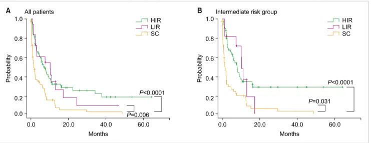
Fig. 2. (A) Survival comparison according to treatment intensity showed that median survivals in the HIR and LIR groups were significantly longer than that in the SC group. There was no difference in median survival between the HIR and the LIR group. (B) In the intermediate risk group, median survivals in the HIR and LIR groups were significantly longer than that in the SC group (P=0.031). There was no difference in median survival between the HIR and the LIR group. Abbreviations: HIR, high-intensity regimen; LIR, low-intensity regimen; SC, supportive care.
Fig. 1. (A) The median survival of all patients was 4.5 months (95% CI: 2.4‒6.7 months). (B) Kaplan-Meier analysis of overall survival according to the cytogenetic risk group.
Table 3. Clinical outcomes according to treatment intensity.
Response N (%)
High-intensity regimen (N=84)
Complete remission/Partial remission 36 (42.9)/13 (15.5)
Resistant 13 (15.5)
Induction mortality (<8 weeks) 12 (14.3)
Not available 10 (11.9)
Low-intensity regimen (N=18)
Complete remission/Partial remission 1 (5.6)/5 (27.8)
Resistant 4 (22.2)
Induction mortality (<8 weeks) 2 (11.1)
Not available 6 (33.3)
Supportive care (N=64)
Complete remission/Partial remission 0 (0)/3 (4.7)
Resistant 21 (32.9)
Early death (<8 weeks) 19 (29.7)
Not available 21 (32.8)
Treatment patterns and outcomes
Half of the patients (N=84, 50%) were treated with a HIR, and cytarabine with idarubicin was used mostly (N=76, 90.5%) among these patients. Further, 18 patients (10.7%) belonged to the LIR group and 66 patients (39.3%) to the SC group (Table 2). The response rate in the HIR group was 58.4% (N=49), with complete remission (CR) in 42.9%
and partial remission (PR) in 15.5% of the patients. The response rate in the LIR group was 33.3% (CR: 5.6%, PR:
27.8%). Only 3 patients in the SC group achieved PR (Table 3). Response rates in the favorable, intermediate, and un- favorable cytogenetic risk groups were 30% (CR: 20%, PR:
10%), 35.8% (CR: 22.6%, PR: 13.2%), and 34.4% (CR: 21.8%, PR: 12.5%), respectively.
The median survival of all patients was 4.5 months (95%
CI: 2.4–6.7 months) (Fig. 1A). Median survival rates in the
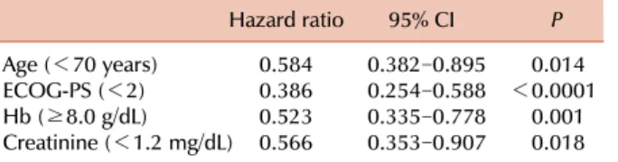
Table 4. Prognostic factors for survival in all patients.
Hazard ratio 95% CI P
Age (<70 years) 0.584 0.382‒0.895 0.014
ECOG-PS (<2) 0.386 0.254‒0.588 <0.0001
Hb (≥8.0 g/dL) 0.523 0.335‒0.778 0.001
Creatinine (<1.2 mg/dL) 0.566 0.353‒0.907 0.018 Abbreviations: ECOG-PS, Eastern Cooperative Oncology Group performance status; Hb, hemoglobin; CI, confidence interval.
Table 5. Prognostic factors for survival in high-intensity regimen group.
Hazard ratio 95% CI P
ECOG-PS (<2) 0.523 0.280‒0.977 0.042
Hb (≥8.0 g/dL) 0.362 0.189‒0.692 0.002
CRP (<10.0 mg/dL) 0.455 0.213‒0.971 0.042 Abbreviations: ECOG-PS, ECOG performance status; CRP, C-reactive protein.
favorable, intermediate, and unfavorable cytogenetic risk groups were 4.1 months (95% CI: 0.8–9.7 months), 5.0 months (95% CI: 1.6–8.4 months), and 4.5 months (95%
CI: 2.4–6.7 months), respectively. There was no significant difference in the survival rates among the cytogenetic risk groups (P=0.394) (Fig. 1B). The median survival rates in the HIR, LIR, and SC treatment groups were 6.8 months (95% CI: 4.4–9.3 months), 10.2 months (95% CI: 0.7–2.5 months), and 1.6 months (95% CI: 1.7–7.4 months), respectively. One-year survival rates in the HIR, LIR, and SC treatment groups were 31%, 26%, and 6%, respectively.
Compared to the SC group, median survival was significantly longer in the HIR and LIR groups (P<0.0001 and P=0.006, respectively) (Fig. 2A). Among the 106 patients in the inter- mediate risk group, the median survival was significantly shorter in the SC treatment group (1.4 months, 95% CI:
0.5–2.3 months) than in the HIR (7.9 months, 95% CI: 5.2–
10.6 months) or LIR treatment group (10.3 months, 95%
CI: 9.2–11.5 months; P<0.0001 and P=0.031, respectively).
There was no survival difference between the HIR and LIR treatment groups (Fig. 2B).
Prognostic factors of survival
Multivariate analysis with age, ECOG-PS, risk group, HCT-CI, CBC count, creatinine (Cr) level, LDH level, CRP level, and BM blast percent was performed for all patients in each treatment group. Age over 70 years, ECOG-PS, Hb level, and Cr level were proven to be significant prognostic factors of survival for all patients (Table 4). ECOG-PS, Hb level, and CRP level were revealed as significant prognostic factors of survival in the HIR group (Table 5).
DISCUSSION
Improvement of survival due to advances in supportive care, chemotherapy regimens, and transplantation techni- ques has been observed in AML patients [2]. However, elderly AML patients show poor prognosis because of characteristics associated with their age. Characteristics such as fragile medi- cal condition leading to the restriction of active treatments, a higher incidence of treatment-related mortality, and a high- er proportion of patients in the high-risk group, including those with high-risk cytogenetic or genetic profiles who may be refractory to treatment, were suggested as the causes of poor clinical outcomes in elderly AML patients [3].
Survival of elderly AML patients has been reported to be only several months [3-7]. In our study, the median survival of elderly AML patients was 4.5 months, which was a result comparable to those obtained in other studies.
In this study, the median survival in the HIR and LIR groups was significantly longer than that in the SC group among all patients and in the intermediate-risk group. This significant difference among treatment groups was con- sistently observed even when meaningful prognostic factors such as ECOG-PS, Hb level, and CRP level were forced into the model (P=0.001). A possible explanation for these observations is that the patients who received HIR had more favorable clinical characteristics than those in the SC group.
Comparing patients who received the HIR treatment to those in the SC group, the former were younger (average age 70.5±3.7 years vs. 75.7±5.7 years, respectively, P=0.01), had better ECOG-PS (0–1 vs. >1, respectively, P=0.049), had lower HCT-CI (0–1 vs. >1, respectively, P=0.049), had high- er average platelet counts (91.0±81.5×10⁹/L vs. 109.0±150.5×
109/L, respectively, P=0.029), and had lower average Cr levels (1.0±0.3 mg/dL vs. 1.3±1.6 mg/dL, respectively, P=0.0333).
This suggests that careful patient selection for high-intensity treatment with a meaningful use of prognostic factors could result in better clinical outcomes even in elderly AML patients.
Various prognostic or predictive models for elderly AML patients have been suggested to better predict clinical outcomes. Many factors, including age, PS, comorbidity, an- tecedent hematologic disorders, adverse cytogenetics, LDH level, blood cell counts, liver or kidney function, other labo- ratory values, and genetic profiles, have been reported as meaningful prognostic or predictive factors [7]. In our multi- variate analysis, age, ECOG-PS, Hb level, and Cr level were defined as prognostic factors (Table 4). Because of the higher incidence of treatment-related mortality in elderly AML pa- tients, selecting the optimal treatment intensity is of primary importance in improving clinical outcomes. Intensive treat- ment could be helpful for selecting patients who are expected to tolerate it. In our study, ECOG-PS, Hb level, and CRP level were suggested to be prognostic factors for the outcomes of intensive treatment (Table 5). However, although other studies have shown significant differences among the survival of general AML patients classified into different cytogenetic risk groups according to the SWOG/MRC criteria, such dif- ferences were not observed (P=0.394) in our study. This might be due to several reasons: the cytogenetic diagnosis
Fig. 3. In the high-intensity regimen group, survival of the patients showing complete remission (CR) was significantly longer than that of patients who did not show CR (non-CR) (P<0.0001).
may not have been accurate enough to enable the classi- fication of each patient into the correct risk group, the lack of a sufficient number of patients, or clinical risk factors such as secondary AML (N=16) and AML with MDS-related changes (N=32), which are not listed in the SWOG/MRC criteria, may have been more common in our elderly AML patients.
Several phase III trials of intensive chemotherapy given to elderly AML patients reported CR rates of 30–50% after induction of treatment [9-11]. In our study, CR rate was 42.9% and the response rate was 58.4% in the HIR group.
The patients with CR in this group had longer survival than non-CR patients (16.3 months vs. 3.2 months, respectively, P<0.0001) (Fig. 3). There was no significant difference in the responses within the LIR group, probably due to the low number of patients. These results imply that a consid- erable length of survival can be expected even in elderly AML patients when they show CR with intensive treatment.
Even though this study has the limitation of a retrospective study, the results are worthy of notice. This study reports the practical aspects of treatment patterns and outcomes in elderly AML patients in 5 Korean institutes. It shows that clinical characteristics and outcomes in the Korean pa- tients are similar to those in patients of western countries, and highlights the role of the same meaningful prognostic factors such as age, ECOG-PS, Hb level, Cr level, and CRP level. It also suggests that careful selection of patients receiv- ing intensive chemotherapy could bring about better out- comes in elderly AML patients.
AuthorsÊ Disclosures of Potential Conflicts of Interest
No potential conflicts of interest relevant to this article were reported.
REFERENCES
1. National Cancer Institute. SEER Cancer Statistics Review, 1975-2009 (Vintage 2009 Populations). Bethesda, MD: National Institutes of Health, 2012. (Accessed March 4 2013 at http://seer.
cancer.gov/csr/1975_2009_pops09/)
2. Pulte D, Gondos A, Brenner H. Improvements in survival of adults diagnosed with acute myeloblastic leukemia in the early 21st century. Haematologica 2008;93:594-600.
3. Dombret H, Raffoux E, Gardin C. Acute myeloid leukemia in the elderly. Semin Oncol 2008;35:430-8.
4. Menzin J, Lang K, Earle CC, Kerney D, Mallick R. The outcomes and costs of acute myeloid leukemia among the elderly. Arch Intern Med 2002;162:1597-603.
5. Lowenberg B, Downing JR, Burnett A. Acute myeloid leukemia.
N Engl J Med 1999;341:1051-62.
6. Shin HC, Na II, Yun T, et al. Acute myelogenous leukemia in the elderly (≥60): retrospective study of 115 patients. Korean J Med 2006;70:196-206.
7. Pollyea DA, Kohrt HE, Medeiros BC. Acute myeloid leukaemia in the elderly: a review. Br J Haematol 2011;152:524-42.
8. Cheson BD, Bennett JM, Kopecky KJ, et al. Revised recommend- ations of the International Working Group for Diagnosis, Standardization of Response Criteria, Treatment Outcomes, and Reporting Standards for Therapeutic Trials in Acute Myeloid Leukemia. J Clin Oncol 2003;21:4642-9.
9. Godwin JE, Kopecky KJ, Head DR, et al. A double-blind placebo-controlled trial of granulocyte colony-stimulating factor in elderly patients with previously untreated acute myeloid leukemia: a Southwest oncology group study (9031). Blood 1998;91:3607-15.
10. Witz F, Sadoun A, Perrin MC, et al. A placebo-controlled study of recombinant human granulocyte-macrophage colony-stim- ulating factor administered during and after induction treatment for de novo acute myelogenous leukemia in elderly patients.
Groupe Ouest Est Leucémies Aiguës Myéloblastiques (GOELAM).
Blood 1998;91:2722-30.
11. Goldstone AH, Burnett AK, Wheatley K, et al. Attempts to improve treatment outcomes in acute myeloid leukemia (AML) in older patients: the results of the United Kingdom Medical Research Council AML11 trial. Blood 2001;98:1302-11.
![Table 2. Patterns of induction treatment. Treatment pattern N (%) High-intensity regimen 84 (50.0) Idarubicin+cytarabine 76 (90.5) [reduced: 19 (22.6)] Daunorubicin+cytarabine 6 (7.1) [reduced: 2 (2.4)] FLAG 1 (1.2) Modified FLAI](https://thumb-ap.123doks.com/thumbv2/123dokinfo/5210059.120318/2.892.80.434.805.1057/induction-treatment-treatment-idarubicin-cytarabine-daunorubicin-cytarabine-modified.webp)