Endocrinol Metab 2013;28:192-198
http://dx.doi.org/10.3803/EnM.2013.28.3.192 pISSN 2093-596X · eISSN 2093-5978
Original Article
The Expression of Tumor-Associated Macrophages in Papillary Thyroid Carcinoma
Seunghwan Kim1, Sun Wook Cho2,3, Hye Sook Min4, Kang Min Kim5, Gye Jeong Yeom6, Eun Young Kim7,8, Kyu Eun Lee7, Yeo Gyu Yun7,8, Do Joon Park2, Young Joo Park2
1Seoul National University College of Medicine; 2Department of Internal Medicine, Seoul National University College of Medicine; 3Department of Internal Medicine, National Medical Center; 4Department of Pathology, Seoul National University College of Medicine; 5Hansung Science High School; 6Biomedical Research Institute, Seoul National University Hospital, Seoul National University College of Medicine; 7Department of Surgery, Seoul National University College of Medicine; 8Department of Surgery, National Medical Center, Seoul, Korea
Background: Tumor-associated macrophages (TAMs) play a tumorigenic role related to advanced staging and poor prognosis in many human cancers including thyroid cancers. Yet, a functional role of TAMs in papillary thyroid carcinoma (PTC) has not been established. The aim of this study was to investigate TAM expression in human PTC with lymph node (LN) metastasis.
Methods: Thirty-six patients who underwent surgery after being diagnosed with PTC with LN metastasis were included. Primary tumor tissues were immunohistochemically stained with an anti-CD68 antibody and clinical characteristics according to TAM density were evaluated.
Results: The TAM densities (CD68+ cells) varied from 5% to 70%, in all tumor areas, while few cells were stained in adjacent normal tissues. TAMs were identified as CD68+ cells with thin, elongated cytoplasmic extensions that formed a canopy structure over tumor cells. Comparing clinicopathologic characteristics between tumors with low (<25%) and high (25% to 70%) TAM densities, primary tumors were larger in the high density group than in the low density group (2.0±0.1 vs. 1.5±0.1; P=0.009).
Conclusion: TAMs were identified in primary PTC tumors with LN metastasis and higher TAM densities were related to larger tumor sizes, suggesting a tumorigenic role of TAMs in human PTCs.
Keywords: Tumor-associated macrophages; Thyroid cancer, papillary; Tumor size
INTRODUCTION
Macrophages are one of the components of bone marrow cells and play roles in innate immunity. Classically, they circulate in the blood and migrate into the inflammatory tissues such as infectious diseases in response to specific, endogenous immune signal [1,2]. Recently their alternative roles in anti-inflamma-
tion, cell clearance and tissue regenerations have also been es- tablished especially in inflammatory metabolic disorders in- cluding obesity and diabetes [1,2]. Therefore, macrophages have been categorized into two subtypes—M1 and M2—de- pending on their distinctive roles. M1 macrophages play pro- inflammatory functions in response to TH1 cytokines and M2 macrophages play anti-inflammatory and imunosuppressive
Received: 18 March 2013, Accepted: 24 May 2013 Corresponding author: Sun Wook Cho
Department of Internal Medicine, National Medical Center, 245 Eulji-ro, Jung-gu, Seoul 100-799, Korea
Tel: +82-2-2276-2305, Fax: +82-2-2269-0750, E-mail: swchomd@gmail.com
Copyright © 2013 Korean Endocrine Society
This is an Open Access article distributed under the terms of the Creative Com- mons Attribution Non-Commercial License (http://creativecommons.org/
licenses/by-nc/3.0/) which permits unrestricted non-commercial use, distribu- tion, and reproduction in any medium, provided the original work is properly cited.
activities in response to Th2 cytokines [3]. However, macro- phages are not fixed to a single M1 or M2 subtype but suspect- ed to rapidly differentiate and redifferentiate in various micro- environments.
Tumor-associated macrophages (TAMs), macrophages ex- isting tumor microenvironment, were first described in the ear- ly 1980s [4,5]. Till now, TAMs have been found to play dual functions, both positively or negatively affect tumor growth through interactions with the microenvironment, and these ac- tions are tissue specific [6,7]. Several clinical studies showed that high density TAMs were present in more advanced stages of cancer with a worse prognosis in breast [8,9], lung [10,11], and bladder cancer [12,13]. Although a high TAM density has been reported in poorly differentiated papillary thyroid carci- noma (PTC) or anaplastic thyroid cancer [14], the distribution and function of TAMs in the most common PTC have not been studied yet. In this study, we have demonstrated the existence of TAMs in PTC tissue with lymph node (LN) metastasis and also used expression distribution and morphological properties to investigate the relationship between TAMs and the frequen- cy of extrathyroidal invasion in PTC.
METHODS
Study subjects
The study was conducted from March to May 2012 at Seoul National University Hospital. The primary PTC lesion and surrounding normal tissues were collected from 36 consecu- tive patients with a previous thyroidectomy and central region LN dissection followed by a pathological diagnosis of PTC with LN metastasis. This diagnosis was defined as having a metastasized lesion in at least one LN from the histological examination of 1 to 37 LNs obtained from a central region LN dissection. All participating patients signed the informed con- sent form after receiving a comprehensive explanation of the study. This study was conducted with the approval of the Insti- tutional Review Board of Seoul National University Hospital (IRB approval No. H-0809-0970-258).
Histologic examination
All sample tissues were fixed in 10% neutral formalin solution for 24 hours and washed with distilled water for 20 minutes before histological observation. Tissues were dehydrated with ethyl alcohol, washed with xylene, embedded in paraffin, and sectioned into 3 µm sections. Sections were deparaffinized, using a standard process of anhydrous ethyl alcohol, washed,
and stained with hematoxilin and eosin. The size of the prima- ry cancer and presence of extrathyroidal invasion (report of other pathological findings) were measured by two or more pathologists.
For immunohistochemistry to demonstrate TAM expression, paraffin-embedded tissues were sectioned at 5 µm thick, depa- raffinized, placed on a slide, dried at 37°C, and deparaffinized with xylene. They were then incubated with antihuman CD68 antibody PG-M1 (1:50, DakoCytomation, Carpinteria, CA, USA). Color staining was achieved with a DAKO EnVision+
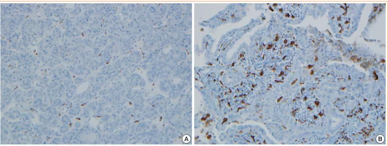
system (DakoCytomation) by using a streptavidin biotin-horse- radish peroxidase process and DAB (3,3´-diaminobenzi dine tetrahydrochloride) as a chromogen. Hematoxilin and eosin staining was used as a control. These sections were observed under an optical microscope. The proportion of CD68+ cells in each tumor on each slide was evaluated after staining. Based on a median value of 25%, weak positive (+) and strong posi- tive (++) staining were defined as <25% and ≥25%, respec- tively (Fig. 1).
Statistical analysis
Patients’ clinical characteristics are presented as mean±SEM.
CD68 immunohistochemistry results were classified accord- ing to the pathological analysis along with a correlation analy- sis of other clinical data. Statistical significance is indicated with the P value, and a significant difference was considered when P values for both were less than 0.05. Statistical analysis was conducted by using SPSS version 17.0 (SPSS Inc., Chica- go, IL, USA).
RESULTS
Clinical characteristics of study subjects
The mean age of the 36 study subjects was 49.3±2.1 years old, and 27 subjects (75%) were female. The mean tumor size was 1.4±0.1 cm, and extrathyroidal invasion was observed in 27 subjects (75%). Ten subjects (28%) had multiple (two or three) tumors and eight of them had tumors in both lobes. LN metastases (median, 2; range, 1 to 19) were pathologically confirmed. Three metastases were found opposite the tumor, and three patients had bilateral LN metastases (Table 1).
Characteristics of TAM staining in PTC
The CD68 immunohistochemistry effectively stained macro- phage cytoplasm and allowed us to observe TAMs in PTC.
The TAM nuclei in PTC tissue were approximately 1/3 to 1/2
the size of the nuclei in surrounding tumor cells, and the cyto- plasm was widely distributed, seeming to wrap around PTC cells. The cytoplasm of TAM and cancer cells appeared to be in close contact (Fig. 2). TAMs with extended nuclei were rare relative to the number of cancer cells due to the multidirec- tional expansion of TAM cytoplasm. The slide preparation may have affected the visibility of the cytoplasm, but not the layer containing the nucleus.
Clinical characteristics for the distribution of TAM expression
CD68+ cells were rare in normal thyroid tissue (Fig. 3A), but with the presence of TAMs, the CD68+ cells were present in
PTC lesions. TAMs accounted 5% to 75% of all cancer cells, and were dispersed throughout the cancer tissues (Fig. 3B) or were grouped together in some lesions (Fig. 3C). This papil- lary arrangement of the thyroid cancer was maintained regard- less of the presence of TAMs. There was no significant differ- ence in clinical characteristics such as age at diagnosis, gen- der, size or number of primary tumor, or presence of extrathy- roidal invasion according to the TAM expression pattern (dif- fuse or localized).
Clinical characteristics by TAM density
The patients’ clinical characteristics were compared in tumors with low (17 patients) and high (19 patients) TAM density. TAM Table 1. Clinical Characteristics according to the Density of Tumor-Associated Macrophage
Characteristic Total (n=36) Low density (n=17) High density (n=19) P value
Age of diagnosis, yr 49.3±2.1 50.0±2.9 48.0±2.8 0.626
Sex, male/female 9/27 5/12 4/15 0.706
Tumor size, cm 1.4±0.1 1.5±0.1 2.0±0.1 0.009
Multiplicity, % 10 (28) 6 (35) 4 (21) 0.255
LN metastasis Number
Location, presence of ipsilateral 2 (1-19)
6 (17) 2 (1-5)
3 (18) 2 (1-19)
3 (16) 1.000
Extrathyroidal invasion, % 27 (75) 12 (71) 15 (79) 0.706
Values are expressed as mean±SD, number (%), or median (range).
LN, lymph node.
Fig. 1. Classification of the density of CD68-positive cells in human papillary thyroid carcinoma. Immunohistochemical staining of CD68 was performed and tumor-associated macrophages (TAMs) were scored by the number of CD68+ cells/total tumor cells under × 400 magnification. Patients were divided into two groups according to TAM density; (A) low (<25%) and (B) high (25% to 70%) TAM density.
A B
Fig. 2. Morphologies of CD68-positive tumor-associated macro- phages (TAMs) in papillary thyroid carcinomas (PTCs). CD68+ cells (brownish) with thin elongated cytoplasmic extensions were intercalated into the papillary structures of PTCs. TAMs formed a distinctive canopy-like structure over cuboidal tumor cells (×400).
Fig. 3. Diverse immunohistochemical staining of CD68 in thyroid tissues. (A) Normal thyroid tissues show negative staining (×200).
(B) Diffuse and (C) focal clustered staining in papillary thyroid carcinomas (×200).
A B C
density was the proportion all cancer cells that were CD68+. The mean age at diagnosis and gender were not significantly different between the two groups, but the primary tumors were statistically larger in the group with high TAM density (1.5±
0.1 vs. 2.0±0.1; P<0.01) (Table 1). The position and number of LN metastases was not analyzed statistically due to the small number of subject, but two patients with six or more LN me- tastases had TAM densities of 50% or higher. The number of tumors and presence of extrathyroidal invasion were similar between the two groups (Table 1).
DISCUSSION
This study demonstrated the presence of TAM in a canopy
structure over thyroid tumor cells in primary tumor tissues from 36 patients with PTC with LN metastasis. In addition, the primary tumors were significantly larger in the group with high TAM density. More LNs had metastases with a higher TAM density, although the difference was not statistically sig- nificant. Based on these findings, TAM density is considered to be related to PTC stage.
Ryder et al. [14] demonstrated a higher TAM density in poor- ly differentiated PTC and anaplastic thyroid cancer than in well-differentiated PTC, by using CD68 and CD163 immuno- histochemistry. Qing et al. [15] also reported a high TAM den- sity in well-differentiated PTC, and the TAM density was sig- nificantly higher in tumors with TNM staging over stage III.
The results from these studies demonstrated that a high TAM density is present in poorly differentiated PTC and is related to poor prognosis, which indicates TAMs may support PTC growth.
However, the role of TAMs in thyroid cancer remains contro- versial. Fiumara et al. [16], using CD68 immunohistochemis- try, found that TAM prevented metastasis in some patients. Of 121 patients with PTC, 15% had TAMs (CD68+) that appeared to have a phagocytic function on cancer cells. These patients had significantly less blood vessel invasion and remote metas- tasis and largely accompanying invasions of lymphocytes and dendritic cells, than patients without an CD68+ cells. These re- sults suggest that these cells may activate the host immune re- sponse to cancer cells. However, in our study, 56% of patients had TAMs without evidence of phagocytosis, and their clinical characteristics were similar to those without TAMs.
This study investigated TAM morphologies and evaluated related clinical characteristics in PTC tissue with relatively advanced LN metastasis. Interestingly, CD68+ TAM cells ob- served in this study appeared to be mostly cytoplasm forming a canopy structure that projected over cancer cells. These can-
opy structures formed over the interior and exterior papillary projections in PTC cells that normally form papillary struc- tures. These morphological characteristics are a standard of the TAM definition and differ from the rounded amoeba shape of inflammatory response cells, which are generally referred to as M1 macrophages [17]. The morphological characteristics of TAMs support cancer cell growth by forming a structural network between cancer cells and the extracellular matrix and are considered to support endothelial cells in forming blood vessels [18,19]. Caillou et al. [20] described the morphological characteristics of TAMs and their adjacency between cells, and these morphological characteristics suggest that TAMs facili- tate cancer cell growth and metastasis. In this study, however, the morphological characteristics of thyroid cancer tissue were maintained regardless of the presence of TAMs, hence we sus- pect that TAMs do not have a huge effect on cancer develop- ment [20].
Interestingly, despite of small number of patients, when we classified patients according to TAM density, the primary tu- mors were larger in the high TAM density group. Hence, we hypothesize that TAMs inhibit tumor growth in advanced PTC.
These results differ from previous findings, where Qing et al.
[15] compared the micropapillary cancer smaller than 1 cm and papillary cancer of typical size (1 cm or larger) and found no difference in TAM density. Apart from the etiological inter- pretation, TAM can be interpreted to be involved in cancer in- vasiveness after mobilization followed by the cancer growth.
Actually, TAM has been related to metastasis in various cancer types [12].
The study has a limitation of not including patients without LN metastasis. This study on TAMs reported that TAMs are rare in PTC, and especially they have only been reported in poorly differentiated cancer or anaplastic thyroid cancer. There- fore, this study only included the subjects with LN metastasis in higher stages of papillary cancer. A further study with more subjects including patients without LN metastasis is warranted.
Studies on cancer treatments targeting TAM have increased since the first report that TAM facilitates cancer growth and metastasis and offers indicates a poor prognosis. Popular treat- ment strategies that have been investigated involve cytokines such as colony stimulating factor (CSF)-1 [21], which mobi- lizes TAM to the tumor; interleukin (IL)-10 [21] and IL-4 [22], which facilitate a cancer’s affinity for TAM through macro- phage differentiation into the M1 and M2 subtypes; and CCR2 [23,24], which facilitates TAM in cancer metastasis. Interest- ingly, a recent animal study was published that suggests that
these strategies might be feasible in treating thyroid cancer [25]. Ryder et al. [25] demonstrated PTC inhibition in a BRAF- mutant, transformed mice model by inhibiting CSF-1 gene ex- pression through genetic modification or use of a CSF-1/CSF- 1 receptor signal pathway inhibitor.
Studies on TAM in thyroid cancer are underway, but clini- cal data and studies on the mechanisms of TAM action are lacking. Thyroid cancer has a relatively favorable prognosis because its growth rate is slow. Therefore, general cancer patho- physiology does not apply to the role of TAMs. Currently the role of TAMs is only known in relatively invasive breast, lung, and pancreatic cancers, but the results of this study indicate that TAMs also exist in PTC, which is generally well differen- tiated with a favorable prognosis. Their role in cancer devel- opment is exciting; especially, the in light of the animal study targeting TAM that found possible inhibition of PTC. Expec- tations are high for treating patients with thyroid cancer since the BRAF mutation rate is higher among the Korean popula- tion than other ethnic groups. Additional study on the relation- ship between the BRAF mutation and TAM in Korean patients is warranted.
This study was conducted with relatively few patients, but it demonstrated the presence of TAMs in clinically common PTC patients, and the presence of a high TAM density in larger pri- mary cancers. However patients without LN metastasis were not included in the study or the comparative analysis. Another limitation arose from not evaluating the characteristics of TAMs in metastasized LNs. Also, there is a fundamental controversy over whether CD68, the TAM marker, is appropriate for the staining M2 macrophages, so the more specific CD 163 should be stained in future analyses.
As a result of immunohistochemistry by using anti-CD68 antibody in PTC tissue with LN metastasis, TAM was present in all tissues with an extended cytoplasm forming a canopy shape over cancer cells. The density varied from 5% to 70%
according to the tissue type. In normal thyroid tissue from the same patients, CD68+ cells were rare. When tumors were clas- sified by TAM density, the primary tumors were statistically significantly larger in the group with a high TAM density. There- fore, TAM is suggested to stimulate cancer growth in PTC tis- sue with LN metastasis.
CONFLICTS OF INTEREST
No potential conflict of interest relevant to this article was re- ported.
ACKNOWLEDGMENTS
This work was supported by a grant from the Next-Generation BioGreen 21 Program (No. PJ00954003), Rural Development Administration, Republic of Korea.
REFERENCES
1. Gordon S. Alternative activation of macrophages. Nat Rev Immunol 2003;3:23-35.
2. Mosser DM. The many faces of macrophage activation. J Leukoc Biol 2003;73:209-12.
3. Mantovani A, Sozzani S, Locati M, Allavena P, Sica A. Ma- crophage polarization: tumor-associated macrophages as a paradigm for polarized M2 mononuclear phagocytes. Trends Immunol 2002;23:549-55.
4. Ladusch M, Schaffner H, Ullmann L, Littmann M, Rei- mann S, Gindl P, Ambrosius H. Pre- and postoperative re- activity of breast cancer patients to tumor associated anti- gens and HEP in the macrophage electrophoresis mobility (MEM) test. Arch Geschwulstforsch 1982;52:469-78.
5. Neumeister B, Hambsch K, Storch H. Macrophage adher- ence inhibition test (MAI) in Wistar rats bearing Jensen tu- mors. I. MAI after incubation with tumor-associated anti- gens. Arch Geschwulstforsch 1983;53:521-8.
6. Bingle L, Brown NJ, Lewis CE. The role of tumour-asso- ciated macrophages in tumour progression: implications for new anticancer therapies. J Pathol 2002;196:254-65.
7. Heusinkveld M, van der Burg SH. Identification and ma- nipulation of tumor associated macrophages in human can- cers. J Transl Med 2011;9:216.
8. Tsutsui S, Yasuda K, Suzuki K, Tahara K, Higashi H, Era S.
Macrophage infiltration and its prognostic implications in breast cancer: the relationship with VEGF expression and microvessel density. Oncol Rep 2005;14:425-31.
9. Campbell MJ, Tonlaar NY, Garwood ER, Huo D, Moore DH, Khramtsov AI, Au A, Baehner F, Chen Y, Malaka DO, Lin A, Adeyanju OO, Li S, Gong C, McGrath M, Olopade OI, Esserman LJ. Proliferating macrophages associated with high grade, hormone receptor negative breast cancer and poor clinical outcome. Breast Cancer Res Treat 2011;
128:703-11.
10. Hirayama S, Ishii G, Nagai K, Ono S, Kojima M, Yamau- chi C, Aokage K, Hishida T, Yoshida J, Suzuki K, Ochiai A. Prognostic impact of CD204-positive macrophages in lung squamous cell carcinoma: possible contribution of
Cd204-positive macrophages to the tumor-promoting mi- croenvironment. J Thorac Oncol 2012;7:1790-7.
11. Sato S, Hanibuchi M, Kuramoto T, Yamamori N, Goto H, Ogawa H, Mitsuhashi A, Van TT, Kakiuchi S, Akiyama S, Nishioka Y, Sone S. Macrophage stimulating protein pro- motes liver metastases of small cell lung cancer cells by affecting the organ microenvironment. Clin Exp Metasta- sis 2013;30:333-44.
12. Zhang QW, Liu L, Gong CY, Shi HS, Zeng YH, Wang XZ, Zhao YW, Wei YQ. Prognostic significance of tumor-asso- ciated macrophages in solid tumor: a meta-analysis of the literature. PLoS One 2012;7:e50946.
13. Takayama H, Nishimura K, Tsujimura A, Nakai Y, Nakaya- ma M, Aozasa K, Okuyama A, Nonomura N. Increased in- filtration of tumor associated macrophages is associated with poor prognosis of bladder carcinoma in situ after in- travesical bacillus Calmette-Guerin instillation. J Urol 2009;
181:1894-900.
14. Ryder M, Ghossein RA, Ricarte-Filho JC, Knauf JA, Fagin JA. Increased density of tumor-associated macrophages is associated with decreased survival in advanced thyroid can- cer. Endocr Relat Cancer 2008;15:1069-74.
15. Qing W, Fang WY, Ye L, Shen LY, Zhang XF, Fei XC, Chen X, Wang WQ, Li XY, Xiao JC, Ning G. Density of tumor- associated macrophages correlates with lymph node me- tastasis in papillary thyroid carcinoma. Thyroid 2012;22:
905-10.
16. Fiumara A, Belfiore A, Russo G, Salomone E, Santonocito GM, Ippolito O, Vigneri R, Gangemi P. In situ evidence of neoplastic cell phagocytosis by macrophages in papillary thyroid cancer. J Clin Endocrinol Metab 1997;82:1615-20.
17. Lawrence T, Natoli G. Transcriptional regulation of mac- rophage polarization: enabling diversity with identity. Nat Rev Immunol 2011;11:750-61.
18. Lewis CE, Pollard JW. Distinct role of macrophages in dif- ferent tumor microenvironments. Cancer Res 2006;66:605- 12.
19. Mosser DM, Edwards JP. Exploring the full spectrum of macrophage activation. Nat Rev Immunol 2008;8:958-69.
20. Caillou B, Talbot M, Weyemi U, Pioche-Durieu C, Al Ghu- zlan A, Bidart JM, Chouaib S, Schlumberger M, Dupuy C.
Tumor-associated macrophages (TAMs) form an intercon- nected cellular supportive network in anaplastic thyroid carcinoma. PLoS One 2011;6:e22567.
21. Verreck FA, de Boer T, Langenberg DM, Hoeve MA, Kra- mer M, Vaisberg E, Kastelein R, Kolk A, de Waal-Malefyt
R, Ottenhoff TH. Human IL-23-producing type 1 macro- phages promote but IL-10-producing type 2 macrophages subvert immunity to (myco)bacteria. Proc Natl Acad Sci U S A 2004;101:4560-5.
22. Stein M, Keshav S, Harris N, Gordon S. Interleukin 4 po- tently enhances murine macrophage mannose receptor ac- tivity: a marker of alternative immunologic macrophage activation. J Exp Med 1992;176:287-92.
23. Penton-Rol G, Cota M, Polentarutti N, Luini W, Bernasco- ni S, Borsatti A, Sica A, LaRosa GJ, Sozzani S, Poli G, Man- tovani A. Up-regulation of CCR2 chemokine receptor ex-
pression and increased susceptibility to the multitropic HIV strain 89.6 in monocytes exposed to glucocorticoid hor- mones. J Immunol 1999;163:3524-9.
24. Geissmann F, Jung S, Littman DR. Blood monocytes con- sist of two principal subsets with distinct migratory prop- erties. Immunity 2003;19:71-82.
25. Ryder M, Gild M, Hohl TM, Pamer E, Knauf J, Ghossein R, Joyce JA, Fagin JA. Genetic and pharmacological tar- geting of CSF-1/CSF-1R inhibits tumor-associated macro- phages and impairs BRAF-induced thyroid cancer progres- sion. PLoS One 2013;8:e54302.