207
치과 환자로부터 발거된 치아에서의 법랑돌기의 발현빈도
배성숙·구 영1†
서울대학교 치과병원 치위생팀
1서울대학교 치과대학 치주과학교실
The Prevalence of Enamel Projection on Molar Teeth Extracted from Dental Patients
Sung-Suk Bae and Young Ku
1†Department of Dental Hygiene, Seoul National University Hospital
1Department of Periodontology, School of Dentistry, Seoul National University 275-1 Yongon-dong, Chongno-gu, Seoul 110-768, Korea
ABSTRACT
Many studies reported that the presence of cervical enamel projection (CEP) in cemento-enamel junction(CEJ) is greatly related to periodontal disease. The aim of this study was to investigate the prevalence of enamel projections in buccal, mesial, distal and lingual(palatal) surface of maxillary and mandibular first and second molars on extracted teeth. Among 660 teeth extracted due to the periodontal disease and dental caries in Seoul National University Dental Hospital was examined, 530 teeth which has distinct CEJ were examined with 8 times x electronic magnifier by one examiner. The prevalence of CEP for maxillary teeth (45.49%) was higher than that of mandible (39.62%). The first molar (45.22%) had more CEP than second (39.89%). Furthermore, buccal surface had highest incidence of CEP than other surfaces. The results of this study imply that the clinicians should take good care of the prevalence of CEP when scaling or root planning, plaque control instruction and periodontal surgery.
Key words
Cervical enamel projection, Cemento-enamel junction, Prevalence
서 론
치아의해부학적또는형태학적특징은임상적으로매우중 요한의미를가지며
,
치아형태의발육이상은치태의축적과성 장에은신처역할을하게된다1). Shiloah
와Kopczyk
는치아의발육이상이치주질환을악화시킨다고하였다2)
.
이미 오래전Master
와Hokins
는 미국인을대상으로 치아발육이상의하나인대구치의법랑경계부에나타나는법랑돌기에대하여보고한 바있으며3)
,
이4)는한국인을대상으로법랑돌기의발현 빈도와 치주질환과의 연관성에 대하여 보고한바 있다. Grew
등은법랑돌기의빈도
,
위치및범위와이로인한치근부착정도와의 관련및 이환된구치부의치주질환에 대한연구에서 상악제1
대구치를제외한모든대구치가법랑돌기와 관련이있다고하 였으나5)
,
조6) 는법랑돌기의발현은다근치인상하악대구치에 있어서치근분지부병변(furcation involvement)
에영향을준다고하였다
.
대구치법랑돌기의발현은결체조직에의한부착이 일어나지않고,
상피세포만의부착을일으켜치태세균의침범을용이하게하는결과에따라부착치은의소실을가져올뿐아니 라
,
대구치치근분지부병변에영향을미치게된다6).
많은 연 구에서 치아형태의발육이상가운데 하나인대구치경계부에 나타나는법랑돌기의발현이치주질환과관련성을가지고있는것을보고하고 있다1-12)
.
한국인을 대상으로한 발거된치아의법랑돌기 발생빈도에대한연구로는
1975
년이4)의보고가거의 유일하며
,
그 후 지난30
년간 이에 관한 조사연구보고는 거의없는실정이다.
이연구의목적은치주질환및 치아우식 등의 원인으로발거된 치아를대상으로법랑돌기를조사하여 상하악대구치별,
치면별 발현빈도를비교분석하는것이다.
재료 및 방법
1. 연구재료
연구재료로는서울대학교치과병원에내원한환자로부터 치 주질환또는치아우식증등의 원인으로발거된
660
개의치아 중,
보철또는보존치료로인해백악법랑경계가불분명한치아 및제1, 2
대구치의구별이어려운치아는조사대상에서제외하 였으며,
총530
개의 치아를대상으로 관찰하였다(Table 1).
2. 연구 방법
발거된치아를포르말린용액에고정시킨다음흐르는물에
†
Corresponding author Tel: 02-2072-2661 Fax: 02-2072-3860
E-mail: [email protected]
잘씻고
,
초음파치석제거기를이용하여치석과이물질들을제 거하고생리식염수로깨끗이세정후건조시켰다.
정확한관찰을 위하여
8
배 전자확대경(S300 II, Tokyo Kinzoku, Japan)
을 이용하였다
.
법랑돌기의 발육이상에대한 등급별 분류는Masters
3)의 방법에 따라 초기상태(incipient), Class I, II, III
로 구분하였다
(Table 2, Fig. 1).
상하악 제1, 2
대구치를 분류하고이들을다시협면,
근심면,
원심면,
설면(
구개면)
으로구분하여 법랑돌기의 발현 정도를 관찰하였다
.
결과 및 고찰
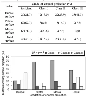
1. 상악 제1대구치의 치면별 법랑돌기의 발현빈도 관찰한상악 제
1
대구치92
개, 368
면의 치아중에서177
개면
(48.09%)
에서 법랑돌기가관찰되었으며,
협면에서78.26%
로 가장 높은 비율을 나타내었다.
상악제
1
대구치에서법랑돌기발현비율은협면에서Class I
이
13.04%, Class II
가23.91%, Class III
가41.30%
로 나타 났다.
구개면에서는Class I
이8.69%, Class II
가16.30%, Class III
가7.60%
였으며,
근심면에서는Class I
이20.65%, Class II
가7.60%
로 나타났으나, Class III
는관찰되지않았다.
원심면에서는
Class I
이15.21%, Class II
가30.43%, Class III
가
7.6%
로 나타났다(Table 3, Fig. 2).
2. 상악 제2대구치의 치면별 법랑돌기의 발현빈도 관찰한상악제
2
대구치229
개치아, 916
치면중에서42.90%
인
393
치면에서법랑돌기가 나타났다.
협면에서가장많이나타났으며
, 186
개의협면(81.22%)
에서법랑돌기의발현이관찰되었다
.
상악 제
2
대구치 협면에서는Class I
이15.72%, Class II
가
37.55%, Class III
가27.94%
로 나타났다.
구개면에서는Class I
이11.35%, Class II
가3.93%, Class III
가2.18%
로 다른 치면에 비해 발현율이 낮았다.
근심면에서는Class I
이20.08%, Class II
가6.55%, Class III
가2.18%
였고,
원심면에 서는Class I
이26.20%, Class II
가13.53%, Class III
가4.36%
로 나타났다
(Table 4, Fig. 3).
Table 1.
Number of examined teeth
Teeth Number of Teeth(EA) Number of tooth
surface (surface) Max. 1st molar
Max. 2nd molar Mand. 1st molar Mand. 2nd molar
22992 108101
368916 432404
Total 530 2,120
Table 4.The prevalence of enamel projection on maxillary 2nd molar Surface Grade of enamel projection (%)
Incipient Class I Class II Class III Buccal
surface 43(18.7) 36(15.7) 86(37.5) 64(27.9) Palatal
surface 189(82.5) 26(11.3) 9(3.9) 5(2.1) Mesial
surface 163(71.1) 46(20.0) 15(6.5) 5(2.1) Distal
surface 128(55.8) 60(26.2) 31(13.5) 10(4.3)
Table 3.The prevalence of enamel projection on maxillary 1st molar Surface Grade of enamel projection (%)
incipient Class I Class II Class III Buccal
surface 20(21.7) 12(13.0) 22(23.9) 38(41.3) Palatal
surface 62(67.3) 8(8.6) 15(16.3) 7(7.6) Mesial
surface 66(71.7) 19(20.6) 7(7.6) 0(0) Distal
surface 43(46.7) 14(15.2) 28(30.4) 7(7.6)
Fig. 1.Classification of examined teeth.
Table 2.Classification of examined teeth
incipient There is no enamel projection
Class I The enamel projection extends from the cementoenamel junction of the tooth toward the furcation entrance Class II The enamel projection approaches the entrance to the furcation. It does not enter the furcation, and there fore no horizontal component is present.
Class III The enamel projection extends horizontally into the furcation.
Fig. 2. The prevalence of enamel projection on the maxillary first molar.
3. 하악 제1대구치의 치면별 법랑돌기의 발현빈도 관찰 하악 제
1
대구치108
개의 치아, 432
치면 중에서42.36%
인183
개의치면에서법랑돌기가나타났으며,
관찰한치면중에서 협면에가장많이나타났고, 83
개의협면(76.85%)
에서법랑돌 기가 나타났다.
하악 제
1
대구치협면에서는Class I
이21.29%, Class II
가
28.70%, Class III
가26.85%
로나타났다.
설면에서는Class I
이43.51%, Class II
가24.07%, Class III
가7.40%
로 나타 났으며,
근심면에서는Class I
이6.48%, Class II
가0.92%, Class III
가0.92%
였고,
원심면에서는Class I
이8.33%, Class II
가0.92%
였으나Class III
는 관찰되지 않았다(Table 5, Fig. 4).
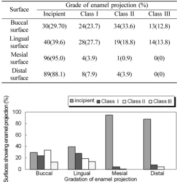
4. 하악 제2대구치의 치면별 법랑돌기의 발현빈도 하악제
2
대구치101
개의치아, 404
개의치면중에서36.88%
인
146
개의 치면에서 법랑돌기가 나타났다.
하악제
2
대구치협면에서가장많이 발현되었으며, 71
개치면
70.29%
에서 법랑돌기가 나타났고,
그 중Class
Ⅰ이23.76%, Class II
가33.66%, Class III
가12.87%
로 나타났다.
설면에서는
Class
Ⅰ이27.72% Class II
가18.81%, Class III
가
13.86%
였으며,
근심면에서는Class I
가3.96 %, Class II
가
0.99%
로나타났으나, Class III
는관찰되지않았다.
원심면 에서는Class I
이7.92%, Class II
가3.96%
로 나타났으나Class III
는 관찰되지 않았다(Table 6, Fig. 5).
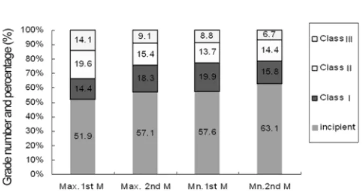
5. 상하악 제1, 2대구치의 치아별 법랑돌기의 발현 상하악제
1, 2
대구치530
개치아의치아별법랑돌기발현빈 도 분석결과,
조사대상전체치아에서초기상태가가장많았고,
상악 제
1
대구치에서는Class II
가,
나머지 대구치에서는ClassI
이 가장 많이 관찰되었다(Table 7, Fig. 6).
이상의결과를종합하여볼때
,
법랑돌기의발생빈도는상악 에서45.49%,
하악에서39.62%
로,
하악에서보다 상악에서높 았으며,
제1
대구치는45.22%,
제2
대구치는39.89%
로,
제2
대구치보다 제
1
대구치에서 더높게나타났다.
또한치면별발현빈 도는 전체대상치아의 협면에서가장 높게나타났으며,
상악 Fig. 3. The prevalence of enamel projection on the maxillarysecond molar.
Table 6. The prevalence of enamel projection on mandibular 2nd molar Surface Grade of enamel projection (%)
Incipient Class I Class II Class III Buccal
surface 30(29.70) 24(23.7) 34(33.6) 13(12.8) Lingual
surface 40(39.6) 28(27.7) 19(18.8) 14(13.8) Mesial
surface 96(95.0) 4(3.9) 1(0.9) 0(0)
Distal
surface 89(88.1) 8(7.9) 4(3.9) 0(0)
Table 5. The prevalence of enamel projection on mandibular 1st molar Surface Grade of enamel projection (%)
Incipient Class I Class II Class III Buccal
surface 25(23.1) 23(21.2) 31(28.7) 29(26.8) Lingual
surface 27(25.0) 47(43.5) 26(24.0) 8(7.4) Mesial
surface 99(91.6) 7(6.4) 1(0.9) 1(0.9)
Distal
surface 98(90.7) 9(8.3) 1(0.9) 0(0)
Fig. 4
.
The prevalence of enamel projection on the mandibular first molar.(Surfaces showing enamel projection (%))Fig. 5. The prevalence of enamel projection on the mandibular second molar.(Surfaces showing enamel projection (%))
Table 7.The prevalence of enamel projection on each molars Tooth Grade number and percenttage of enamel projection
incipient Class I Class II Class III Maxillary 1st
Molar 191(51.9) 53(14.4) 72(19.6) 52(14.1) Maxillary
2nd Molar 523(57.1) 168(18.3) 141(15.4) 84(9.1) Mandibular
1st Molar 249(57.6) 86(19.9) 59(13.7) 38(8.8) Mandibular
2nd Molar 255(63.1) 64(15.8) 58(14.4) 27(6.7)
제
1
대구치협면에서78.27%,
상악제2
대구치협면에서81.23%,
하악 제
1
대구치협면에서76.86%,
하악제2
대구치협면에서70.3%
의 발현율을 보였다.
법랑돌기는치관과치근의경계부위인치경부에나타나는이 상형태이며
,
치주질환과의관련성에 대하여많은연구가진행되어왔다1-12)
. Masters
등의 연구에의하면,
상악에서28.6%,
하악에서
17%
의분포율을보고하였으나3),
우리의 연구결과는 상악에서45.49%,
하악에서39.62%
로 높게나타났다.
미국인 을대상으로 연구한Grewe
등 5)의보고에서는상악 제1
대구치에서
8.2%,
하악 제2
대구치에서35.5%
였음을보고 하였으 며, Tsatsas
등7)은1,608
개의 상하악대구치를대상으로조사 한 결과 상악 제2
대구치에서24.4%,
하악 제2
대구치에서34.8%
의 발현율을 보고하였다.
한국인을 대상으로 한 이4)의연구에서도하악 제
2
대구치에서35%,
상악 제1
대구치에서는27.5%
의출현율을보고하였는데,
우리들의연구결과는하악제2
대구치에서36.88%,
상악 제1
대구치에서48.09%
로 상반된 결과를 나타내었다.
권 등1)에 의하면, Class III
의 출현은 대 부분협면에서 관찰되었다고보고하였는데,
우리들의연구결 과에서도대상치아모두에서협면의법랑돌기발현빈도가가장 높았으며, Grade III
의출현은상악제2
대구치협면에서27.94%,
하악제
1
대구치협면에서26.85%
의발현율을 보였다.
우리들의 연구결과가다른연구와비교해상대적으로 높은법랑돌기발 현이관찰되었는데,
이는대부분의연구가 육안으로관찰하였거나
, 2~3
배정도의확대경을이용한관찰인데비하여,
우리들의 연구는
8
배 전자확대경을 이용하여 관찰하여 법랑돌기의발현
,
특히ClassI
이 많이 관찰된 것으로 생각된다.
법랑돌기는치근분지부병소와의 상관관계등
,
치주질환과 의연관성에관한연구가다수진행되었으나,
명확한인과관계나상호 연관성에 대하여는 상반된의견들이보고되었다10-12)
.
그러나임상적으로구강위생이불량한 경우결체조직의 부착 이없는 법랑돌기부위의치주질환이 보다빠르게진행할가 능성은매우높다할 것이므로 치석제거술및 외과적치주치 료시법랑돌기의존재여부를세심히관찰하여야할것이다
.
특히
Class III
의 법랑돌기가존재할경우에는치태조절교육을더욱 철저히 시행하거나
,
치주치료시 법랑질성형술(enam-
eloplasty)
등을통하여이를근본적으로제거해줌으로써치주질환의 진행을 억제시키는 등의접근이 필요하다하겠다
.
법랑돌기의발현빈도와 분지부병소와의 관계에대한여러 연구들이보고되었으나아직까지명확한상관관계가밝혀지지는 않은상태이다
.
특히현재까지사용되고 있는등급분류는법 랑돌기가치근분지부로의침범정도로만구별하고있으나,
동일한 등급이라할지라도돌기의두께의 차이가임상적으로어떤 영향을 미치는 가에대한추가적인 연구가필요하다하겠다
.
요 약
많은연구에서대구치의백악법랑경계부에나타나는법랑돌 기의발현이치주질환과밀접한연관성을보인다고하였다
.
이 연구는서울대학교 치과병원에 내원한환자에서발거된치아660
개중 법랑돌기의관찰이가능하고,
제1, 2
대구치의구별 이 가능한530
개의치아를대상으로치면별,
치아별법랑돌기의 발현빈도를알아보고자시행하였다
.
법랑돌기의관찰이용 이하도록 깨끗이 세정한 후, 8
배 전자확대경을이용하여 상,
하악 제
1, 2
대구치의협면,
근심면,
원심면,
설면(
구개면)
에 나타나는 법랑돌기를 조사하였다.
법랑돌기의발현빈도는상악에서
45.49%,
하악에서39.62%
로
,
하악에서보다상악에서 높았으며,
제1
대구치는45.22%,
제2
대구치는39.89%
로,
제2
대구치보다 제1
대구치에서 더 높게 나타났다.
또한치면별 발현빈도는전체대상치아의협면에서 가장 높게나타났으며,
상악제1
대구치협면에서78.27%,
상악 제
2
대구치 협면에서81.23%,
하악 제1
대구치 협면에서76.86%,
하악제2
대구치협면에서70.3%
의 발현율을보였다.
이 연구의결과는이전의보고에비해높은법랑돌기의 발 현빈도를보여주고 있으며
,
이는8
배 전자확대경을이용한 관 찰에 의한 결과로 생각된다.
감사의 글
이연구를 위하여도움을주신서울대학교치과병원치주과 교수님과 김수환전임의선생님
,
중앙기공실의 박윤우선생님께 감사의 말씀을 드립니다
. 참고문헌
1. Kwon YH: The Prevalance and Distribution of Cervical Enamel Projections of Extracted Molars Due to Periodontal Disease.
Theses Collection Kyung Hee Univ. Seoul, Korea 14: 329-338.
1985.
2. Shiloah J, Kopczyk RA: Developmental variations of tooth morphology and periodontal disease. J Am Dent Assoc 99: 627- 630, 1979
3. Master DH, Hoskins SW: Projection of cervical enamel into molar furcations. J Periodontol 35: 49-53, 1964.
4. Lee MS: A statistical Study of the Prevalence of the Cervical Enamel Projection and Periodontal Implications on the Korean Molar Teeth. J New Medical 18: 675-693, 1975.
5. Grewe JM, Meskin LH, Miller T: Cervical enamel projections:
prevalence, location, and extent: With associated periodontal implication. J Periodonol 36: 460-465, 1965.
6. Cho KY, Choi SM: Prevalence of cervical enamel projections and its relationship to furcation involvement. J Korean Acad Periodontol 16(1): 96-97, 1986.
7. Tsatsas B, Mandi F, Kerani S: Cervical enamel projections in the molar teeth. J Periodontol 44: 312-314, 1973.
8. Kim YC, Lee MS: The Relation between Developmental Variations of Tooth Morphology and Periodontal Disease. J Korean Acad of Periodontol 12(1): 187-191, 1982.
9. Zee KY, Chiu ML, Holmgren CJ, Walker RT, Corbet EF.
Fig. 6. The percentage of enamel projection on each molars.