Korean J Gastroenterol Vol. 76 No. 3, 171-173 https://doi.org/10.4166/kjg.2020.76.3.171 pISSN 1598-9992 eISSN 2233-6869
IMAGE OF THE MONTH
Korean J Gastroenterol, Vol. 76 No. 3, September 2020 www.kjg.or.kr
미만성 미세 결절 형태로 나타난 점막슈반세포과오종
장한별, 김종옥
1, 강상범
가톨릭대학교 의과대학 대전성모병원 내과, 병리과1
Mucosal Schwann Cell Hamartoma Presenting as Diffuse Fine Nodularities
Han Beol Jang, Jong Ok Kim1 and Sang-Bum Kang
Departments of Internal Medicine and Pathology1, Daejeon St. Mary’s Hospital, College of Medicine, The Catholic University of Korea, Daejeon, Korea
Received July 31, 2020. Revised September 8, 2020. Accepted September 15, 2020.
CC This is an open access article distributed under the terms of the Creative Commons Attribution Non-Commercial License (http://creativecommons.org/licenses/
by-nc/4.0) which permits unrestricted non-commercial use, distribution, and reproduction in any medium, provided the original work is properly cited.
Copyright © 2020. Korean Society of Gastroenterology.
교신저자: 강상범, 34943, 대전시 중구 대흥로 64, 가톨릭대학교 의과대학 대전성모병원 소화기내과
Correspondence to: Sang-Bum Kang, Division of Gastroenterology, Department of Internal Medicine, Daejeon St. Mary’s Hospital, College of Medicine, The Catholic University of Korea, 64 Daeheung-ro, Jung-gu, Daejeon 34943, Korea. Tel: +82-42-220-9501 Fax: +82-42-252-6807, E-mail: dxandtx@catholic.ac.kr, ORCID:
https://orcid.org/0000-0002-1946-7896 Financial support: None. Conflict of interest: None.
Fig. 1. Colonoscopy shows diffuse fine nodularities around the rectosigmoid junction (red circle).
Fig. 2. Microscopic findings. Hematoxylin and eosin (200) staining shows the scattered proliferation of bland spindle cells in the lamina propria.
증례: 47세 남자 환자가 검진 대장 내시경을 시행하였다.
환자는 평상 시 간헐적인 소화불량을 호소하였고 배변횟수 가 하루 3-4회로 잦은 편이었다. 하루 2-3병, 주 3회의 음주 력이 있었고 음주 시 배변횟수가 증가한다고 하였다. 대장 암의 가족력은 없었으며 유전성 폴립증 증후군, 신경섬유종 증(neurofibromatosis), Cowden 증후군, 염증성 장질환 등 의 기왕력도 없었다.
활력징후는 혈압 114/77 mmHg, 맥박수 78회/분, 호흡수 20회/분이었다. 말초혈액 검사에서 백혈구 5,900/μL, 혈색소 14.9 g/dL, 혈소판 281,000/μL였고, 생화학 검사에서 모두 정
상 범위 내로 측정되었다. CEA는 1.01 ng/mL (정상 범위 0-3.8 ng/mL), CA 19-9는 0.6 U/mL (정상 범위 0-39 U/mL) 였다. 시행한 대장 내시경에서 직장구불결장이행부에서 미만 성 미세결절 소견(diffuse fine nodularities)이 관찰되어 조직
172
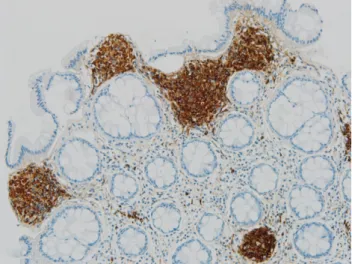
장한별 등. 미만성 미세 결절 형태로 나타난 점막슈반세포과오종The Korean Journal of Gastroenterology Fig. 3. Microscopic findings. Strong positive immunoreactivity for
S-100 (200).
Fig. 4. Microscopic findings. Focal positive immunoreactivity for CD56 (200).
Fig. 5. Microscopic findings. Negativity for CD68 (200).
검사를 시행하였다(Fig. 1).
조직 검사상 방추세포(spindle cells)가 세포소(nest) 형성 이 관찰되었고 이형성이나 악성을 시사하는 소견은 관찰되지 않았다(Fig. 2). 면역조직화학염색 검사에서는 S-100 양성, CD 56은 부분 양성, CD 68은 음성이 확인되었다(Figs. 3-5).
이를 바탕으로 점막슈반세포과오종(mucosal Schwann cell hamartoma, MSCH)으로 진단 내릴 수 있었으며 환자는 추 가 검사나 치료 없이 경과 관찰 중이다.
진단: 미만성 미세 결절 형태로 나타난 MSCH
MSCH는 대장 고유판층의 S-100 양성을 보이는 슈반세포의 증식으로 이루어진 용종으로 1형 신경섬유종증 환자에서 나타 나는 결장 신경섬유종(neurofibroma)과는 구별되며 Gibson과 Hornick1이 26개의 증례를 모아 2009년 처음 보고하였다. 이후 현재까지 16 증례가 추가로 보고되었고,2-13 우리나라에서도 Bae 등2,3과 Han 등4이 보고한 바 있다. 중년 여성에서 가장 빈도가 높게 나타났으며 대부분 무증상이지만 일부 위장관 출 혈, 설사 등의 증상이 동반된 경우도 있었다.12 빈도가 매우 드물기 때문에 정확한 발생률과 유병률을 알기는 어렵다.
현재까지 보고된 MSCH의 내시경적 소견을 살펴보면 궤양 이나 괴사가 동반되지 않은 1-8 mm 크기의 작은 무경성의 용종 형태로 주로 직장과 구불결장에서 나타는 것으로 알려져 있다.12 대부분 단일 용종 형태로 나타나지만 다발성 용종형 점막 병변,6 미만성의 작은 흰색 결절 형태14도 보고된 바 있 다. 본고에서는 흔하게 나타나는 단일 용종 형태가 아닌 미만 성 미세결절 소견이 확인되었다. 육안적 내시경 소견만으로는 판단이 어렵기 때문에 진단을 위해서는 조직 검사 및 면역조 직화학염색 검사가 필수적이다.
조직학적으로 MSCH는 점막고유층 내의 방추세포의 증식 이 관찰되며 세포형태는 이형성 소견 없이 비교적 균일하고 호산구성 세포질을 동반한다. 면역조직화학염색 검사상 S-100 양성을 보이며 CD34, KIT (CD117), 평활근 액틴(smooth muscle actin), 상피막항원(epithelial membrane antigen)에 는 음성을 보인다.13진단에 있어서 방추세포 증식을 보이거나 면역조직화학염색 검사에서 S-100 양성을 보이는 다른 질환들 의 감별이 중요하다.
위장관기질종양은 위장관에서 나타나는 가장 흔한 방추세포 종양이다.10 면역조직화학염색 검사에서 KIT (CD117) 양성이 약 95%에서 나타나 이를 통해 MSCH와 감별할 수 있다. CD34 는 60-80% 양성을 보이며 5%에서 S-100 양성이 나타난다.15 위장관 신경초종(gastrointestinal Schwannoma)은 소화 기계 종양의 0.2%의 빈도를 차지하는 드문 종양16으로 조직학 적으로 방추형 세포다발이 관찰되며, 경계가 비교적 명확하 다. MSCH에서는 나타나지 않는 주변부의 두꺼운 림프구층10
Jang HB, et al. MSCH Presenting as Diffuse Fine Nodularities
173
Vol. 76 No. 3, September 2020
과 치밀한 세포구조가 있는 Antoni A 부위 및 세포가 적고 점액 기질로 구성된 Antoni B 부위를 확인할 수 있다.17
결장의 신경섬유종은 신경섬유종증 1형과 동반되어 나타 나며 대부분 다발성이지만 드물게 피부 병변 없이 단발성으로 나타나는 경우도 있어9 MSCH와 감별이 필요하다. 비교적 균 일하게 방추세포의 증식을 보여주는 MSCH와 다르게 신경섬 유종은 슈반세포, 신경모세포, 말초신경세포, 축삭돌기 등 다 양한 세포의 집합으로 나타나며 그 결과로 면역조직화학염색 검사에서도 S-100은 일부에서만 양성을 보이며 CD34, neu- rofilament 단백, 상피막항원(epithelial membrane antigen) 등에서도 양성이 나타난다.12
위장관 신경절신경종(ganglioneuroma)은 용종성 신경절신 경종(polypoid ganglioneuroma), 신경절신경종성 용종증 (ganglioneuromtous polyposis), 미만성 신경절신경종(diffuse ganglioneuromaosis)으로 나눌 수 있으며,18용종성 신경절신 경종은 전신 질환과 연관성이 없고 결장에 단발성으로 나타날 수 있어 MSCH와 감별이 필요하다. 조직학적으로 S-100 양성 인 슈반세포와 섬유기질이 관찰되며 MSCH와는 다르게 neu- ron specific enolase 양성인 신경절세포가 관찰된다.9
현재까지 MSCH는 양성 종양으로 생각되며 악성을 보이거 나 유전적 질환과 연관성은 보고된 바 없으며 특별한 치료를 요하지 않는다. 본고의 환자의 경우도 다른 검사나 치료 없이 경과 관찰 중에 있다. 다만 진단 과정에서 방추세포 증식을 보이거나 S-100 양성을 보이는 다른 질환의 감별이 중요한데, 위장관기질종양처럼 악성화 가능성이 있는 질환도 포함되므 로 진단 과정이 그만큼 중요하다고 할 수 있겠다. 빈도가 매우 드물기에 추적 대장 내시경 검사 등에 대한 가이드라인은 현 재까지 정립된 바 없으며 추가적인 연구가 필요할 것으로 보 인다.
본고는 검진 내시경에서 확인된 MSCH 증례로 현재까지 보고된 대부분의 MSCH 증례들이 작은 크기의 용종 형태였 던 것과 비교하여 미만성 미세결절 형태를 보인다는 점에서 이전의 증례들과는 차이를 보인다. 현재까지 미만성 결절 형 태로 나타난 MSCH는 일본에서 1예 보고된 바 있으며 국내 에서는 최초의 증례로 판단되어 간단한 문헌고찰과 함께 보고 하는 바이다.
REFERENCES
1. Gibson JA, Hornick JL. Mucosal Schwann cell "hamartoma": clin- icopathologic study of 26 neural colorectal polyps distinct from neurofibromas and mucosal neuromas. Am J Surg Pathol 2009;
33:781-787.
2. Bae MN, Lee JE, Bae SM, et al. Mucosal Schwann-cell hamarto- ma diagnosed by using an endoscopic snare polypectomy. Ann Coloproctol 2013;29:130-134.
3. Bae JM, Lee JY, Cho J, Lim SA, Kang GH. Synchronous mucosal Schwann-cell hamartomas in a young adult suggestive of mu- cosal Schwann-cell harmatomatosis: a case report. BMC Gastro- enterol 2015;15:128.
4. Han J, Chong Y, Kim TJ, Lee EJ, Kang CS. Mucosal Schwann cell hamartoma in colorectal mucosa: a rare benign lesion that re- sembles gastrointestinal neuroma. J Pathol Transl Med 2017;51:
187-189.
5. Feng X, Xu H, Dela Cruz N. Mucosal Schwann cell hamartoma in sigmoid colon – a rare case report and review of literature.
Human Pathology: Case Reports 2020;19:200337.
6. Vaamonde-Lorenzo M, Elorriaga K, Montalvo I, Bujanda L.
Colonic mucosal Schwann cell hamartoma. J Dig Dis 2020;21:
475-477.
7. Chintanaboina J, Clarke K. Case of colonic mucosal Schwann cell hamartoma and review of literature on unusual colonic polyps.
BMJ Case Rep 2018;2018:bcr2018224931.
8. Pasquini P, Baiocchini A, Falasca L, et al. Mucosal Schwann cell
"hamartoma": a new entity?. World J Gastroenterol 2009;15:
2287-2289.
9. Rocco EG, Iannuzzi F, Dell'Era A, et al. Schwann cell hamartoma:
case report. BMC Gastroenterol 2011;11:68.
10. Neis B, Hart P, Chandran V, Kane S. Mucosal Schwann cell ha- martoma of the colon in a patient with ulcerative colitis.
Gastroenterol Hepatol (N Y) 2013;9:183-185.
11. Kanar O, Nakshabendi R, Berry AC. Colonic mucosal Schwann cell hamartoma on incidental screening colonoscopy. J Gastro- intestin Liver Dis 2015;24:411.
12. Gaspar R, Santos-Antunes J, Marques M, et al. Endoscopic sub- mucosal dissection of a schwann cell hamartoma mimicking a lateral spreading tumor of the rectum. Acta Gastroenterol Belg 2017;80:429.
13. Hashimoto H, Usui G, Sakai E, Ohata K, Morikawa T. Mucosal Schwann cell hamartoma of the rectosigmoid junction: a rare le- sion mimicking mucosal prolapse syndrome and other neural lesions. Int J Surg Pathol 2019;27:515-517.
14. Sagami S, Fukumoto A, Amano M, et al. A case of mucosal Schwann cell hamartoma. Nihon Shokakibyo Gakkai Zasshi 2012;109:1776-1783.
15. Kang YK, Koo DH. Diagnosis and treatment of gastrointestinal stromal tumor. Korean J Med 2013;85:341-353.
16. Jacobson BC, Hirsch MS, Lee JH, Van Dam J, Shoji B, Farraye FA.
Multiple asymptomatic plexiform schwannomas of the sigmoid colon: a case report and review. Gastrointest Endosc 2001;53:
801-804.
17. Moon SY, Chang YW, Lee CK, et al. A case of gastric Schwannoma in a male patient. Clin Endosc 2007;34:210-213.
18. Srinivasan R, Mayle JE. Polypoid ganglioneuroma of colon. Dig Dis Sci 1998;43:908-909.