상피하 종양의 형태로 나타난 진행 위암
전체 글
수치


관련 문서
Primary hepatocytes were incubated with T090 for 24 h following Res pretreatment (1 h). Cells were fixed and stained with Oil red O and observed under a microscope. The Oil
:Continuous new-bone formation (asterisk) is identified around the defect margin (arrows).(H-E stain, × 40) Higher magnification demonstrates some new-bone formation (asterisks)
[r]
[r]
이번 STEAM R&E STEAM R&E에 참여한 경험을 통해 어떻게 연구를 진행해 나가야 하는지 알 수 있었고, STEAM R&E STEAM R&E에 참여하며 얻은
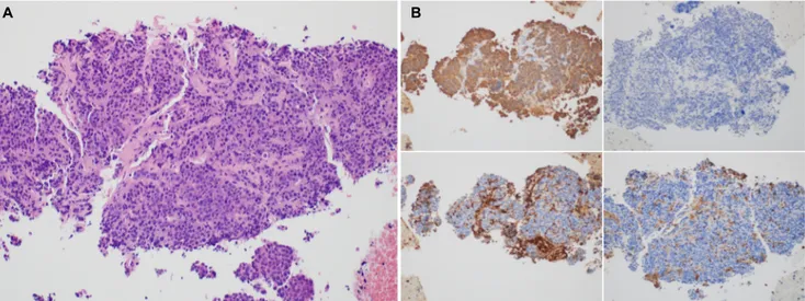
Relationship between lymph node metastasis and lymphatic invasion, diagnosed by immunohistochemical staining and H&E staining in gastric
Ri s pe ns ,Mc Br i de -Chang,& Re i st ma,2008) .선행연구( Ape l& Thomas -Tat e , 2009 ;Cho& Mc br i de -Chang,& Par k,20 07) 에서는
To investigate whether oncogenic H-ras might modulate mdr1b expression in NIH3T3 cells, V12-ras-NIH3T3 and pcDNA3-NIH3T3 cells were transiently transfected with
