Korean J Gastroenterol Vol. 74 No. 3, 183-186 https://doi.org/10.4166/kjg.2019.74.3.183 pISSN 1598-9992 eISSN 2233-6869
IMAGE OF THE MONTH
Korean J Gastroenterol, Vol. 74 No. 3, September 2019 www.kjg.or.kr
상피하 종양의 형태로 나타난 진행 위암
양효준, 도인구
1, 류창학
2성균관대학교 의과대학 강북삼성병원 내과, 병리과1, 외과2
Advanced Gastric Cancer Resembling Subepithelial Tumor
Hyo-Joon Yang, In-Gu Do1 and Chang Hak Yoo2
Departments of Internal Medicine, Pathology1 and Surgery2, Kangbuk Samsung Hospital, Sungkyunkwan University School of Medicine, Seoul, Korea
CC This is an open access article distributed under the terms of the Creative Commons Attribution Non-Commercial License (http://creativecommons.org/licenses/
by-nc/4.0) which permits unrestricted non-commercial use, distribution, and reproduction in any medium, provided the original work is properly cited.
Copyright © 2019. Korean Society of Gastroenterology.
교신저자: 양효준, 03181, 서울시 종로구 새문안로 29, 성균관대학교 의과대학 강북삼성병원 내과
Correspondence to: Hyo-Joon Yang, Department of Internal Medicine, Kangbuk Samsung Hospital, Sungkyunkwan University School of Medicine, 29 Saemunan-ro, Jongno-gu, Seoul 03181, Korea. Tel: +82-2-2001-8330, Fax: +82-2-2001-8360, E-mail: hyojoonyang@gmail.com, ORCID: https://orcid.org/0000-0002-0265-672X Financial support: None. Conflict of interest: None.
Fig. 1. Esophagogastroduodenoscopy findings. A large subepithelial tumor with a normal overlying epithelium larger than 5 cm in size occupying cardia and mid to high body lesser curvature side.
증례: 78세 여자 환자가 한 달 전부터 시작된 복통을 주소 로 내원하였다. 외부 병원에서 시행한 복부 전산화단층촬영에서 위 외장성(exophytic) 종괴가 관찰되었다. 환자는 내원 7개월 전 본원에서 시행한 상부위장관 내시경에서 위염 이외에 특이 소견은 없었다. 환자는 고혈압과 당뇨 병력이 있었다.
내원 당시 활력징후는 혈압 131/67 mmHg, 맥박수 67회/분, 체온은 36.9°C로 측정되었다. 복부 검진에서 종괴는 촉지되지 않았으며, 복부 압통도 없었다. 말초혈액 검사에서 백혈구 7,680/μL (정상 3,700-9,800), 혈색소 11.7 g/dL (정상 11.0-15.0), 혈소판 241,000/μL (정상 160,000-362,000), 혈액응고 검사 에서 프로트롬빈 시간은 11.4초(정상 9.5-12.1)였다. 생화학 검사에서 혈액요소질소 14.6 mg/dL (정상 8-20), 크레아티닌 1.29 (정상 0.5-0.9), 알부민 4.2 mg/dL (정상 4.1-5.1)였다.
다시 시행한 상부위장관 내시경에서 유문부와 상체부 및 중체부 소만에 이르는 5 cm 이상의 크기가 정확하게 추정되 지 않는 상피하 종괴 소견이 관찰되었으며, 병변의 상피에서 는 발적, 미란 혹은 함몰과 같은 이상 소견은 관찰되지 않았다 (Fig. 1). 복부 전산화단층촬영 영상 재판독에서 6.7 cm 크기 의 종괴는 위 외벽에서 외장성 성장 소견을 보이고 있었으며, 대동맥 주위(paraaortic) 림프절 종대 소견이 동반되어 있었 다(Fig. 2). 또한, 양전자단층촬영에서 위 종괴와 대동맥 주위
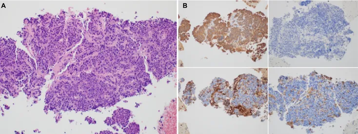
림프절에 18F-fluorodeoxyglucose 섭취 증가 소견이 관찰되 었다(Fig. 2). 병변의 조직학적 진단을 위하여 내시경 초음파 미세침 흡인 검사를 시행하였다. 흡인 검체는 상피세포양 세 포 군집을 보이는 분화가 나쁜 암(poorly differentiated car- cinoma with small epithelioid cell nests)으로 면역조직검 사에서 C-kit, Dog-1, vimentin 음성, chromogranin A, syn- aptophysin, CD56 음성, LCA 음성 소견으로 위장관 기질
184
양효준 등. 상피하 종양의 형태로 나타난 진행 위암The Korean Journal of Gastroenterology
Fig. 4. Pathology findings of a surgical specimen. (A) Gross appearance of the resected stomach shows a gray white to pale yellowish solid mass involving the submucosa to serosa. The mucosa surface is intact. (B) H&E stain revealed intact mucosa and poorly differentiated small epithelioid tumor cells involving the submucosa to serosa (upper left: H&E, ×4). Tumor cells are positive for CK (AE1/AE3) and negative for LCA, CD56, and vimentin. Based on the H&E and immunohistochemistry findings, tumor is consistent with a poorly differentiated adenocarcinoma (upper right: CK (AE1/AE3), ×4; lower left: LCA, ×4; lower right: CD 56, ×4).
Fig. 2. Abdominal computed tomography (CT) and positron emission tomography (PET). CT images show (A) a 6.7 cm sized large mass arising from the left side of the gastric body (arrow) and (B) left paraaortic lymph node enlargement (arrow). PET images show increased 18F-fluorodeoxyglucose uptake in (C) left gastric mass and (D) left paraaortic lymph node.
종양, 신경내분비 종양, 림프종이 배제되었다. 한편, CK7 양 성, CK20 음성으로 저분화 위선암 또는 원발 부위 미상의 암 이 의심되었다(Fig. 3).
환자는 상피하 종괴 형태를 보이는 위선암 가능성이 높다 고 판단되는 가운데 진단 및 치료 목적의 위전 절제술, 비장 절제술 및 림프절 절제술을 시행하였다. 조직학적으로 종괴는 저응집암종(poorly cohesive carcinoma)으로 췌장을 침범하 고, 32개 중 4개의 림프절에 전이된 진행 위암으로 진단되었 다(Fig. 4). 환자는 잔여 대동맥 주위 림프절 종대에 대하여 추적 관찰하면서 항암화학요법을 하며 4개월째 추적 관찰 중 이다.
진단: 상피하 종양의 형태로 나타난 진행 위암
위암은 드물게 상피하 종양의 형태로 나타날 수 있는데, 기존 문헌보고에 따르면 전체 위암 증례 중 약 0.5% 미만으로 보고되 고 있으며,1국내 문헌에도 10예 미만의 보고가 있다.2조직학적
으로는 저분화선암(poorly differentiated adenocarcinoma), 림프양 기질을 동반하는 위암(carcinoma with lymphoid stro- ma), 점액성 선암(mucinous adenocarcinoma), 점막하 이소성 위점막선관(submucosal heterotopia of the gastric glands) 에서 발생한 위암이 상피하 종양의 형태로 나타날 수 있다고 제시되었다.3
위 상피하 종양 중 악성 혹은 악성화 가능성으로 인하여 감별 진단이 필요한 원인에는 위장관 기질종양, 신경내분비 종양 및 림프종이 있다.4 위장관 기질종양이 가장 흔한 위 상 피하 종양으로 알려져 있으며, 림프절 전이는 거의 발생하지 않는 것으로 알려져 있다. 본 증례는 상부위장관 내시경에서 명확하게 위 상피하 종양으로 진단되었으나 복부 전산화단층 촬영에서 림프절 전이를 동반하고 있어 위장관 기질종양으로 진단하기 어려웠던 증례이다. 다른 감별 진단, 특히 림프종은 수술적 치료의 역할이 적기 때문에 조직학적 진단을 위하여 내시경 초음파를 통한 미세침 흡인 검사를 시행하였다.
A B
C D
Yang HJ, et al. Advanced Gastric Cancer Resembling Subepithelial Tumor
185
Vol. 74 No. 3, September 2019
Fig. 4. Pathology findings of a surgical specimen. (A) Gross appearance of the resected stomach shows a gray white to pale yellowish solid mass involving the submucosa to serosa. The mucosa surface is intact. (B) H&E stain revealed intact mucosa and poorly differentiated small epithelioid tumor cells involving the submucosa to serosa (upper left: H&E, ×4). Tumor cells are positive for CK (AE1/AE3) and negative for LCA, CD56, and vimentin. Based on the H&E and immunohistochemistry findings, tumor is consistent with a poorly differentiated adenocarcinoma (upper right: CK [AE1/AE3], ×4; lower left: LCA, ×4; lower right: CD 56, ×4).
Fig. 3. Pathology findings of a fine needle aspiration/biopsy specimen. (A) Microscopic findings of the specimen show poorly differentiated small epithelioid cell nests (H&E, ×20). (B) Immunohistochemical staining findings were compatible with the diagnosis of a poorly differentiated carcinoma (upper left: CK [AE1/AE3], ×20; upper right: CD 56, ×20; lower left: LCA, ×20; lower right: Vimentin, ×20).
상피하 종양 형태의 위암은 상피하 종양과 비교하여 내시 경 소견에서 차이가 있는 경우가 있다.5상피하 종양은 일반적 으로 병변의 상피층에 미란이나 발적이 잘 동반되지 않고, 종 양 내부의 함몰이 있더라도 변연부까지는 정상 점막 소견인 반면, 상피하 종양 형태의 위암은 상피에 불규칙한 발적, 미 란, 함몰 및 변연부 백태 등을 관찰할 수 있다. 이러한 소견이 관찰되는 경우 해당 부위에서 조직 검사를 시행하여 진단이 가능할 수 있다. 그러나 본 증례와 같이 병변의 상피에 해당 소견이 관찰되지 않는 경우에는 미세침 흡인 검사가 진단 방 법이 될 수 있다.6본 증례는 미세침 흡인 검사를 통하여 위장
관 기질종양, 신경내분비 종양 및 림프종을 배제할 수 있었고, 위선암의 가능성이 높다는 판단으로 위 절제술 및 림프절 절 제술을 시행할 수 있었다.
요약하면, 본 증례는 위 상피하 종양의 형태로 나타난 위암 으로 전체 위암 증례의 0.5% 미만을 차지하는 드문 증례이나 위 상피하 종양의 감별 진단 과정에서 이러한 형태의 위암을 고려할 필요가 있다. 특히, 병변 상피의 불규칙한 변화나 위 주변 림프절 종대를 동반한 경우 의심할 수 있고, 내시경 조직 생검을 통하여 진단이 어려운 경우 미세침 흡인 검사를 고려 할 수 있다.
A B A B
186
양효준 등. 상피하 종양의 형태로 나타난 진행 위암The Korean Journal of Gastroenterology
REFERENCES
1. Umehara Y, Kimura T, Okubo T, et al. Gastric carcinoma resem- bling submucosal tumor. Gastric Cancer 1999;2:191-193.
2. Kim HI, Shim KN, Yoon SY, et al. A case of early gastric ad- enocarcinoma resembling subepithelial tumor. Korean J Helicobacter Up Gastrointest Res 2013;13:60-63.
3. Ohara N, Tominaga O, Uchiyama M, Nakano H. A case of ad- vanced gastric cancer resembling submucosal tumor of the stomach. Jpn J Clin Oncol 1997;27:423-426.
4. Standards of Practice Committee, Faulx AL, Kothari S, et al. The
role of endoscopy in subepithelial lesions of the GI tract.
Gastrointest Endosc 2017;85:1117-1132.
5. Fujiyoshi A, Kawamura M, Ishitsuka S. Gastric adenocarcinoma mimicking a submucosal tumor: case report. Gastrointest Endosc 2003;58:633-635.
6. Yamane H, Ishida M, Banzai S, et al. Advanced gastric cancer with features of a submucosal tumor diagnosed by endoscopic ultra- sound-guided fine needle aspiration and boring biopsy pre- operatively: a case report and literature review. Int J Surg Case Rep 2019;55:223-226.