ISSN 0378-6471 (Print)⋅ISSN 2092-9374 (Online)
http://dx.doi.org/10.3341/jkos.2015.56.11.1728
Original Article
결절맥락막혈관병증에서 유리체강내 애플리버셉트 주입술의 단기 효과
Short-Term Efficacy of Intravitreal Aflibercept for Polypoidal Choroidal Vasculopathy
양 헌⋅전혜민⋅김상원⋅윤희성⋅최우석
Heon Yang, MD, Hye Min Jeon, MD, Sang Won Kim, MD, Hee Seong Yoon, MD, Woo Seok Choae, MD
성모안과병원
Sungmo Eye Hospital, Busan, Korea
Purpose: To evaluate the short-term effect of intravitreal aflibercept (Eylea®; Regeneron Pharmaceuticals Inc., Tarrytown, NY, USA and Bayer, Basel, Switzerland) on the visual outcomes and retinal anatomic changes of patients with polypoidal choroidal vasculopathy (PCV).
Methods: Intravitreal Eylea® was injected into 16 eyes of 16 patients with PCV in this retrospective case study. After therapy, the patients were followed up for over 3 months. Changes in best-corrected visual acuity (BCVA) and central foveal thickness (CFT) using optical coherence tomography (OCT) and abnormal vasculature on indocyanine green angiography (ICGA) were evaluated.
Results: The mean log MAR BCVA was 0.75 ± 0.60 at baseline, 0.74 ± 0.60 and 0.71 ± 0.63 at 1 and 2 months, respectively (p
> 0.05) and 0.57 ± 0.53 at 3 months (p < 0.05) after treatment. The mean CFT was 379 ± 130 μm at baseline, 281 ± 92 μm, 247
± 54 μm, and 231 ± 51 μm at 1, 2, and 3 months, respectively, after treatment (p < 0.05). Complete resolution was 43%, 55%, and 50% at 1, 2, and 3 months, respectively in pigment epithelial detachment (PED), 67%, 83%, and 92% at 1, 2, and 3 months, re- spectively in subretinal fluid (SRF) and 33%, 60%, and 60% at 1, 2, and 3 months, respectively in intraretinal fluid (IRF) using OCT. The polypoidal lesions in ICGA decreased in 12 of 14 eyes (86%).
Conclusions: Intravitreal injection of Eylea® with PCV reduced CFT due to decreased retinal PED, SRF, IRF and occluded effec- tively the polypoidal lesion leaking. Compared with baseline, mean BCVA at the 3-month follow-up was significantly improved.
J Korean Ophthalmol Soc 2015;56(11):1728-1735
Key Words: Aflibercept, Intravitreal injection, Polypoidal choroidal vasculopathy
■Received: 2015. 1. 16. ■ Revised: 2015. 6. 30.
■Accepted: 2015. 8. 21.
■Address reprint requests to Woo Seok Choae, MD
Sungmo Eye Hospital, #409-1 Haeun-daero, U-dong, Haeundae-gu, Busan 48064, Korea
Tel: 82-51-743-0775, Fax: 82-51-743-0776 E-mail: choiooseok@hanmail.net
* This study was presented as an e-poster at the 112th Annual Meeting of the Korean Ophthalmological Society 2014.
ⓒ2015 The Korean Ophthalmological Society
This is an Open Access article distributed under the terms of the Creative Commons Attribution Non-Commercial License (http://creativecommons.org/licenses/by-nc/3.0/) which permits unrestricted non-commercial use, distribution, and reproduction in any medium, provided the original work is properly cited.
결절맥락막혈관병증(polypoidal choroidal vasculopthy, PCV) 은 내측 맥락막혈관(inner choroidal vascular network of
vessels)에 분지하는 혈관망과 말단의 결절 형태의 확장을 특징으로 하는 질환이다.1,2 일반적으로 맥락막신생혈관에 비해 양호한 자연경과를 보이지만,2,3 반복적인 출혈이나 삼 출 변화로 만성, 재발성 경과를 거치게 되어 결국 시력저하 를 보이게 되는 경우도 많다.4,5
PCV는 삼출성 나이관련황반변성(age-related macular de- generation, ARMD) 환자 중 서양에서는 8-13%의 빈도를 보이는 반면,6,7 흑인과 히스패닉, 동양인 등 유색인종에서 유병률이 높으며, 일본에서 23.0-54.7%, 중국에서 22.3%, 우리나라에서 24.6%의 빈도가 보고되기도 하였다.8-11
PCV의 병인은 아직 완전히 밝혀져 있지 않으며, 일부에 서는 ARMD의 한 변형이라고 보기도 하나,12 임상경과와 시력 예후, 치료에 대한 반응이 다르기 때문에 ARMD와는 구분된다는 주장도 있다.13,14 최근 PCV 환자에서 ARMD 환자와 비교하여 방수의 혈관내피세포증식인자(vascular endothelial growth factor, VEGF)가 높은 농도로 확인되었 으며, PCV를 가진 눈의 subfoveal fibrovascular membranes 에서 VEGF가 강하게 표현되는 것이 관찰되어 상호 관련성이 있는 것으로 보고된 바 있다.15,16
기존의 anti-VEGF 제제인 bevacizumab이나 ranibizumab 유리체강내 주입술은 최대교정시력의 상승과 중심와두께 의 감소를 보였으나 분지하는 혈관망의 감소나 결절과 pig- ment epithelial detachment (PED)의 regression과 같은 구조 적인 변화에는 효과적이지 못한 결과를 보여주었다.17,18 반 면 aflibercept (Eylea®; Regeneron Pharmaceuticals Inc., Tarrytown, NY, USA and Bayer, Basel, Switzerland)는 기 존의 약제들과 달리 해부학적인 변화에도 효과적임이 보고 되었으며,19 국내보다 앞서 미국, 일본 등에서 PCV 환자에게 적용되어 왔다.
이에 저자들은 이전에 치료한 적이 없는 PCV 환자에 대 해aflibercept 유리체강내 주입술이 시력과 망막의 해부학 적 구조에 미치는 효과를 알아보고자 하였다.
대상과 방법
2014년 5월부터 2014년 9월까지 본원에서 PCV로 진단 받고 이전에 다른 치료를 받은 적이 없는 환자에서 유리체 강 내 aflibercept 주입술을 시행한 16명, 16안을 대상으로 하였다. 남자는 14명, 여자는 2명이었으며, 연령 분포는 53 세에서 80까지로 평균 70세였다.
PCV의 진단은 안저소견 및 빛간섭단층촬영(optical co- herence tomography, OCT), F10 (Nidek, Gamagori, Japan) 을 이용하여 촬영한 형광안저혈관조영(fluorescin angiog- raphy, FA) 및 인도시아닌그린혈관조영(indocyanine green angiography, ICGA) 소견을 바탕으로 이루어졌다. 진단은 분지하는 혈관망과 그 말단부의 결절 병변을 보이는 특징 적 소견이 있는 경우로 제한하도록 노력하였으나, 출혈이 나 삼출, 위축 병변으로 인해 분지하는 혈관망을 파악하기 힘든 경우에는 결절 병변만 있어도 그 모양이 특징적이면 포함시켰다.
주입술은 3명의 술자가 시행하였고 모든 환자에서 동의 (informed consent)하에, 술 안을 Proparacaine hydrochloride 0.5% (Alcaine®, Alcon, Fort Worth, TX, USA)로 점안 마취 후 5% 베타딘 용액으로 안검과 결막소독을 하였고, 개검기
를 착용 후 결막낭에 점안하였다. 2.5 mg (0.1 mL)의 afli- bercept를 30 gauge 주사바늘을 이용하여 수정체안에서는 윤부에서 3.5 mm, 인공수정체안 또는 무수정체안에서는 3.0 mm 떨어진 하이측 섬모체 평면부를 통해 유리체강에 주입하였다.
주입술 전과 주입술 후 1개월, 2개월 및 3개월째 최대교 정시력과 Stratus OCT (Zeiss Humphrey Instruments, Dubin, CA, USA)를 이용하여 한 명의 연구자가 중심와두께(central foveal thickness, CFT), 망막색소상피박리(pigment epithelial detachments, PED), 망막하액(subretinal fluid, SRF), 망막내 액(intraretinal fluid, IRF) 여부를 측정하였고, 3개월에 인도 시아닌그린혈관조영술상의 이상혈관 변화를 술 전과 비교 하였다. 빛간섭단층촬영을 이용한 fluid의 변화정도는 완전 관해(complete resolution), 부분관해(partial resolution)와 호 전 없음으로 분류하였으며 망막색소상피박리, 망막하액, 망 막내액 각각의 완전한 사라짐은 완전관해, 일부 감소하였 으나 잔존하는 경우 부분관해로 판단하였다.
모든 조사는 의무기록을 통하여 후향적으로 시행하였다.
시력과 황반 두께에 대한 통계 분석은 paired t-test를 이용 하였으며 p-value가 0.05보다 작은 경우를 유의한 것으로 간주하였다.
결 과
술 후 경과관찰 기간은 평균 15주였으며, aflibercept 유 리체강내 주입 횟수는 평균 2.88회였다. 이 중 2안은 2회 주사 이후 추적관찰이 되지 않았으며 각각 상기도 감염으 로 인한 입원치료와 연락두절 때문이었다. 환자들의 특징 은 Table 1에 요약하였다.
최대교정시력은 경과관찰 기간 동안 주입술 전 평균 logMAR 0.75 ± 0.60에서 1개월째 0.74 ± 0.64, 2개월째 0.71 ± 0.63, 3개월째 0.57 ± 0.53으로 점차 개선되는 양상 을 보였으며, 술 후 3개월에서 통계학적으로 유의하게 호전 되었다(p<0.05) (Fig. 1). 주입술 후 시력이 호전되는 비율 을 조사한 결과 1개월째 0.2 logMAR 이상 호전이 18.8%, 유지가 62.5%, 0.2 logMAR 이상 악화가 18.7%였고, 2개월 째 0.2 logMAR 이상 호전이 25%, 유지가 58.3%, 0.2 logMAR 이상 악화가 16.7%였으며, 3개월째는 0.2 logMAR 이상 호전 이 28.6%, 유지가 64.3%, 0.2 logMAR 이상 악화가 7.1%였 다(Fig. 2).
빛간섭단층촬영상 중심와두께는 주사 전 평균 379 ± 130 μm 에서 주사 후 1개월에 281 ± 92 μm로 통계학적으로 유의하 게 감소하였으며(p<0.05), 2개월에 247 ± 54 μm (p<0.05), 3개월에 232 ± 51 μm로 측정되어(p<0.05), 지속적으로 유
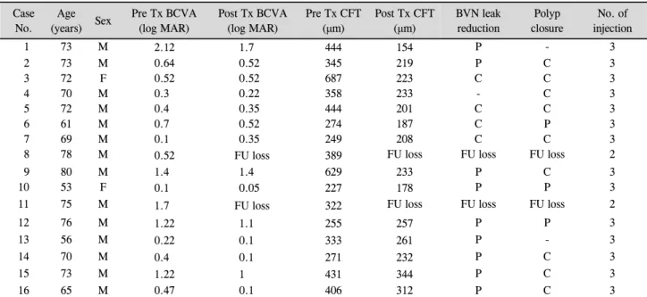
Table 1. Patient characterstics and clinical data before and after intravitreal injection of aflibercept for PCV
CaseNo.
Age
(years) Sex Pre Tx BCVA (log MAR)
Post Tx BCVA (log MAR)
Pre Tx CFT (μm)
Post Tx CFT (μm)
BVN leak reduction
Polyp closure
No. of injection
1 73 M 2.12 1.7 444 154 P - 3
2 73 M 0.64 0.52 345 219 P C 3
3 72 F 0.52 0.52 687 223 C C 3
4 70 M 0.3 0.22 358 233 - C 3
5 72 M 0.4 0.35 444 201 C C 3
6 61 M 0.7 0.52 274 187 C P 3
7 69 M 0.1 0.35 249 208 C C 3
8 78 M 0.52 FU loss 389 FU loss FU loss FU loss 2
9 80 M 1.4 1.4 629 233 P C 3
10 53 F 0.1 0.05 227 178 P P 3
11 75 M 1.7 FU loss 322 FU loss FU loss FU loss 2
12 76 M 1.22 1.1 255 257 P P 3
13 56 M 0.22 0.1 333 261 P - 3
14 70 M 0.4 0.1 271 232 P C 3
15 73 M 1.22 1 431 344 P C 3
16 65 M 0.47 0.1 406 312 P C 3
PCV = polypoidal choroidal vasculopathy; Tx = treatment; BCVA = best corrected visual acuity; CFT = central foveal thickness; BVN = branching vascular network; FU = follow-up; P = partial; C = complete.
Figure 1. Change in logarithm of the minimum angle of reso-
lution (log MAR), best-corrected visual acuity with time (months). Decrease at 1, 2, and 3 months was observed. A sig- nificant decrease was found at 3 months (p < 0.05). Case 8, 11 were loss to FU after 1 month. FU = follow-up.Figure 2. Change in percentage (%), in proportion of more
than 0.2 log MAR with time (months). Increase in proportion of gain more than 0.2 log MAR, decrease in proportion of loss more than 0.2 log MAR with time was observed. log MAR = logarithm of the minimum angle of resolution.의한 두께 감소를 보였다(Fig. 3).
치료 후 빛간섭단층촬영상 PED의 부분/완전 관해율은 1 개월에 29/43%, 2개월에 36/55%, 3개월에 50/50%였고, SRF 의 부분/완전 관해율은 1개월에 33/67%, 2개월에 17/83%, 3 개월에 8/92%였으며, IRF의 부분/완전 관해율은 1개월에 50/33%, 2개월에 40/60%, 3개월에 40/60%의 관해율을 보 여 첫 주입술 후 3개월째에는 모든 대상 환자에서 빛간섭
단층촬영 결과, 부분 또는 완전하게 해부학적으로 호전 된 소견을 보였다(Table 2).
Gain more than 0.2 log MAR Time (months)
Time (months)
Table 2. Change in percentage (%), in proportion of partial or complete solution in PED, SRF, IRF by OCT
N Partial solutionat 1 month (n, %)
Complete solution at 1 month (n, %)
Partial solution at 2 months (n, %)
Complete solution at 2 months (n, %)
Partial solution at 3 months (n, %)
Complete solution at 3 months (n, %)
PED 14 4/14 (29) 6/14 (43) 4/11 (36) 6/11 (55) 6/12 (50) 6/12 (50)
SRF 15 5/15 (33) 10/15 (67) 2/12 (17) 10/12 (83) 1/13 (8) 12/13 (92)
IRF 6 3/6 (50) 2/6 (33) 2/5 (40) 3/5 (60) 2/5 (40) 3/5 (60)
All cases were significantly improved partially or completely within 3 months.
PED = pigment epithelial detachments; SRF = subretinal fluid; IRF = intraretinal fluid; OCT = optical coherence tomography.
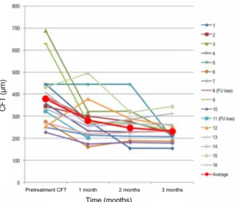
Figure 3. Change in CFT (μm) with time (months). A sig-
nificant decrease was found at 1, 2, and 3months (p < 0.05).Case 8, 11 were loss to FU after 1 month. CFT = central fo- veal thickness; FU = follow-up.
주사 전 보였던 인도시아닌그린혈관조영술상 결절은 주 입술 후 3개월째 86%에서 감소되는 모습이 관찰되었다. 첫 주입술 후 3개월 형광안저혈관조영에서 누출이 있었던 14안 중 9안에서 부분적으로(partial reduction), 4안에서 완전하 게(complete reduction) 누출이 사라졌다. 또한 3개월 인도 시아닌그린혈관조영술에서 결절이 완전히 사라지거나 작 은 과형광 반점으로 크기가 감소하는 것을 관찰하였는데, 결절이 존재하였던 14안 중 3안에서 부분적으로(partial clo- sure), 9안에서 완전하게(complete closure) 결절이 폐쇄되었 다. 분지하는 선형의 혈관(branching vascular network, BVN) 은 모두에서 잔존하는 것이 관찰되었다.
경과관찰 중 안내염이나 안내출혈, 망막박리와 같은 안 내 합병증이나 혈전증과 같은 전신적 합병증은 발생하지 않았다.
증례 5 환자에서는 치료 전 시력이 logMAR 0.4에서 세 번의 aflibercept 주입술 후 0.45로 저하되었고, 중심와두께 는 치료 전 444 μm에서 세 번의 주입술 후 201 μm로 감소 하였다. 주입술 후 빛간섭단층촬영에서 PED, SRF가 완전
히 사라지고 3개월째 인도시아닌그린혈관조영술에서 결절 이 폐쇄되었으나, 분지혈관망은 잔존하는 것이 관찰되었다 (Fig. 4, 5).
고 찰
PCV는 ARMD에 동반된 맥락막신생혈관과는 다르게 비 교적 좋은 자연 예후를 보이는 질환으로 알려져 있다. 환자 중 거의 절반은 병변이 퇴행되는 양호한 예후를 보이며, 반 면 나머지 절반은 진행하여 시력을 잃게 될 수도 있다.4
PCV의 치료로는 과거에는 활동성 결절에 대해 선택적으 로 레이저를 시행하여 왔으나 장기간 추적 시 남아있는 혈 관망에서 새로운 결절이 형성되어 재발성, 만성적 경과를 보이는 것이 보고되었다. 따라서 병변 전체를 치료하는 것 이 장기적으로 효과적이지만, 황반에 인접한 경우에는 적용 이 불가능하다. 또한 레이저 치료는 심한 망막하출혈, 시세 포 손상을 유발할 수 있고, 전형적 맥락막신생혈관이 발생 하여 결국은 원반반흔을 형성할 수 있는 문제점이 있다.5,7 반면 photodynamic therapy (PDT)는 망막의 시세포 손상 을 최소화하면서 이상혈관을 선택적으로 치료할 수 있어 결절이 중심와 근처에 위치하거나 여러 개 있을 때 유용하 게 사용될 수 있고, 병변 전체를 치료함으로써 현재의 활동 성 결절뿐 아니라 분지혈관망까지 폐쇄를 기대할 수 있다. 여러 연구에서 유의한 시력 상승과 결절 폐쇄, 그리고 인도 시아닌그린혈관조영술상의 호전을 보고하였으나,20,21 결절 에 비해 분지혈관망의 폐쇄에는 효과가 낮아 2-3년 후 새로 운, 또는 재발성 결절의 발생으로 장기간의 결과는 좋지 않 았다.22,23
최근 연구에서 ARMD와 PCV의 혈관내피세포와 망막색 소상피에서 신생혈관생성 촉진자인 VEGF가 면역조직화학 염색에서 강하게 표현되었고,16 방수에서 VEGF의 증가도 확인되어,15 VEGF가 ARMD뿐 아니라 PCV와도 관련되었 을 것으로 생각됨에 따라, bevacizumab이나 ranibizumab과 같은 anti-VEGF 제제를 치료에 적용하고 있다. 관련된 보 고로 Gomi et al24이 PCV 환자에 시행한 유리체강내 bev- acizumab 주입술 시행의 결과 polyp regression에 대해서는
Pretreatment CFT
Time (months)
1 month 2 months 3 months
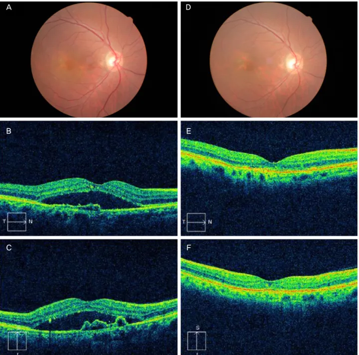
Figure 4. Case No. 5 (A) Baseline fundus photograph shows hard exudates, pigmentations around macula. (B) Baseline horizontal
OCT image and (C) vertical OCT image shows protruding polyps and macular edema. (D-F) One month after the third injection, PED, SRF was completely resolved. OCT = optical coherence tomography; PED = pigment epithelial detachment; SRF= sub- retinal fluid.9%의 부분 혹은 완전 관해, PED의 75% 부분적 관해를 보 고하였으나 분지혈관망에 대해서는 변화하지 않은 결과를 보였고, Kokame et al25이 시행한 유리체강내 ranibizumab 주입 시행의 결과 polyp regression에 대해서는 33%의 부분 혹은 완전 관해, PED의 17%의 완전 관해와 50%의 부분적 관해를 보고하였으며, 분지혈관망은 역시 감소를 보이지 않았다.
Aflibercept는 bevacizumab이나 ranibizumab보다 VEGF
에 높은 결합력을 가지는 약제로 여러 연구에서 삼출성 나 이관련황반변성의 치료에 있어 그 효용성이 보고된 바 있
다.26-28 최근에는 Aflibercept를 PCV 치료에도 적용하여 그
효과들이 보고되고 있는데, Aflibercept의 높은 결합력뿐 아 니라 vascular endothelial growth factor-A (VEGF-A) 외에 도 VEGF-B, placental growth factor (PlGF) 등에도 작용하 는 특성으로 인해 VEGF-A에만 결합하는 ranibizumab이나 bevacizumab보다 PCV의 치료에 있어 효과가 우수할 것이
A D
B E
C F
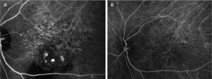
Figure 5. Case No. 5 (A) Baseline ICGA shows polypoidal dilatation of choroidal vessels and branching vascular network. (B) One
month after the third injection, polyps are resolved. But branching vascular network remains. ICGA = indocyanine green angiography.라 기대할 수 있다.27
Inoue et al19은 이전에 치료 받지 않은 PCV 환자 군에서 6개월간의 유리체강내 aflibercept의 주입술 시행 후 교정시 력의 유의한 호전과 황반부와 맥락막 평균 두께의 유의한 감소, 93.3%의 SRF 완전 관해, 75%의 polyp regression, 89%의 PED 부분 및 완전 관해를 보였으나, 분지혈관망은 주입술 전과 비교하여 모든 안에서 변화가 없음을 보고하 였으며, Ijiri and Sugiyama29도 이전에 치료 받지 않은 PCV 환자 군에서 3개월간의 유리체강내 aflibercept의 주입술이 평균 교정시력의 유의한 호전과 평균 황반부 두께의 유의 한 감소 및 polyp regression에 대해서는 48%의 완전 관해, 27%의 부분 관해와 PED의 71% 부분 및 완전 관해가 있었 으나, 분지혈관망 병변의 최대 직경의 감소는 보이지 않음 을 보고하였다.
본 연구에서는 Aflibercept 주입술 후 3개월째 빛간섭단 층촬영상 황반부 부종, 특히 SRF (100%)와 PED (100%)의 부분 및 완전 감소와 함께 인도시아닌그린혈관조영술에서 맥락막 결절 폐쇄(86%)를 보여, 이전 연구 결과에서 보고 된 바와 같이 단기간의 형태적 개선에 대해 효율적인 양상 을 보였으나 분지혈관망의 감소에는 효과가 없음을 알 수 있었다. 또한 최대교정시력은 1차 치료 후 점차 회복되는 양상을 보였으며, 주입술 후 3개월째에는 통계적으로 유의 한 시력호전이 관찰되어 이전의 보고들과 유사한 결과를 관찰할 수 있었다.
Kawashima et al30은 이전 ranibizumab 치료로 호전이 없 었던 ARMD와 PCV의 경우에 유리체강내 aflibercept의 주 입술이 최대교정시력 향상에 효과적이라고 보고한 바 있으 며, 이는 이전에 치료 받은 병력이 없는 환자들뿐 아니라,
ranibizumab이나 bevacizumab 주입술로 치료 효과가 적었 던 환자군에 대해서도 aflibercept 치료의 필요성에 대한 근 거가 될 것이다.
이상에서 살펴본 바로 aflibercept 유리체강내 주사 치료 는 이전에 치료한 적 없는 PCV에서 단기간의 형태적 개선 과 시력 회복을 유도하는 매우 효과적이고 안전한 결과를 보여주었다. 관찰된 14안 중 12안에서의 결절의 폐쇄는 aflibercept로 인한 효과일 수도 있으나, 일부는 질병의 자연 경과의 가능성을 배제할 수 없다. 반면 aflibercept 단독 유 리체강내 주입술은 기존의 anti-VEGF 제제인 bevacizumab 과 ranibizumab 주입술에서 보고된 바와 같이 분지혈관망 의 폐쇄에 효과가 없는 결과를 보여주었으며, 이는 PCV에 서 향후 재발의 가능성을 보여주는 결과이다. 따라서 어느 정도의 분지혈관망의 폐쇄에 치료 효과가 확인된 PDT와의 병합 치료가 더 좋은 결과를 나타낼 수 있는지에 대해서도 연구가 필요할 것으로 보인다.
본 연구는 대상 안이 적어 통계분석에 한계가 있고, 경과 관찰 기간이 짧아 장기간 효과는 알 수 없는 문제점이 있다.
따라서 보다 많은 수의 환자를 대상으로 한 장기적인 afli- bercept 치료 경과에 대한 연구가 필요하겠다.
참고문헌
1) Yannuzzi LA, Sorenson J, Spaide RF, Lipson B. Idiopathic poly- poidal choroidal vasculopathy (IPCV). Retina 1990;10:1-8.
2) Ciardella AP, Donsoff IM, Huang SJ, et al. Polypoidal choroidal vasculopathy. Surv Ophthalmol 2004;49:25-37.
3) Uyama M, Matsubara T, Fukushima I, et al. Idiopathic polypoidal choroidal vasculopathy in Japanese patients. Arch Ophthalmol 1999;117:1035-42.
A B
4) Uyama M, Wada M, Nagai Y, et al. Polypoidal choroidal vasculop- athy: natural history. Am J Ophthalmol 2002;133:639-48.
5) Lee WK, Kwon SI. Polypoidal choroidal vasculopathy. J Korean Ophthalmol Soc 2000;41:2573-84.
6) Scassellati-Sforzolini B, Mariotti C, Bryan R, et al. Polypoidal choroidal vasculopathy in Italy. Retina 2001:21:121-5.
7) Lafaut BA, Leys AM, Snyers B, et al. Polypoidal choroidal vascul- opathy in Caucasians. Graefes Arch Clin Exp Ophthalmol 2000;
238:752-9.
8) Yannuzzi LA, Wong DW, Sforzolini BS, et al. Polypoidal choroi- dal vasculopathy and neovascularized age-related macular degen- eration. Arch Ophthalmol 1999;117:1503-10.
9) Sho K, Takahashi K, Yamada H, et al. Polypoidal choroidal vascul- opathy: incidence, demographic features, and clinical characte- ristics. Arch Ophthalmol 2003;121:1392-6.
10) Wen F, Chen C, Wu D, Li H. Polypoidal choroidal vasculopathy in elderly Chinese patients. Graefes Arch Clin Exp Ophthalmol 2004;242:625-9.
11) Byeon SH, Lee SC, Oh HS, et al. Incidence and clinical patterns of polypoidal choroidal vasculopathy in Korean patients. Jpn J Ophthalmol 2008;52:57-62.
12) Rosa RH Jr, Davis JL, Eifrig CW. Clinicopathologic reports, case reports, and small case series: clinicopathologic correlation of idio- pathic polypoidal choroidal vasculopathy. Arch Ophthalmol 2002;120:502-8.
13) Kuroiwa S, Tateiwa H, Hisatomi T, et al. Pathological features of surgically excised polypoidal choroidal vasculopathy membranes.
Clin Experiment Ophthalmol 2004;32:297-302.
14) Yuzawa M, Mori R, Kawamura A. The origins of polypoidal cho- roidal vasculopathy. Br J Ophthalmol 2005;89:602-7.
15) Tong JP, Chan WM, Liu DT, et al. Aqueous humor levels of vas- cular endothelial growth factor and pigment epithelium-derived factor in polypoidal choroidal vasculopathy and choroidal neovascularization. Am J Ophthalmol 2006;141:456-62.
16) Matsuoka M, Ogata N, Otsuji T, et al. Expression of pigment epi- thelium derived factor and vascular endothelial growth factor in choroidal neovascular membranes and polypoidal choroidal vasculopathy. Br J Ophthalmol 2004;88:809-15.
17) Cho HJ, Kim JW, Lee DW, et al. Intravitreal bevacizumab and ranibizumab injections for patients with polypoidal choroidal vasculopathy. Eye (Lond) 2012;26:426-33.
18) Cho HJ, Baek JS, Lee DW, et al. Short-term effectiveness of intravitreal bevacizumab vs. ranibizumab injections for patients with polypoidal choroidal vasculopathy. Korean J Ophthalmol 2012;26:
157-62.
19) Inoue M, Arakawa A, Yamane S, Kadonosono K. Short-term effi- cacy of intravitreal aflibercept in treatment-naive patients with pol- ypoidal choroidal vasculopathy. Retina 2014;34:2178-84.
20) Yuzawa M, Mori R, Haruyama M. A study of laser photocoagul- ation for polypoidal choroidal vasculopathy. Jpn J Ophthalmol 2003;47:379-84.
21) Lee SC, Seong YS, Kim SS, et al. Photodynamic therapy with ver- teporfin for polypoidal choroidal vasculopathy of the macula.
Ophthalmologica 2004:218:193-201.
22) Lee PY, Kim KS, Lee WK. Photodynamic therapy with verteporfin in polypoidal choroidal vasculopathy. J Korean Ophthalmol Soc 2004;45:216-27.
23) Silva RM, Figueira J, Cachulo ML, et al. Polypoidal choroidal vas- culopathy and photodynamic therapy with verteporfin. Graefes Arch Clin Exp Ophthalmol 2005;243:973-9.
24) Gomi F, Sawa M, Sakaguchi H, et al. Efficacy of intravitreal bev- acizumab for polypoidal choroidal vasculopathy. Br J Ophthalmol 2008;92:70-3.
25) Kokame GT, Yeung L, Lai JC. Continuous anti-VEGF treatment with ranibizumab for polypoidal choroidal vasculopathy: 6-month results. Br J Ophthalmol 2010;94:297-301.
26) Stewart MW, Rosenfeld PJ. Predicted biological activity of intra- vitreal VEGF Trap. Br J Ophthalmol 2008;92:667-8.
27) Papadopoulos N, Martin J, Ruan Q, et al. Binding and neutraliza- tion of vascular endothelial growth factor (VEGF) and related li- gands by VEGF Trap, ranibizumab and bevacizumab. Angiogen- esis 2012;15:171-85.
28) Heier JS, Brown DM, Chong V, et al. Intravitreal aflibercept (VEGF trap-eye) in wet age-related macular degeneration. Ophthalmology 2012;119:2537-48.
29) Ijiri S, Sugiyama K. Short-term efficacy of intravitreal aflibercept for patients with treatment-naïve polypoidal choroidal vasculo- pathy. Graefes Arch Clin Exp Ophthalmol 2015;253:351-7.
30) Kawashima Y, Oishi A, Tsujikawa A, et al. Effects of aflibercept for ranibizumab-resistant neovascular age-related macular degen- eration and polypoidal choroidal vasculopathy. Graefes Arch Clin Exp Ophthalmol 2014 Nov 13. [Epub ahead of print]
= 국문초록 =
결절맥락막혈관병증에서 유리체강내 애플리버셉트 주입술의 단기 효과
목적: 이전에 치료한 적 없는 결절맥락막혈관병증(polypoidal choroidal vasculopathy, PCV) 환자에 유리체강내 aflibercept (EyleaⓇ; Regeneron Pharmaceuticals Inc., Tarrytown, NY, USA and Bayer, Basel, Switzerland)를 주입한 후 단기간 시력 및 망막의 해부학 적 구조에 미치는 변화를 확인하고자 하였다.
대상과 방법: 2014년 5월부터 9월 사이에 본원에서 PCV로 진단 받고 aflibercept 주입술을 받은 환자 중 과거 치료 받은 적 없었던 16명 16안에 대해 의무기록을 후향적으로 분석하였다. Aflibercept 주입술 전과 후 1, 2, 3개월째 최대교정시력 및 빛간섭단층촬영 (optical coherence tomography, OCT) 상 소견과 3개월째 인도시아닌그린혈관조영술(indocyanine green angiography, ICGA) 상 변화를 관찰하였다.
결과: 최대교정시력(logarithm of the minimum angle of resolution, logMAR)은 치료 전 0.75 ± 0.60에서 치료 후 1, 2, 3개월에 0.74
± 0.64, 0.71 ± 0.63, 0.57 ± 0.53으로 점차 호전되는 양상을 보였고, 3개월째에 유의한 시력 상승을 보였다(p<0.05). OCT 상 중심와두께는 치료 전 379 ± 130 μm에서 치료 후 281 ± 92, 247 ± 54, 232 ± 51 μm로 유의하게 감소되었다(p<0.05). OCT 상 망막색소상피박리 완전 관해율은 치료 후 43, 55, 50%, 망막하액은 67, 83, 92%, 망막내액은 33, 60, 60%였다. ICGA상 결절은 치료 후 3개월에 86%에서 감소되었다.
결론: PCV에서 aflibercept 주입술은 단기간 형태학적 개선을 유도하는 효과적이고 안전한 방법으로 보인다. 최대교정시력은 해부학 적 지표들의 호전소견에 비하여 술 후 2개월까지 의의 있는 차이가 보이지 않았으나, 3개월째에는 유의한 상승을 보였다.
<대한안과학회지 2015;56(11):1728-1735>