https://doi.org/10.5468/ogs.2020.63.3.293 pISSN 2287-8572 · eISSN 2287-8580
Introduction
While most fetuses experience the physiologic fetal-to-neo- natal transition to air breathing after birth at term, 10% of newborns require some degree of active resuscitation to acti- vate breathing and 1% require extensive care [1]. According to the most recent neonatal resuscitation protocol endorsed by the American Academy of Pediatrics (AAP) and supported by published studies, it was reported that approximately 3% of newborns needed positive-pressure ventilation, 2%
needed endotracheal intubation, and 0.1% needed cardiac
Umbilical cord arterial blood gas analysis in term singleton pregnancies: a retrospective analysis over 11 years
Ji Hee Lee, MD, Jihee Jung, MD, Hyea Park, MD, Seo-yeon Kim, MD, Do youn Kwon, MD, Suk-Joo Choi, MD, PhD, Soo-young Oh, MD, PhD, Cheong-Rae Roh, MD, PhD
Department of Obstetrics and Gynecology, Samsung Medical Center, Sungkyunkwan University School of Medicine, Seoul, Korea
Objective
Given that the large volume of data on cord arterial blood gas analysis (ABGA) have been rarely addressed in Korean population, we aimed to examine the incidence, associated factors, and neonatal outcomes in cases of low cord pH, and investigate the incidence of cerebral palsy (CP).
Methods
From data of all consecutive term singleton pregnancies delivered in our institution from 2006 to 2016 (n=15,701), cases with cord ABGA (n=14,221) available were included. We collected information on maternal clinical characteristics and delivery outcomes and also examined neonatal and infant outcomes, including neonatal intensive care unit (NICU) admission and CP, in cases with low cord pH, defined as a pH <7.1.
Results
Rates of low Apgar scores at 1 minute (<4) and 5 minutes (<7) were 0.6% (n=79) and 0.4% (n=58), respectively. Rates of cord pH <7.2, <7.1, and <7.0 were 7.1% (n=1,011), 1.1% (n=163), and 0.3% (n=38), respectively. Among cases with low cord pH, 30.1% (n=49/163) were admitted to the NICU and 11.0% (n=18/163) required ventilator support.
Ultrasonography of the brain was performed in 28.8% (n=47/163), with abnormal findings observed in 27.7%
(n=13/47). Among cases with low cord pH, 1.8% (n=3/163) were subsequently diagnosed with CP, including 2 cases of spastic CP and 1 of ataxic CP.
Conclusion
Although low cord pH was a relatively frequent finding observed in 1 out of every 87 cases, hypoxic-ischemic encephalopathy-related CP was found in only 1 out of 7,111 term singleton deliveries over 11 years in our institution.
Keywords: Umbilical cord; Analysis, blood gas; Term birth; Cerebral palsy
Received: 2019.09.19. Revised: 2019.11.12. Accepted: 2019.11.17.
Corresponding author: Soo-young Oh, MD, PhD
Department of Obstetrics and Gynecology, Samsung Medical Center, Sungkyunkwan University School of Medicine, 81 Ilwon- ro, Gangnam-gu, Seoul 06351, Korea
E-mail: [email protected]
https://orcid.org/0000-0003-3002-0048
The abstract of this study was presented as an oral presentation at 103rd Annual Meeting of the Korean Society of Obstetrics and Gynecology (22 September 2017).
Articles published in Obstet Gynecol Sci are open-access, distributed under the terms of the Creative Commons Attribution Non-Commercial License (http://creativecommons.
org/licenses/by-nc/3.0/) which permits unrestricted non-commercial use, distribution, and reproduction in any medium, provided the original work is properly cited.
Copyright © 2020 Korean Society of Obstetrics and Gynecology
compressions or epinephrine administration [2-4].
Difficulty in initiating respiration, and depression of tone and reflexes are common symptoms of neonatal encepha- lopathy (NE), which is a clinically defined syndrome of dis- turbed neurologic function in the earliest days of life in ba- bies born at or beyond 35 weeks of gestation manifested by a subnormal level of consciousness or seizures. Historically, the incorrect assumption that most NE results from hypoxia during the intrapartum period has hindered serious research into other possible causes of NE. Recent epidemiologic stud- ies have identified several preconception demographic, and maternal medical conditions (advanced maternal age, family history of seizure, family history of neurologic disorder, ma- ternal thyroid disease, and infertility treatment) and antepar- tum risk factors (preeclampsia, moderate to severe vaginal bleeding, advancing pregnancy beyond 39 weeks gestation, late or no prenatal care, and low neonatal birth weight per- centile) as independent risk factors for NE [5-7]. Of note, it was reported that 70% of NE cases were likely the result of events arising before the onset of labor [8].
Hypoxic ischemic encephalopathy (HIE) comprises a cause- specific subset of all NE. Inaccurate prediction of fetal acide- mia by intrapartum fetal heart rate (FHR) monitoring can lead to the presumptive diagnosis of HIE as the direct cause of depressed neonates in cases that are otherwise unexplained, thereby overestimating the true incidence of HIE. According to the American College of Obstetricians and Gynecologists (ACOG) and the AAP, a low cord arterial blood pH <7.0 and/
or a base excess (BE) <−12 mmol/L are the generally accept- ed cutoff values for pathological acidosis, which increases the risk of seizures, HIE, and cerebral palsy (CP) [9]. As um- bilical cord blood gas and acid-base assessment are the most objective predictors of fetal acidemia [10,11], it is usually recommended by the ACOG to obtain a cord arterial blood gas analysis (ABGA) in certain clinical circumstances such as a low 5-minute Apgar score, severe growth restriction, or abnormal FHR tracing [12,13], although there is no specific guideline in Korea.
Meanwhile, as a tertiary center, our institution has adopted the routine performance of cord blood ABGA at birth for every delivery since 2006 for a variety of reasons, includ- ing medicolegal considerations. Surprisingly, we could not find any study with a large volume of data including Korean population regarding cord ABGA at birth and the association analysis between cord pH and other clinical factors such as
meconium staining, nuchal cord, and mode of delivery. This led us to initiate a comprehensive analysis of cord blood pH in consecutive singleton pregnancies delivered at term in our hospital population.
The specific questions that we pursued here were as fol- lows. First, what are the distribution of cord blood pH and the incidence of low cord pH? Second, what are the as- sociations between low cord blood pH and clinical factors including the grade of meconium, number of nuchal cord cases, and mode of delivery? Thirdly, among cases with low cord pH defined as a pH <7.1, what is the actual frequency of adverse neonatal outcomes including neonatal intensive care unit (NICU) admission, ventilator support, need for brain ultrasonography, and abnormal sonographic findings? Lastly, how many CP cases were subsequently diagnosed among neonates with low cord pH at term?
Materials and methods
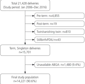
During the study period from January 2006 to December 2016, there were 21,428 deliveries in our institution includ- ing 4,855 preterm deliveries, 16,554 term deliveries, and 19 post-term deliveries. Fig. 1 shows the inclusion and exclu-
Fig. 1. Study population with inclusion and exclusion criteria.
FDIU, Fetal death in utero; ABGA, arterial blood gas analysis;
VBGA, venous blood gas analysis.
Total 21,428 deliveries (Study period: Jan 2006~Dec 2016)
Term, Singleton deliveries n=15,701
Final study population n=14,221 (90.6%)
Unavailable ABGA: n=1,480 (9.4%) Pre-term: n=4,855
Post-term: n=19
Twin/vanishing twin: n=810 Stillbirth/FDIU:n=43
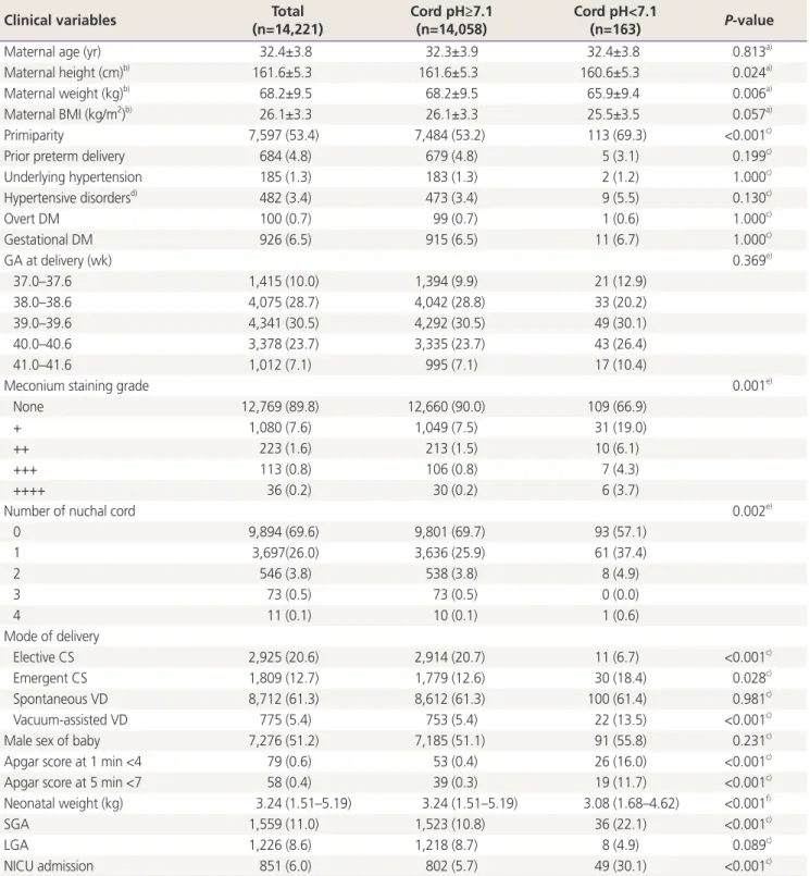
Table 1. Comparison of clinical characteristics according to cord blood pH at birth
Clinical variables Total
(n=14,221)
Cord pH≥7.1 (n=14,058)
Cord pH<7.1
(n=163) P-value
Maternal age (yr) 32.4±3.8 32.3±3.9 32.4±3.8 0.813a)
Maternal height (cm)b) 161.6±5.3 161.6±5.3 160.6±5.3 0.024a)
Maternal weight (kg)b) 68.2±9.5 68.2±9.5 65.9±9.4 0.006a)
Maternal BMI (kg/m2)b) 26.1±3.3 26.1±3.3 25.5±3.5 0.057a)
Primiparity 7,597 (53.4) 7,484 (53.2) 113 (69.3) <0.001c)
Prior preterm delivery 684 (4.8) 679 (4.8) 5 (3.1) 0.199c)
Underlying hypertension 185 (1.3) 183 (1.3) 2 (1.2) 1.000c)
Hypertensive disordersd) 482 (3.4) 473 (3.4) 9 (5.5) 0.130c)
Overt DM 100 (0.7) 99 (0.7) 1 (0.6) 1.000c)
Gestational DM 926 (6.5) 915 (6.5) 11 (6.7) 1.000c)
GA at delivery (wk) 0.369e)
37.0–37.6 1,415 (10.0) 1,394 (9.9) 21 (12.9)
38.0–38.6 4,075 (28.7) 4,042 (28.8) 33 (20.2)
39.0–39.6 4,341 (30.5) 4,292 (30.5) 49 (30.1)
40.0–40.6 3,378 (23.7) 3,335 (23.7) 43 (26.4)
41.0–41.6 1,012 (7.1) 995 (7.1) 17 (10.4)
Meconium staining grade 0.001e)
None 12,769 (89.8) 12,660 (90.0) 109 (66.9)
+ 1,080 (7.6) 1,049 (7.5) 31 (19.0)
++ 223 (1.6) 213 (1.5) 10 (6.1)
+++ 113 (0.8) 106 (0.8) 7 (4.3)
++++ 36 (0.2) 30 (0.2) 6 (3.7)
Number of nuchal cord 0.002e)
0 9,894 (69.6) 9,801 (69.7) 93 (57.1)
1 3,697(26.0) 3,636 (25.9) 61 (37.4)
2 546 (3.8) 538 (3.8) 8 (4.9)
3 73 (0.5) 73 (0.5) 0 (0.0)
4 11 (0.1) 10 (0.1) 1 (0.6)
Mode of delivery
Elective CS 2,925 (20.6) 2,914 (20.7) 11 (6.7) <0.001c)
Emergent CS 1,809 (12.7) 1,779 (12.6) 30 (18.4) 0.028c)
Spontaneous VD 8,712 (61.3) 8,612 (61.3) 100 (61.4) 0.981c)
Vacuum-assisted VD 775 (5.4) 753 (5.4) 22 (13.5) <0.001c)
Male sex of baby 7,276 (51.2) 7,185 (51.1) 91 (55.8) 0.231c)
Apgar score at 1 min <4 79 (0.6) 53 (0.4) 26 (16.0) <0.001c)
Apgar score at 5 min <7 58 (0.4) 39 (0.3) 19 (11.7) <0.001c)
Neonatal weight (kg) 3.24 (1.51–5.19) 3.24 (1.51–5.19) 3.08 (1.68–4.62) <0.001f)
SGA 1,559 (11.0) 1,523 (10.8) 36 (22.1) <0.001c)
LGA 1,226 (8.6) 1,218 (8.7) 8 (4.9) 0.089c)
NICU admission 851 (6.0) 802 (5.7) 49 (30.1) <0.001c)
Data are presented as the mean±standard deviation or number (%) or median (range).
BMI, body mass index; DM, diabetes mellitus; GA, gestational age; CS, caesarean section; VD, vaginal delivery; SGA, small for gestational age;
LGA, large for gestational age; NICU, neonatal intensive care unit.
a)P-value by Student’s t-test; b)Cases unavailable with maternal height (n=3,149), weight (n=2,939), and BMI (n=3,242) were excluded from this analysis; c)P-value by chi-squared test; d)Hypertensive disorders include gestational hypertension, mild pre-eclampsia, severe pre-eclampsia, superimposed pre-eclampsia on chronic hypertension, HELLP syndrome (hemolysis, elevated liver enzymes, and a low platelet count), and ec- lampsia; e)P-value by linear-by-linear association; f)P-value by Mann-Whitney U test; P<0.05: statistically significant.
sion protocols of this study population. We excluded cases with preterm birth, post-term birth, twin pregnancy includ- ing vanishing twin, stillbirth and fetal death in utero. After the exclusions, there were 15,701 term singleton pregnan- cies. Among them, the results of umbilical cord ABGA were available in 14,221 cases (90.6%), which constituted the final population of this study. We collected data on clini- cal variables including maternal age, height, weight, parity, history of prior preterm delivery, gestational age at delivery, the presence of meconium staining, the number of nuchal cord, mode of delivery, sex of baby, cord blood pH, Apgar scores at 1 minute and 5 minutes, neonatal birth weight, and NICU admission. Data were acquired from the electronic medical record system in our institution. The grade of me- conium staining was divided into the following 5 groups by experienced labor and delivery nurses: none, 1+, 2+, 3+, and 4+; nuchal cord was graded on a scale of 0 to 4. Mode of delivery was divided into 4 groups: elective caesarean section delivery, emergent caesarean section delivery, spontaneous vaginal delivery, and vacuum-assisted vaginal delivery. Apgar scores were assigned by experienced labor and delivery nurs- es or pediatric doctors. The cord blood was sampled imme- diately after delivery with heparinized syringes and analyzed within 30 minutes in most cases. We defined low cord pH as a pH <7.1. Neonatal weight was categorized into 3 groups:
small for gestational age (SGA), appropriate for gestational age (AGA), and large for gestational age (LGA) according to national data from the Korean Health Insurance Review and Assessment Service 2009.
In cases of low cord pH, we also examined additional neo- natal and infant outcomes including ventilator support, brain ultrasonographic findings, major anomaly, and CP by thor- ough review of medical records. The major anomaly category included heart anomaly, central nervous system anomaly, chromosomal anomaly, specific syndromes, and complicated cleft palate. CP was identified based on the diagnosis by the pediatric or rehabilitation departments during the infant’s follow-up period. We also searched for all cases of neonates born with normal cord pH (≥7.1) during the same study pe- riod who were diagnosed with CP during their follow-up pe- riod by screening the International Classification of Diseases, Tenth Revision, Clinical Modification (ICD-10-CM) infant codes (G80; CP) in our electronic medical system. Case con- firmation was based on the clinical diagnosis made by pedi- atric or rehabilitation doctors as documented in the medical
Table 2.Mean value and distribution of cord blood pH according to meconium staining grade, the number of nuchal cord, and mode of delivery Variables Meconium staining gradeNumber of nuchal cordMode of delivery None (n=12,769)+ (n=1,080)++ (n=223)+++ (n=113)++++ (n=36)P-value0 (n=9,894)1 (n=3,697)2 (n=546)3 (n=73)4 (n=11)P-valueElective CS (n=2,925)
Emer- gent CS (n=1,809) Sponta- neous VD (n=8,712)
Vacuum- assisted VD (n=775)P-value Mean pH 7.28±0.06 7.26±0.07 7.24±0.08 7.24±0.08 7.23±0.12<0.001a) 7.28±0.06 7.28±0.06 7.28±0.06 7.28±0.05 7.25±0.11<0.001a) 7.29±0.04 7.29±0.06 7.28±0.06 7.26±0.07<0.001a) Mean BEb)−3.48±2.57−4.68±3.09−5.86±3.46−5.82±3.79−6.38±5.11<0.001a)−3.58±2.67−3.76±2.71−3.63±2.80−3.61±2.85−4.55±4.43 0.024a)−2.45±1.92−3.16±2.82−3.97±2.70−5.38±2.88<0.001a) pH ≥7.211,986 (93.87)939 (86.94)171 (76.68)88 (77.88)26 (72.22)<0.001c)9,243 (93.42)3,388 (91.64)503 (92.12)68 (93.15)8 (72.73)<0.001c)2,850 (97.44)1,695 (93.70)8,010 (91.94)655 (84.52)<0.001d) pH <7.2e)783 (6.13)141 (13.06)52 (23.32)25 (22.12)10 (27.78)<0.001c)651 (6.58)309 (8.36)43 (7.88)5 (6.85)3 (27.27)<0.001c)75 (2.56)114 (6.30)702 (8.06)120 (15.48)<0.001d) pH <7.1e)109 (0.85)31 (2.87)10 (4.48)7 (6.19)6 (16.67)<0.001c)93 (0.94)61 (1.65)8 (1.47)0 (0)1 (9.09) 0.002c)11 (0.38)30 (1.66)100 (1.15)22 (2.84)<0.001d) pH <7.023 (0.18)5 (0.46)5 (2.24)2 (1.77)3 (8.33)<0.001c)21 (0.21)14 (0.38)3 (0.55)0 (0)0 (0) 0.073c)0 (0)12 (0.66)21 (0.24)5 (0.65)<0.001d) Data are presented as the mean±standard deviation or number (%). CS, caesarean section; VD, vaginal delivery; BE, base excess. a) P-value by ANOVA; b) Cases unavailable with no meconium staining (n=3), 1+ meconium staining (n=1), and 2+ meconium staining (n=2), cord neck 0 (n=5), cord neck 1 (n=1), emergent cesarean section (n=2), spontaneous vaginal delivery (n=1), and vacuum-assisted vaginal delivery (n=3) were excluded from this analysis; c) P-value by linear-by-linear association; d) P-value by chi-square test; P<0.05: statistically significant; e) Cases of cord pH <7.2 include those of cord pH <7.1 or pH <7.0. Cases of cord pH <7.1 include those of cord pH <7.0.
record. Statistical analysis was done using Mann-Whitney U tests, analysis of variance tests, Student’s t-tests, and linear- by-linear association tests using SPSS software (version 25.0;
IBM Corp., Armonk, NY, USA).
Results
Table 1 shows a comparison of the clinical characteristics ac- cording to cord blood pH at birth. The low cord pH group was characterized by lower maternal height and weight and a higher rate of primiparity. The low cord pH group was associated with a higher grade of meconium staining and higher number of nuchal cord compared to the control group. In terms of the mode of delivery, the low cord pH group showed a significantly lower rate of elective caesarean section and higher rates of emergent caesarean section and vacuum-assisted delivery compared to the control group.
Overall, rates of low Apgar scores at 1 minute (<4) and 5 minutes (<7) were 0.6% (n=79) and 0.4% (n=58), respec- tively. The low cord pH group was also associated with a higher rate of low Apgar scores at 1 minute and 5 minutes and NICU admission compared to the control group. Of
note, the low cord pH group showed a lower mean neo- natal weight (3.08 [1.68, 4.62] kg vs. 3.24 [1.51, 5.19] kg;
P<0.001) and a higher rate of SGA compared to the control group (22.1% vs. 10.8%; P<0.001).
In our study population of term singleton pregnancies, the mean cord blood pH was 7.28±0.06, ranged 6.75–7.53 and the mean BE was −3.63±2.69. Rates of cord pH <7.2, <7.1, and <7.0 were 7.1% (n=1,011), 1.1% (n=163), and 0.3%
(n=38), respectively. Next, we analyzed the mean value and distribution of cord blood pH according to the grade of me- conium staining, number of nuchal cord, and mode of deliv- ery (Table 2). There were significant differences in the mean cord pH based on the grade of meconium staining, number of nuchal cord, and mode of delivery (P<0.001 for all), but all mean values were within the normal range. Severe grades of meconium staining were associated with higher rates of cord pH <7.2, <7.1, and <7.0. The incidence of nuchal cord was also associated with a higher rate of cord pH <7.2 and <7.1 but not with cord pH <7.0. There were also significant differ- ences in the rates of cord pH <7.2, <7.1, and <7.0 according to the mode of delivery.
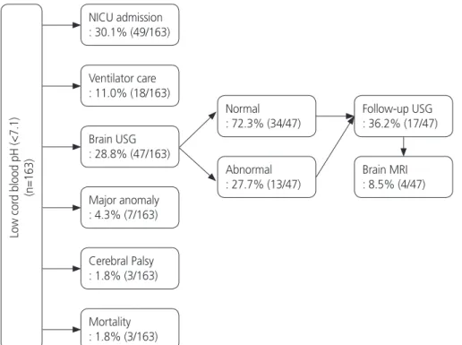
Fig. 2 shows the clinical course information in cases with low cord blood pH. Among 163 cases, 49 babies (30.1%)
Fig. 2. Clinical course and information in cases with low cord blood pH. NICU, neonatal intensive care unit; USG, ultrasonography; MRI, magnetic resonance imaging.
Low cord blood pH (<7.1) (n=163)
NICU admission : 30.1% (49/163)
Ventilator care : 11.0% (18/163)
Brain USG : 28.8% (47/163)
Major anomaly : 4.3% (7/163)
Cerebral Palsy : 1.8% (3/163)
Mortality : 1.8% (3/163)
Normal : 72.3% (34/47)
Abnormal : 27.7% (13/47)
Follow-up USG : 36.2% (17/47) Brain MRI : 8.5% (4/47)
were admitted to the NICU and 18 (11.0%) needed ventila- tor support. Ultrasonography of the brain was performed in 28.8% (n=47) of cases, and significant abnormal findings were observed in 27.7% (n=13) as follows: diffuse brain edema (n=7), grade 2 intraventricular hemorrhage (n=2), basal ganglia vasculopathy (n=2), Dandy–Walker complex (n=1), and myelination disorder (n=1).
Table 3 presents detailed clinical information on 3 cases of presumed CP with low cord pH. The baby in the first case was born at 39.0 weeks by prompt emergent caesarean sec- tion due to a non-reassuring FHR pattern detected at the time of admission. The neonatal weight was 3.234 kg and the meconium staining grade was 4+. The Apgar scores at 1 minute and 5 minutes were 0 and 4, respectively, but in- creased to 7 at 10 minutes. Cord blood pH was 7.021, and BE was −15.00 mmol/L. The baby was admitted into the NICU immediately and placed on a ventilator. An ultrasound examination of the brain on the first day of life revealed profound HIE findings, and the baby was diagnosed with moderate-severe cerebral coordination disturbance 1 month later. The baby was discharged 23 days after birth and, un- fortunately, was found dead at home 6 months later. The
second case was born by emergent caesarean section due to a non-reassuring FHR pattern developed during the second stage. The neonatal weight was 3.392 kg, and meconium staining grade was 1+. The Apgar scores at 1 minute and 5 minutes were 2 and 5, respectively. Cord blood pH was 6.887, and the BE was −18.30 mmol/L. The baby was admitted to the NICU and placed on a ventilator. Magnetic resonance imaging (MRI) of the brain checked on the 9th day showed a selective deep gray or white matter injury probably due to profound asphyxia. The electroencephalogram finding was also suggestive of diffuse cerebral dysfunction. The baby was lost to follow-up but was presumed to have CP. The third baby was born by spontaneous vaginal delivery without any remarkable event in the perinatal period. The baby had rou- tine nursery care and was discharged uneventfully. However, after 4 months, the baby visited the pediatric clinic in our hospital due to difficulty with head tilting. Unfortunately, the baby suffered severe developmental delays and was finally diagnosed with ataxic CP at age 2.5 years, which was not HIE-related CP.
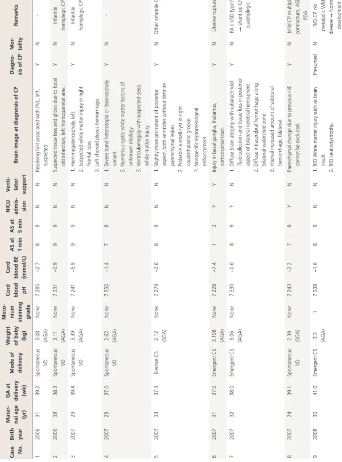
Lastly, we summarized the perinatal characteristics of the CP cases (n=18) with normal cord pH (≥7.1) at birth during
Table 3. CP cases among low cord pH at birth
Clinical variables Case 1 Case 2 Case 3
Birth year 2006 2007 2014
Maternal age (yr) 26 29 33
GA at delivery (wk) 39.0 41.4 40.0
Mode of delivery Emergent CS Emergent CS Spontaneous VD
Weight of baby (kg) 3.234 (AGA) a) 3.392 (AGA) a) 3.280 (AGA)a)
Meconium staining grade 4+ 1+ None
Cord blood pH 7.021 6.887 6.969
Cord blood base excess (mmol/L) −15.00 −18.30 −7.30
Apgar score at 1 min 0 2 7
Apgar score at 5 min 4 5 9
Apgar score at 10 min 7 NA NA
NICU admission Yes Yes No
Ventilator support Yes Yes No
Brain USGb) Done Done NA
Diagnosis of CP Yes Presumed Yes (Ataxic CP)
Mortality Yes No No
GA, gestational age; CS, caesarean section; VD, vaginal delivery; AGA, appropriate for gestational age; NA, not assessed; NICU, neonatal in- tensive care unit; USG, ultrasonography; CP, cerebral palsy.
a)Neonatal weight category; b)Brain USG performed at perinatal period.
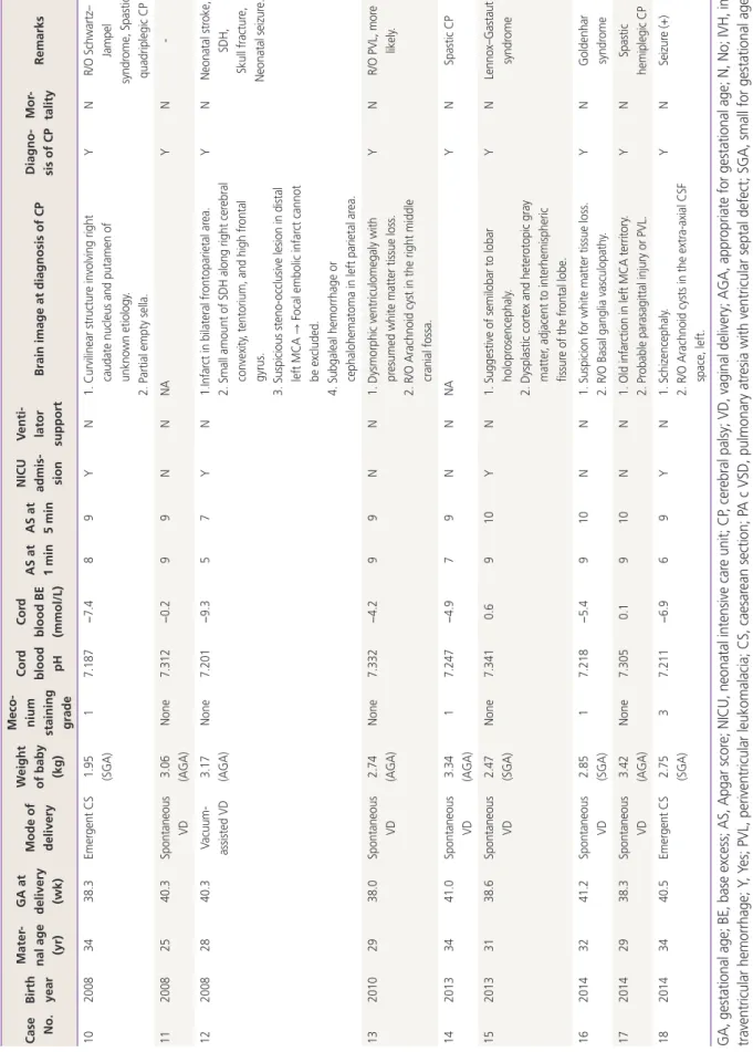
Table 4.CP cases among normal cord pH at birth during the same study period Case No.Birth year
Mater- nal age (yr)
GA at delivery (wk)
Mode of delivery
Weight of baby (kg)
Meco- nium staining grade
Cord blood pH
Cord blood BE (mmol/L) AS at 1 minAS at 5 min
NICU admis- sion
Venti- lator supportBrain image at diagnosis of CPDiagno- sis of CPMor- talityRemarks 120063139.2Spontaneous VD3.08 (AGA)None7.285−2.789NNResolving IVH associated with PVL, left, suspected.YN- 220063838.3Spontaneous VD3.11 (AGA)None7.331−0.999NNSuspected tissue loss and gliosis due to focal old infarction, left frontoparietal area.YNInfantile hemiplegic CP 320072939.4Spontaneous VD3.39 (AGA)None7.241−5.999NN1. Hemimegalencephaly, left. 2. Suspected white matter injury in right frontal lobe. 3. Left choroid plexus hemorrhage.
YNInfantile hemiplegic CP 420072337.0Spontaneous VD2.62 (AGA)None7.350−1.478NN1. Severe band heterotopia or lissencephaly variant. 2. Numerous cystic white matter lesions of unknown etiology. 3. Ventriculomegaly with suspected deep white matter injury.
YN- 520073337.3Elective CS2.12 (SGA)None7.279−2.689NN1. Slightly more prominent of posterior aspect, both ventricles without definite parenchymal lesion. 2. Probable a small cyst in right caudothalamic groove. 3. Nonspecific leptomeningeal enhancement.
YNOther infantile CP 620073137.0Emergent CS3.198 (AGA)None7.228−7.413YYInjury in basal ganglia, thalamus, corticospinal tract.YNUterine rupture 720073238.0Emergent CS3.06 (AGA)None7.330−0.689YN1. Diffuse brain atrophy with subarachnoid fluid collection and tissue loss in posterior aspect of bilateral cerebral hemisphere. 2. Diffuse intracerebral hemorrhage along bilateral watershed zone. 3. Interval increased amount of subdural hemorrhage, bilateral.
YNPA c VSD type IV → shunt op CP, quadriplegic 820072439.1Spontaneous VD2.39 (SGA)None7.243−2.278YNParenchymal change due to previous HIE cannot be excluded.YNMild CP multiple contracture, ASD, PDA 920083041.0Emergent CS3.3 (AGA)17.338−1.689NN1. R/O White matter injury such as brain insult. 2. R/O Leukodystrophy.
Presumed NR/O CP, r/o metabolic WM disease → Normal development
Case No.Birth year
Mater- nal age (yr)
GA at delivery (wk)
Mode of delivery
Weight of baby (kg)
Meco- nium staining grade
Cord blood pH
Cord blood BE (mmol/L) AS at 1 minAS at 5 min
NICU admis- sion
Venti- lator supportBrain image at diagnosis of CPDiagno- sis of CPMor- talityRemarks 1020083438.3Emergent CS1.95 (SGA)17.187−7.489YN1. Curvilinear structure involving right caudate nucleus and putamen of unknown etiology. 2. Partial empty sella.
YNR/O Schwartz– Jampel syndrome, Spastic quadriplegic CP 1120082540.3Spontaneous VD3.06 (AGA)None7.312−0.299NNNAYN- 1220082840.3Vacuum- assisted VD 3.17 (AGA)None7.201−9.357YN1.Infarct in bilateral frontoparietal area. 2. Small amount of SDH along right cerebral convexity, tentorium, and high frontal gyrus. 3. Suspicious steno-occlusive lesion in distal left MCA → Focal embolic infarct cannot be excluded. 4. Subgaleal hemorrhage or cephalohematoma in left parietal area.
YNNeonatal stroke, SDH, Skull fracture, Neonatal seizure. 1320102938.0Spontaneous VD2.74 (AGA)None7.332−4.299NN1. Dysmorphic ventriculomegaly with presumed white matter tissue loss. 2. R/O Arachnoid cyst in the right middle cranial fossa.
YNR/O PVL, more likely. 1420133441.0Spontaneous VD3.34 (AGA)17.247−4.979NNNAYNSpastic CP 1520133138.6Spontaneous VD2.47 (SGA)None7.3410.6910YN1. Suggestive of semilobar to lobar holoprosencephaly. 2. Dysplastic cortex and heterotopic gray matter, adjacent to interhemispheric fissure of the frontal lobe.
YNLennox–Gastaut syndrome 1620143241.2Spontaneous VD2.85 (SGA)17.218−5.4910NN1. Suspicion for white matter tissue loss. 2. R/O Basal ganglia vasculopathy.YNGoldenhar syndrome 1720142938.3Spontaneous VD3.42 (AGA)None7.3050.1910NN1. Old infarction in left MCA territory. 2. Probable parasagittal injury or PVL.YNSpastic hemiplegic CP 1820143440.5Emergent CS2.75 (SGA)37.211−6.969YN1. Schizencephaly. 2. R/O Arachnoid cysts in the extra-axial CSF space, left.
YNSeizure (+) GA, gestational age; BE, base excess; AS, Apgar score; NICU, neonatal intensive care unit; CP, cerebral palsy; VD, vaginal delivery; AGA, appropriate for gestational age; N, No; IVH, in- traventricular hemorrhage; Y, Yes; PVL, periventricular leukomalacia; CS, caesarean section; PA c VSD, pulmonary atresia with ventricular septal defect; SGA, small for gestational age; ASD, atrial septal defect; PDA, patent ductus arteriosus; R/O (and r/o), rule out; WM, white matter; NA, not assessed; SDH, subdural hemorrhage; MCA, middle cerebral artery; CSF, cerebrospinal fluid.
Table 4.Continued