Korean Circulation Journal
Introduction
The presence of myocardial ischemia causes various symptoms in patients and is predictive of future events
1)2)and revasculariza- tion of those lesions is important since it has the potential to im- prove patient outcomes.
2-4)However, revascularization of stenotic lesions that do not lead to myocardial ischemia is not beneficial and can rather be harmful. Therefore, the decision to revascularize a coronary artery stenosis should be guided by the evidence of myo- cardial ischemia.
Coronary angiography is limited in its ability to determine the physiologic significance of coronary stenosis.
5)6)Especially in pa- tients with intermediate stenosis, angiographic information does not correlate well with the functional significance of a lesion.
7-9)This uncertainty may result in unnecessary revascularization of insignifi- cant lesions or failure to revascularize the clinically significant ones.
As a result, fractional flow reserve (FFR) was introduced and has prov- en to be a reliable method for determining the functional signifi-
Print ISSN 1738-5520 • On-line ISSN 1738-5555
Fractional Flow Reserve: The Past, Present and Future
Jeong-Eun Kim, MD and Bon-Kwon Koo, MD
Department of Internal Medicine and Cardiovascular Center, Seoul National University Hospital, Seoul, Korea
Revascularization of coronary artery stenosis should be based on the objective evidence of ischemia. It is common practice for physicians to make decisions on revascularization in the cardiac catheterization laboratory based on the results of angiography, despite the fact that angiographic information does not correlate well with the functional significance of a coronary lesion. Fractional flow reserve (FFR) is a physiologic parameter which can be measured easily during the invasive procedure and can assess the functional significance of coronary stenosis. FFR-guided revascularization strategy is reported to be more effective than angiography-guided strategy in patients with coro- nary artery disease. Moreover, novel technologies based on FFR have been developed and will soon be incorporated into clinical practice.
(Korean Circ J 2012;42:441-446)
KEY WORDS: Coronary artery disease; Fractional flow reserve, myocardiol; Ischemia.
Correspondence: Bon-Kwon Koo, MD, Department of Internal Medicine, Cardiovascular Center, Seoul National University Hospital, 101 Daehak-ro, Jongno-gu, Seoul 110-744, Korea
Tel: 82-2-2072-2062, Fax: 82-2-3675-0805 E-mail: [email protected]
• The authors have no financial conflicts of interest.
This is an Open Access article distributed under the terms of the Creative Commons Attribution Non-Commercial License (http://creativecommons.
org/licenses/by-nc/3.0) which permits unrestricted non-commercial use, distribution, and reproduction in any medium, provided the original work is properly cited.
cance of coronary stenosis. FFR expresses the maximal achievable blood flow in a coronary vessel as a fraction of normal maximal blood flow to the same myocardial territory.
10)In other words, FFR repre- sents the extent to which maximal myocardial blood flow is limited by the presence of epicardial stenosis and can be easily measured by the ratio of distal coronary pressure to aortic pressure during maxi- mum hyperemia (Fig. 1). This index is independent of changes in he- modynamic conditions such as systemic blood pressure, heart rate, or myocardial contractility.
11)As the clinical benefit of an FFR-guided revascularization strategy has been proven in several studies with different lesion subsets, this strategy has become more popular in recent years (Fig. 2).
Fractional Flow Reserve: The Past
In the very early period of percutaneous coronary intervention (PCI), clinical application of intracoronary pressure was tried in pa- tients with coronary artery stenosis but failed. However, the cause of failure at that time was due to the fact that intracoronary pres- sure was measured with a large over-the-wire balloon catheter wi- thout hyperemia (minimal microvascular resistance). Since then, clinical application of intracoronary pressure had been almost for- gotten until the concept of myocardial FFR was developed and in- troduced by N. Pijls and B. De Bruyne in the early 1990s.
The concept was first validated in an animal study
12)and later in
humans using a positron emission tomography scan.
13)Given that
FFR is a continuous variable, a certain cutoff value was necessary to
determine the presence of myocardial ischemia (dichotomous vari-
able). In 1996, Pijls et al.
10)performed a clinical study to define the cutoff value of FFR to determine the presence of ischemia using non-invasive tests and sequential Bayesian considerations. In this study, an FFR cutoff value of 0.75 had a positive predictive value of 100% and a negative predictive value of 88% to determine the presence of ischemia. Due to a small zone of uncertainty between 0.75 and 0.80 (grey zone) and the results of the FFR versus Angiogra- phy for Multivessel Evaluation (FAME) study,
3)many clinicians now use the FFR cutoff value of 0.80 as a guide to perform revascularization.
After validation of a cutoff value, the clinical benefit of FFR-guid- ed revascularization was tested in the DEFER study (FFR to Deter- mine the Appropriateness of Angioplasty in Moderate Coronary Ste- noses).
4)This study included 325 patients referred for PCI of a single, de novo stenosis of intermediate severity. PCI was performed in all patients with an FFR <0.75 (reference group, n=144). If the FFR was
≥0.75, patients were randomized to either medical treatment (de- fer group, n=91) or PCI (perform group, n=90). After 5 years of fol- low-up, event free survival did not differ between the defer and PCI groups (80% and 73%, respectively) and the percentage of patients free from chest pain at follow-up was not different between the 2 groups. The composite rate of death and acute myocardial infarction
(MI) in the defer group was only 3.3% during the period of 5 years.
This study showed that patient outcomes with deferral of PCI ac- cording to FFR was excellent and the risk of death or acute MI was
<1% per year which could not be further decreased by stenting.
Since then, the benefit of FFR-guided revascularization strategy was tested and confirmed in more complex scenarios involving multiple lesions, multivessel disease, in-stent restenosis, post-stent- ing, left main disease, bifurcation lesions and patients with MI.
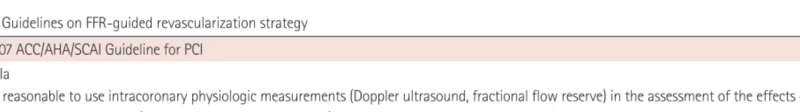
14-19)These results culminated in a Class IIA recommendation of FFR in 2007 American College of Cardiology/American Heart Association Society for Cardiac Angiography and Interventions PCI Guidelines on myo- cardial revascularization: “It is reasonable to use intracoronary physiologic measurements (Doppler ultrasound, fractional flow re- serve) in the assessment of the effects of intermediate coronary ste- noses in patients with angina symptoms” (Table 1).
20)Fractional Flow Reserve: The Present
A nuclear substudy of the Clinical Outcomes Utilizing Revascu- larization and Aggressive Drug Evaluation trial showed that PCI could improve the outcome of patients with coronary artery disease (CAD) which resulted in the relief of myocardial ischemia.
2)Investi- gators of the FAME study addressed the hypothesis that an FFR- guided PCI approach with drug-eluting stents would be superior to the current practice of conventional angiography-guided PCI in patients with multivessel CAD. The FAME protocol directed the in- vestigators to stent a lesion with at least 50% stenosis and if the
FFR= Qmax = (Pd-Pv)/R= Pd
Qmax (Pa-Pv)/R Pa s
N
Fig. 1. The concept of fractional flow reserve (FFR). Qsmax: hyperemic myo- cardial blood flow in the presence of a stenosis, QNmax: normal hyperemic myocardial blood flow, Pd: distal coronary pressure, Pa: aortic pressure, Pv:
venous pressure, R: hyperemic myocardial resistance.
Fig. 2. Clinical application of FFR to a patient with multiple lesions and multi-vessel disease. By coronary angiography, 11 stenoses (arrow) were found, yet none of those in the left anterior descending and left circumflex arteries were functionally significant by FFR. FFR values measured in the right coronary artery was FFR 0.65 and pullback pressure tracing revealed the lesion at the mid right coronary artery (*) was hemodynamically the most significant steno- sis. After one stent implantation at the mid right coronary artery, the FFR was 0.81. FFR: fractional flow reserve.
investigators thought that stenting was warranted on the basis of available clinical information. The patients were then randomized 1 : 1 to either standard PCI as planned (n=496) or to FFR-guided PCI (n=509). Although the number of angiographically significant ste- noses was identical between the 2 groups (2.7±0.9 vs. 2.8±1.0), the FFR group used fewer stents per patient (1.9±1.3 vs. 2.7±1.2, p<
0.001) and less contrast medium (272 mL vs. 302 mL, p<0.001). More importantly, at 1-year follow-up, the FFR group had fewer total cli- nical events (13.2% vs. 18.4%, p=0.02) and fewer combined death or MI (7.3% vs. 11%, p=0.04) compared to the angiography-guided PCI group. At 2 years, the rate of combined mortality or MI was still in favor of the FFR group (8.4% vs. 12.9%, p=0.02).
21)Further analy- sis showed that an FFR-guided strategy is not only cost-effective but also cost-saving compared to an angiography-guided strategy.
22)Another important finding of the FAME study is that assessment by FFR in patients with multivessel disease can lead to a reduction in the number of diseased coronary arteries and change in the treat- ment strategy. Of all patients with angiographic triple vessel disease (VD) in the FFR group, only 14% of the patients had functionally sig- nificant triple VD and 86% had ≤2 functionally significant diseased coronary arteries (2-VD=43%, 1-VD=34%, 0-VD=9%).
8)Furthermore, the functional SYNTAX score (SYNTAX score only by ischemia-in- ducing lesions as determined by FFR) was shown to decrease the number of high-risk patients and better discriminate the risk for fu- ture adverse events in patients with multivessel CAD.
23)With the results of FAME study and its substudies, the FFR-guided revascularization strategy has become more popular and was clas- sified as a Class IA recommendation in the 2010 European Guide- lines on myocardial revascularization (Table 1).
24)Fractional Flow Reserve: The Future
Although FFR has become the gold standard invasive assessment to detect the ischemia-related lesion, it requires an invasive proce- dure, expensive devices and pharmacologic intervention to induce
maximal hyperemia. Therefore, further development is still necessary to expand the clinical applications of FFR.
Novel hyperemic stimuli
Continuous infusion of adenosine via the central vein has been considered as the gold-standard method of hyperemia for FFR mea- surement.
25)However, this method requires relatively large doses of adenosine resulting in high cost, an additional procedure for femoral vein access and is practically not feasible during transradial coronary catheterization procedures. Furthermore, the adenosine adminis- tration itself is associated with adverse systemic effects such as AV block, dyspnea and chest pain.
26)27)To overcome the complexity of central vein infusion of adenosine, the feasibility and efficacy of peripheral infusion of adenosine were tested in recent studies.
28)29)Seo et al.
29)compared the hyperemic efficacy between continuous IV infusion (140 μg/min/kg) via the femoral vein and the forearm vein and found that the hyperemic ef- ficacy of the forearm vein infusion (FFR: 0.80±0.11) was not inferi- or (p for non-inferiority=0.01) to the femoral vein infusion (FFR: 0.80±
0.10) of adenosine. The number of functionally significant stenoses was not different between the 2 methods {femoral vein vs. forearm vein; 17 (25.0%) vs. 17 (25.0%), p=1.0}. Therefore, this method can be used for FFR measurement, especially during transradial coronary catheterization procedures.
Novel hyperemic agents for invasive physiologic assessment were also introduced. Nair et al.
30)compared the hyperemic effica- cy between a selective A
2Areceptor antagonist, regadenoson (400 ug, IV bolus) and adenosine in 25 patients with intermediate coro- nary stenosis and found that a single IV bolus of regadenoson was as effective as an IV infusion of adenosine. Jang et al.
31)compared the hyperemic efficacy of a bolus administration of nicorandil (intra- coronary, 2 mg) with continuous infusion of adenosine in 210 pa- tients. In this study, hyperemic efficacy of nicorandil was not inferior to that of adenosine (0.82±0.10 vs. 0.82±0.10; p for non-inferiority
<0.001) and there was a strong linear correlation between the FFR
Table 1. Guidelines on FFR-guided revascularization strategy 2005, 2007 ACC/AHA/SCAI Guideline for PCI
Class IIa
It is reasonable to use intracoronary physiologic measurements (Doppler ultrasound, fractional flow reserve) in the assessment of the effects of inter- mediate coronary stenoses (30% to 70% luminal narrowing) in patients with anginal symptoms. Coronary pressure or Doppler velocimetry may also be useful as an alternative to performing noninvasive functional testing (e.g., when the functional study is absent or ambiguous) to determine wheth- er an intervention is warranted (level of evidence: B).
2010 ESC/EACTS Guideline on myocardial revascularization Class IA
FFR-guided PCI is recommended for detection of ischemia-related lesion(s) when objective evidence of vessel-related ischemia is not available.
FFR: fractional flow reserve, ACC/AHA/SCAI: American College of Cardiology/American Heart Association Society for Cardiac Angiography and Interven- tions, PCI: percutaneous coronary intervention, ESC/EACTS: European Society of Cardiology/European Association of Cardio-Thoracic Surgery
measured by IV infusion of adenosine and nicorandil (R
2=0.934).
Moreover, nicorandil caused less changes in mean blood pressure, he- art rate, PR interval and less severe chest pain than adenosine (p<
0.05). While transient AV block occurred in 16 patients with adenos- ine, none were detected with nicorandil.
These novel agents and methods of adenosine administration will cause less discomfort in patients and reduce the complexity of in- vasive physiologic assessment.
Novel physiologic index without hyperemia
A new physiologic index, instantaneous wave-free ratio (iFR) wi- thout the requirement for hyperemia was introduced and tested in Adenosine Vasodilator Independent Stenosis Evaluation (ADVISE) study.
32)From the meticulous investigation on coronary flow and re- sistance, the investigators found that there is a certain period in the cardiac cycle during which the resistance at rest is similar in vari- ability and magnitude to those during hyperemia. In the ADVISE study, the distal-to-proximal pressure ratio during this period, which is also known as iFR, was compared with FFR in 157 stenoses. In this study, iFR had a good correlation with FFR (r=0.9, p<0.001) with ex- cellent diagnostic performance (Fig. 3). This novel concept, iFR, has great appeal as it may provide a faster and easier invasive physio- logic assessment for CAD. However, this concept still awaits further validation.
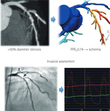
Non-invasive assessment of fractional flow reserve using CT scan
Recent advancements of CT technologies have enabled several novel methods to assess the functional significance of coronary stenosis in addition to anatomical information. One of these is the application of computational fluid dynamics to coronary CT angiog- raphy (CCTA) images.
33)With this technology, FFR can be computed using images from conventional CCTA (CT-derived computed FFR;
Fig. 3. Correlation between iFR and FFR according to the coronary artery (data from ADVISE study, courtesy of Justin Davies, MD). iFR: instantaneous wave-free ratio, FFR: fractional flow reserve.
1
0.8
0.6
0.4
0.2
0
FFR
iFR
0 0.2 0.4 0.6 0.8 1
Regression coefficient y=1.022x+0.03 r=0.90
Left coronary artery Right coronary artery
>50% diameter stenosis
>50% diameter stenosis
Non-invasive assessment
Invasive assessment
FFRCT0.74 → ischemia
FFR0.74 → ischemia 0.74
Fig. 4. A case example of CT-derived computed FFR. By coronary CT angi- ography, significant stenosis was found at the proximal left anterior de- scending coronary artery. When this lesion was assessed by CT-derived computed FFR, FFRG was 0.74 and found to be functionally significant. This information derived from non-invasive assessment matched very well with invasive angiography and invasive FFR measurement (FFR=0.74). FFR: frac- tional flow reserve.
Fig. 5. Comparison of the diagnostic performance between FFRCT and CCTA (from DISCOVER FLOW study, per-vessel analysis, n=159). PPV: positive predictive value, NPV: negative predictive value, FFRCT: CT-derived comput- ed FFR, CCTA: coronary CT angiography, DISCOVER FLOW: Diagnosis of Ischemia-Causing Stenoses Obtained Via Noninvasive Fractional Flow Re- serve, FFR: fractional flow reserve.
100 80 60 40 20
0 Sensitivity Specificity PPV NPV Accuracy 88 91
82
40
74 47
92 89 84
59 FFRCT ≤0.80 CCTA ≥50%
FFR
CT) without any invasive procedure and without hyperemia (Fig.
4). A prospective, multicenter clinical trial, Diagnosis of Ischemia- Causing Stenoses Obtained Via Noninvasive Fractional Flow Reserve (DISCOVER-FLOW) study, was performed to assess the diagnostic performance of FFR
CTin the prediction of the functional significance of stenosis.
34)In this study, 103 patients (159 vessels) with stenosis in a major epicardial coronary artery who had diagnostic quality CT images from 64 or more detector row CT scanners were consecu- tively enrolled and the diagnostic accuracy of CCTA (≥50% steno- sis) and FFR
CTwere compared. On a per-vessel basis, accuracy, sen- sitivity, specificity, positive predictive value, and negative predictive value for FFR
CTand CCTA were 84.3%, 87.9%, 82.2%, 73.9%, 92.2%, respectively, and 58.5%, 91.4%, 39.6%, 46.5%, 88.9%, respectively (Fig. 5). This study showed that noninvasive FFR derived from CCTA (FFR
CT) had a high diagnostic performance for the detection and exclusion of coronary lesions that lead to ischemia. Clinical applica- tion of this novel technology may potentially reduce unnecessary invasive procedures. Moreover, combination of virtual intervention and this technology can help to determine the treatment strategy in complex lesions prior to the invasive procedure. Although the concept and results of the DISCOVER-FLOW study are very encour- aging, further studies with a larger number of patients are needed to validate the clinical usefulness of this novel technology. A larger, prospective multicenter clinical trial, Determination of Fractional Flow Reserve by Anatomic Computed Tomographic Angiography study, has completed patient enrollment and the results will soon be available.
35)Conclusion
Fractional flow reserve has become the gold standard to define the functional significance of coronary stenosis. Novel hyperemic stimuli and novel physiologic indices without hyperemia will reduce the barriers and expand the clinical application of FFR. Furthermore, non-invasive assessment of the functional significance of coronary stenosis such as CT-derived computed FFR, can be helpful in optimiz- ing the interventional treatment strategy for patients with CAD prior to the invasive procedure.
References
1. Shaw LJ, Iskandrian AE. Prognostic value of gated myocardial perfu- sion SPECT. J Nucl Cardiol 2004;11:171-85.
2. Shaw LJ, Berman DS, Maron DJ, et al. Optimal medical therapy with or without percutaneous coronary intervention to reduce ischemic bur- den: results from the Clinical Outcomes Utilizing Revascularization and Aggressive Drug Evaluation (COURAGE) Trial Nuclear Substudy. Cir- culation 2008;117:1283-91.
3. Tonino PA, De Bruyne B, Pijls NH, et al. Fractional flow reserve versus angiography for guiding percutaneous coronary intervention. N Engl J Med 2009;360:213-24.
4. Pijls NH, van Schaardenburgh P, Manoharan G, et al. Percutaneous coronary intervention of functionally nonsignificant stenosis: 5-year follow-up of the DEFER Study. J Am Coll Cardiol 2007;49:2105-11.
5. White CW, Wright CB, Doty DB, et al. Does visual interpretation of the coronary arteriogram predict the physiologic importance of a coronary stenosis? N Engl J Med 1984;310:819-24.
6. Vogel RA. Assessing stenosis significance by coronary arteriography:
are the best variables good enough? J Am Coll Cardiol 1988;12:692-3.
7. Koo BK, Park KW, Kang HJ, et al. Physiological evaluation of the provi- sional side-branch intervention strategy for bifurcation lesions using fractional flow reserve. Eur Heart J 2008;29:726-32.
8. Tonino PA, Fearon WF, De Bruyne B, et al. Angiographic versus func- tional severity of coronary artery stenoses in the FAME Study frac- tional flow reserve versus angiography in multivessel evaluation. J Am Coll Cardiol 2010;55:2816-21.
9. Yong AS, Ng AC, Brieger D, Lowe HC, Ng MK, Kritharides L. Three-di- mensional and two-dimensional quantitative coronary angiography, and their prediction of reduced fractional flow reserve. Eur Heart J 2011;
32:345-53.
10. Pijls NH, De Bruyne B, Peels K, et al. Measurement of fractional flow reserve to assess the functional severity of coronary-artery stenoses.
N Engl J Med 1996;334:1703-8.
11. De Bruyne B, Bartunek J, Sys SU, Pijls NH, Heyndrickx GR, Wijns W. Si- multaneous coronary pressure and flow velocity measurements in hu- mans: feasibility, reproducibility, and hemodynamic dependence of co- ronary flow velocity reserve, hyperemic flow versus pressure slope index, and fractional flow reserve. Circulation 1996;94:1842-9.
12. Pijls NH, van Son JA, Kirkeeide RL, De Bruyne B, Gould KL. Experimen- tal basis of determining maximum coronary, myocardial, and collateral blood flow by pressure measurements for assessing functional stenosis severity before and after percutaneous transluminal coronary angio- plasty. Circulation 1993;87:1354-67.
13. De Bruyne B, Baudhuin T, Melin JA, et al. Coronary flow reserve calcu- lated from pressure measurements in humans: validation with posi- tron emission tomography. Circulation 1994;89:1013-22.
14. Bech GJ, Droste H, Pijls NH, et al. Value of fractional flow reserve in making decisions about bypass surgery for equivocal left main coro- nary artery disease. Heart 2001;86:547-52.
15. Lindstaedt M, Yazar A, Germing A, et al. Clinical outcome in patients with intermediate or equivocal left main coronary artery disease after deferral of surgical revascularization on the basis of fractional flow reserve measurements. Am Heart J 2006;152:156. e1-9.
16. Potvin JM, Rodés-Cabau J, Bertrand OF, et al. Usefulness of fractional flow reserve measurements to defer revascularization in patients with stable or unstable angina pectoris, non-ST-elevation and ST-elevation acute myocardial infarction, or atypical chest pain. Am J Cardiol 2006;
98:289-97.
17. Fischer JJ, Wang XQ, Samady H, et al. Outcome of patients with acute coronary syndromes and moderate coronary lesions undergoing defer- ral of revascularization based on fractional flow reserve assessment.
Catheter Cardiovasc Interv 2006;68:544-8.
18. Berger A, Botman KJ, MacCarthy PA, et al. Long-term clinical outcome after fractional flow reserve-guided percutaneous coronary interven- tion in patients with multivessel disease. J Am Coll Cardiol 2005;46:
438-42.
19. Wongpraparut N, Yalamanchili V, Pasnoori V, et al. Thirty-month out- come after fractional flow reserve-guided versus conventional multi- vessel percutaneous coronary intervention. Am J Cardiol 2005;96:
877-84.
20. Smith SC Jr, Feldman TE, Hirshfeld JW Jr, et al. ACC/AHA/SCAI 2005 guideline update for percutaneous coronary intervention--summary article: a report of the American College of Cardiology/American Heart Association Task Force on Practice Guidelines (ACC/AHA/SCAI Writing Committee to Update the 2001 Guidelines for Percutaneous Coronary Intervention). Circulation 2006;113:156-75.
21. Pijls NH, Fearon WF, Tonino PA, et al. Fractional flow reserve versus angiography for guiding percutaneous coronary intervention in pa- tients with multivessel coronary artery disease: 2-year follow-up of the FAME (Fractional Flow Reserve Versus Angiography for Multives- sel Evaluation) Study. J Am Coll Cardiol 2010;56:177-84.
22. Fearon WF, Bornschein B, Tonino PA, et al. Economic evaluation of fractional flow reserve-guided percutaneous coronary intervention in patients with multivessel disease. Circulation 2010;122:2545-50.
23. Nam CW, Mangiacapra F, Entjes R, et al. Functional SYNTAX score for risk assessment in multivessel coronary artery disease. J Am Coll Cardiol 2011;58:1211-8.
24. Wijns W, Kolh P, Danchin N, et al. Guidelines on myocardial revascular- ization. Eur Heart J 2010;31:2501-55.
25. De Bruyne B, Pijls NH, Barbato E, et al. Intracoronary and intravenous adenosine 5’-triphosphate, adenosine, papaverine, and contrast medi- um to assess fractional flow reserve in humans. Circulation 2003;107:
1877-83.
26. Wilson RF, Wyche K, Christensen BV, Zimmer S, Laxson DD. Effects of adenosine on human coronary arterial circulation. Circulation 1990;82:
1595-606.
27. Kern MJ, Deligonul U, Tatineni S, Serota H, Aguirre F, Hilton TC. Intrave-
nous adenosine: continuous infusion and low dose bolus administra- tion for determination of coronary vasodilator reserve in patients with and without coronary artery disease. J Am Coll Cardiol 1991;18:718-29.
28. Lindstaedt M, Bojara W, Holland-Letz T, et al. Adenosine-induced maxi- mal coronary hyperemia for myocardial fractional flow reserve mea- surements: comparison of administration by femoral venous versus antecubital venous access. Clin Res Cardiol 2009;98:717-23.
29. Seo MK, Shin DH, Yang HM, et al. Comparison of hyperemic efficacy between central and peripheral veous adenosine infusion for fractional flow reserve measurement. Circulation 2010;122:A18620. Abstract.
30. Nair PK, Marroquin OC, Mulukutla SR, et al. Clinical utility of regadeno- son for assessing fractional flow reserve. JACC Cardiovasc Interv 2011;
4:1085-92.
31. Jang HJ, Koo BK, Kim JH, et al. Safety and efficacy of a novel hyperemic agent, nicorandil, for invasive physiologic assessment in a catheteriza- tion laboratory: a prospective multicenter study. Korean Circ J 2011;
550608A. Abstract.
32. Sen S, Escaned J, Malik IS, et al. Development and Validation of a New Adenosine-Independent Index of Stenosis Severity From Coronary Wave-Intensity Analysis: results of the ADVISE (ADenosine Vasodila- tor Independent Stenosis Evaluation) Study. J Am Coll Cardiol 2012;
59:1392-402.
33. Kim HJ, Vignon-Clementel IE, Coogan JS, Figueroa CA, Jansen KE, Tay- lor CA. Patient-specific modeling of blood flow and pressure in human coronary arteries. Ann Biomed Eng 2010;38:3195-209.
34. Koo BK, Erglis A, Doh JH, et al. Diagnosis of ischemia-causing coro- nary stenoses by noninvasive fractional flow reserve computed from coronary computed tomographic angiograms. Results from the pro- spective multicenter DISCOVER-FLOW (Diagnosis of Ischemia-Caus- ing Stenoses Obtained Via Noninasive Fractional Flow Reserve) study.
J Am Coll Cardiol 2011;58:1989-97.
35. Min JK, Berman DS, Budoff MJ, et al. Rationale and design of the De- FACTO (Determination of Fractional Flow Reserve by Anatomic Com- puted Tomographic AngiOgraphy) Study. J Cardiovasc Comput Tomogr 2011;5:301-9.