• 검색 결과가 없습니다.

Catheter Ablation of Parahisian Premature Ventricular ComplexJun Kim, MD, Jeong Su Kim, MD, Yong Hyun Park, MD, June Hong Kim, MD, and Kook Jin Chun, MD

N/A
N/A
Protected

Academic year: 2022

Share "Catheter Ablation of Parahisian Premature Ventricular ComplexJun Kim, MD, Jeong Su Kim, MD, Yong Hyun Park, MD, June Hong Kim, MD, and Kook Jin Chun, MD"

Copied!
4
0
0

로드 중.... (전체 텍스트 보기)

전체 글

(1)

766

Print ISSN 1738-5520 / On-line ISSN 1738-5555 Copyright © 2011 The Korean Society of Cardiology CASE REPORT

http://dx.doi.org/10.4070/kcj.2011.41.12.766

Open Access

Catheter Ablation of Parahisian Premature Ventricular Complex

Jun Kim, MD, Jeong Su Kim, MD, Yong Hyun Park, MD, June Hong Kim, MD, and Kook Jin Chun, MD Department of Internal Medicine, Pusan National University School of Medicine, Cardiovascular Center,

Pusan National University Yangsan Hospital, Yangsan, Korea ABSTRACT

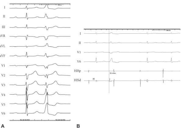
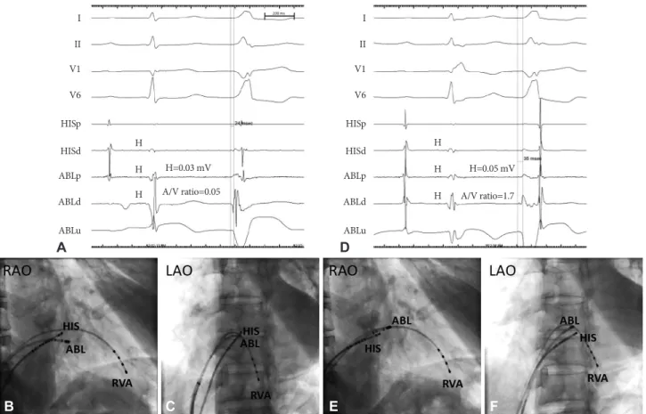
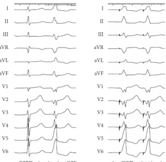
Catheter ablation is performed in selected patients with a symptomatic premature ventricular complex (PVC) or PVC-induc- ed cardiomyopathy. Ablation of PVC from the His region has a high risk of inducing a complete atrioventricular block. Here we report successful catheter ablation of a parahisian PVC in a 63-year-old man. (Korean Circ J 2011;41:766-769)

KEY WORDS: Premature ventricular complexes; Bundle of His; Catheter ablation.

Received: April 25, 2011 Accepted: May 18, 2011

Correspondence: Jun Kim, MD, Cardiovascular Center, Pusan Nation- al University Yangsan Hospital, 20 Geumo-ro, Mulgeum-eup, Yangsan 626-770, Korea

Tel: 82-55-360-2357, Fax: 82-55-360-2204 E-mail: [email protected]

• The authors have no financial conflicts of interest.

cc

This is an Open Access article distributed under the terms of the Cre- ative Commons Attribution Non-Commercial License (http://creativecom- mons.org/licenses/by-nc/3.0) which permits unrestricted non-commer- cial use, distribution, and reproduction in any medium, provided the origi- nal work is properly cited.

Introduction

Catheter ablation of the premature ventricular complex (PVC) is performed in highly symptomatic patients whose conditions are refractory to medical treatment.

1)2)

Frequent PVC can cause ventricular dysfunction, and PVC catheter ablation improves ventricular function.

3-6)

Selective PVC ab- lation suppresses ventricular fibrillation in selected pa- tients.

7)8)

PVC from the parahisian region is rare and catheter ablation has a high risk of heart block.

9)10)

Here we report a case of successful PVC catheter ablation in the parahisian region.

Case

A 63-year-old man with symptomatic frequent PVCs was admitted for catheter ablation. His symptomatic PVCs were refractory to class I antiarrhythmic drugs for 1 year. The pa- tient’s medial history was unremarkable. Physical examina- tion was normal except for an irregular heart rhythm. A 12- lead electrocardiogram (ECG) of PVC showed a monophasic

R wave in leads 1 and aVL and precordial transition between leads V3 and V4 (Fig. 1A). Ambulatory ECG monitoring sh- owed frequent PVC (55,882 beats; 30.1% of the total beats) and a type I second-degree atrioventricular (AV) block dur- ing sleep. Echocardiography showed normal biventricular size and function.

After informed consent was obtained from the patient, a cardiac electrophysiological study was performed under a fasting state. Sedation and analgesia were performed using midazolam and morphine. Baseline ECG showed sinus rhy- thm and PVC with the same configuration as clinical PVC.

Programmed ventricular stimulation with or without iso- proterenol did not induce sustained ventricular tachycardia.

Activation mapping was performed using a 7-Fr convention- al ablation catheter with a 4-mm distal tip. Local activation time at the His recording area was 18 ms ahead of PVC onset (Fig. 1B). Mapping of the right ventricle localized the PVC focus in the region of retroventricular (RV) inflow. The tar- get site was initially mapped where the His potential had low amplitude and AV ratio is <1 to decrease the risk of a com- plete AV block. A site with local ventricular activation that preceded PVC onset by 24 ms was found anterior and inferior to the His recording catheter (Fig. 2A, B and C). The proce- dure was continued after we discussed the potential risk of a complete AV block with the patient.

After radiofrequency (RF) energy was applied, a complete

right bundle branch block occurred without affecting the

PVC. Mapping of the His recording area more proximally re-

vealed optimal mapping site with local ventricular activation

ahead of PVC onset by 35 ms and the QS pattern on unipolar

electrogram (Fig. 2D, E and F). Application of RF energy (esc-

alated from 15 to 30 watts with a target temperature of 55°C)

(2)

Jun Kim, et al. 767

abolished the PVC within 4 seconds (Fig. 3). There was no junctional beat during the RF energy application. Pace map- ping at the ablation site showed QRS configuration matches in all 12 leads (Fig. 4). PVC did not recur during the 60 minutes waiting period. Neither the PR nor the AH intervals changed before and after ablation. Total procedural time and fluoro- scopic time was 180 and 20 minutes, respectively. The proce- dure was completed without further complications. Ambu- latory monitoring showed a PVC burden of 0.3% and 0.0% at 1 month and 1 year of follow-up, respectively. A maximal heart rate of 140 beats per minute with normal AV conduc- tion was noted during a treadmill exercise test 1 year after the procedure. The patient had no symptoms and did not require an antiarrhythmic drug during the 1-year follow-up period.

Discussion

Here we reported successful catheter ablation of a PVC from the proximal parahisian region without the creation of a complete AV block. Several investigators have developed al- gorithms to predict the origin of premature ventricular ta- chycardia and PVCs from the ventricular outflow tract using a 12-lead ECG.

11)12)

Based on this schema, the PVC origin in this patient is presumed to be located near the His bundle.

Characteristics of PVC from the RV inflow tract include monophasic R waves in lead I, R, or RR in the aVL, the QS pat- tern in lead V1, and large R-wave amplitudes in leads V5-6.

9)

Ashikaga et al.

10)

reported a case of parahisian PVC that was successfully treated by catheter ablation. Compared with previous reports, the configuration of PVC differed in the aspects of vertical PVC axis, deep S wave in lead III, a QRS pattern in leads V1 and V2, and a RR pattern in leads V4-6.

This finding may be related to the PVC origin being from the more proximal His bundle region.

Successful ablation of PVC wherein the His potential is recorded from the right side suggests that the PVC origi- nates from the myocardium in the vicinity of His at the right AV septum. Even though the AV ratio >1, absence of an AV block during and after ablation suggests that the PVC origin is very close to the distal penetrating bundle, which is surro- unded by the central fibrous body. The central fibrous body may protect the His bundle from thermal injury caused by RF energy. Because the proximal penetrating bundle is not sur- rounded by the central fibrous body, ablation at this site may cause an AV block.

13)

The existence of an AV block at night and the disappear- ance of the AV block during exercise suggests that the AV block is due to increased vagal tone. Normal AH and HV in- tervals also support the hypothesis that AV block is due to increased vagal tone. The idea that PVC induced the AV block by a mechanism of concealed conduction is less likely because there was no PVC at the time of the AV block and the second-degree AV block persisted after PVC ablation.

Catheter ablation in patients with parahisian PVC should

I II

III aVR

aVL

aVF

V1

V2

V3

V4

V5 V6

I II V1 V6 HISp HISd

A B

Fig. 1. A 12-lead electrocardiogram of the patient (A). Premature ventricular complex in bigeminal pattern is shown. Surface electrocardio-

gram I, II, V 1, V6 together with intracardiac electrograms from the proximal and distal bipolar electrograms in the His region (HIp HIS d) are

displayed from top to bottom (B). Local ventricular activation in the His recording area is earlier than onset of PVC by 18 ms.

(3)

768 Parahisian Premature Ventricular Complex

be performed only after careful evaluation of PVC-related symptoms, a trial of maximally tolerated antiarrhythmic drug therapy, and full discussion about ablation-related catastroph- ic complications of the AV block. We performed the proce- dure because the patient’s condition did not respond to stan- dard antiarrhythmic drug treatment and he accepted the

potential procedural risks.

Catheter ablation of arrhythmias originating in the vicinity of the bundle of His requires special attention to decrease the risk of iatrogenic AV block. In addition to careful mapping of the optimal target site, escalation of power from low energy, and immediate discontinuation of RF energy delivery imme- Fig. 2. Surface electrocardiogram I, II, V 1, V6 together with intracardiac electrograms from the proximal and distal bipolar electrograms in the His region (HIp HIS d) and proximal, distal bipolar and unipolar electrogram andablation catheter (ABLp, ABLd and ABLuni) are dis- played from top to bottom. Local ventricular activation was earlier at the unsuccessful ablation site than the onset of PVC by 24 ms (A).

Right anterior oblique (RAO) and left anterior oblique (LaO) fluoroscopic views of unsuccessful ablation site. Ablation catheter (ABL) is lo- cated a few millimeter anterior and inferior to the His recording catheter (HIS) (B and C). Also shown is a catheter in the right ventricular apex (RVA). Local ventricular activation was earliest at the successful ablation site than the onset of PVC by 35 ms (D). RAO and LAO fluoro- scopic views of successful ablation site (E and F). Compared with B and C, ablation catheter (ABL) is located a few millimeter anterior and superior to the His recording catheter (HIS).

I II V1 V6

HISp HISd ABLp ABLd

ABLu

I II V1 V6

HISp HISd ABLp ABLd

ABLu H

H=0.03 mV H=0.05 mV

A/V ratio=0.05 A/V ratio=1.7

H H

H H H

D A

C F

B E

Fig. 3. A 12-lead electrocardiogram during application of radiofrequency energy. After application of RF energy there was immediate disap- pearance of premature ventricular complex.

I

II

III

aVR

aVL

aVF

V1

V2

V3

V4

V5

V6

(4)

Jun Kim, et al. 769

diately after the appearance of junctional rhythm are prereq- uisites to successful ablation.

14)

As in catheter ablation of the parahisian AV pathway, same precautions should be applied to avoid AV blocks during the ablation of parahisian PVC. Cry- oablation can be considered a safer source of energy compar- ed with RF energy in such patients at high risk of AV blocks.

15)

There are several limitations in this study. We did not per- form electroanatomic mapping, which might have revealed a ventricular substrate abnormality, or alternative site map- ping from the left AV septum or right coronary cusp, which might have shown a more optimal site and had a lower risk of an AV block. We also did not map the anteroseptal region us- ing the superior approach, which may have provided more stable and optimal target sites as did ablation of the antero- septal accessory pathway.

15)

In conclusion, we performed a successful catheter ablation of a PVC from the proximal His region under activation mapp- ing guidance. Detailed mapping and careful delivery of RF energy are essential for successful ablation without AV block creation.

Acknowledgments

This work was supported by a 2011 research grant from Pusan Na- tional University Yangsan Hospital.

REFERENCES

1) Zhu DW, Maloney JD, Simmons TW, et al. Radiofrequency catheter ablation for management of symptomatic ventricular ectopic activity.

J Am Coll Cardiol 1995;26:843-9.

2) Seidl K, Schumacher B, Hauer B, et al. Radiofrequency catheter ab- lation of frequent monomorphic ventricular ectopic activity. J Car- diovasc Electrophysiol 1999;10:924-34.

3) Takemoto M, Yoshimura H, Ohba Y, et al. Radiofrequency catheter ablation of premature ventricular complexes from right ventricular outflow tract improves left ventricular dilation and clinical status in patients without structural heart disease. J Am Coll Cardiol 2005;45:

1259-65.

4) Yarlagadda RK, Iwai S, Stein KM, et al. Reversal of cardiomyopathy in patients with repetitive monomorphic ventricular ectopy originating from the right ventricular outflow tract. Circulation 2005;112:1092-7.

5) Bogun F, Crawford T, Reich S, et al. Radiofrequency ablation of fre- quent, idiopathic premature ventricular complexes: comparison with a control group without intervention. Heart Rhythm 2007;4:863-7.

6) Kanei Y, Friedman M, Ogawa N, Hanon S, Lam P, Schweitzer P. Fre- quent premature ventricular complexes originating from the right ventricular outflow tract are associated with left ventricular dysfunc- tion. Ann Noninvasive Electrocardiol 2008;13:81-5.

7) Haïssaguerre M, Extramiana F, Hocini M, et al. Mapping and abla- tion of ventricular fibrillation associated with long-QT and Brugada syndromes. Circulation 2003;108:925-8.

8) Noda T, Shimizu W, Taguchi A, et al. Malignant entity of idiopathic ventricular fibrillation and polymorphic ventricular tachycardia ini- tiated by premature extrasystoles originating from the right ventricu- lar outflow tract. J Am Coll Cardiol 2005;46:1288-94.

9) Yamauchi Y, Aonuma K, Takahashi A, et al. Electrocardiographic characteristics of repetitive monomorphic right ventricular tachycar- dia originating near the His-bundle. J Cardiovasc Electrophysiol 2005;

16:1041-8.

10) Ashikaga K, Tsuchiya T, Nakashima A, Hayashida K. Catheter abla- tion of premature ventricular contractions originating from the His bundle region. Europace 2007;9:781-4.

11) Callans DJ, Menz V, Schwartzman D, Gottlieb CD, Marchlinski FE.

Repetitive monomorphic tachycardia from the left ventricular outflow tract: electrocardiographic patterns consistent with a left ventricular site of origin.J Am Coll Cardiol 1997;29:1023-7.

12) Ito S, Tada H, Naito S, et al. Development and validation of an ECG algorithm for identifying the optimal ablation site for idiopathic ven- tricular outflow tract tachycardia. J Cardiovasc Electrophysiol 2003;

14:1280-6.

13) Anderson RH, Ho SY, Becker AE. Anatomy of the human atrioven- tricular junctions revisited. Anat Rec 2000;260:81-91.

14) Haïsaguerre M, Marcus F, Poquet F, Gencel L, Le Métayer P, Clémen- ty J. Electrocardiographic characteristics and catheter ablation of parahissian accessory pathways. Circulation 1994;90:1124-8.

15) Di Biase L, Al-Ahamad A, Santangeli P, et al. Safety and outcomes of cryoablation for ventricular tachyarrhythmias: results from a multi- center experience. Heart Rhythm 2011;8:968-74.

16) Choi YS, Sung JD, Song JM, et al. Radiofrequency catheter ablation of anteroseptal accessory pathways from right atrium. Korean Circ J 1999;29:1089-96.

Fig. 4. Pacemapping at the successful ablation site. Morphology of QRS complex during pacing by ablation catheter at the ablation site is same with morphology of PVC in all 12 leads.

I II III aVR aVL aVF V1 V2 V3 V4 V5 V6

I

II

III

aVR

aVL

aVF

V1

V2

V3

V4

V5

V6

수치

Fig. 1. A 12-lead electrocardiogram of the patient (A). Premature ventricular complex in bigeminal pattern is shown
Fig. 3. A 12-lead electrocardiogram during application of radiofrequency energy. After application of RF energy there was immediate disap- disap-pearance of premature ventricular complex.
Fig. 4. Pacemapping at the successful ablation site. Morphology of  QRS complex during pacing by ablation catheter at the ablation  site is same with morphology of PVC in all 12 leads.

참조

관련 문서