Impaired Coronary Flow Reserve Is the Most Important Marker of Viable Myocardium in the Myocardial Segment-Based Analysis of Dual-Isotope Gated Myocardial Perfusion Single-Photon Emission Computed Tomography
9
0
0
전체 글
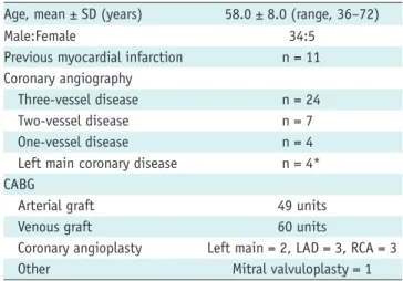
(2) Lee et al.. INTRODUCTION Myocardial viability is an important issue in the field of cardiology, because revascularization in the ischemic viable area improves regional wall motion, left ventricular (LV) function, and ultimately, outcomes of patients (1). The dysfunctional myocardium which is not viable should be managed by medical treatment, whereas the ischemic viable myocardium should be re-vascularized by coronary artery bypass graft (CABG) surgery or percutaneous coronary intervention. Therefore, it is important to predict the presence of viable myocardium in the management of patients with coronary artery disease (CAD) patients and particular in those with LV dysfunction (2). In the field of nuclear cardiology are several tools available to detect viable myocardium. First, 201Tl scintigraphy has been the most widely-investigated nuclear imaging tool (3, 4). Preserved 201Tl uptake could indicate viable myocardium (4), because the degree of 201 Tl uptake is well correlated with the functioning mass of viable myocytes (5). The presence of early (1–6 hours post-injection) (3) or delayed (8–24 hours post-injection) (4) redistribution of 201Tl has been the hallmark of viable myocardium. Furthermore, the low detectability of the 201 Tl redistribution protocol for viable myocardium could be improved through an additional investigation of 201Tl re-injection (6, 7). Second, 99mTc-labeled agents such as 99m Tc-methoxyisobutylisonitrile (99mTc-MIBI) are promising radiopharmaceuticals for not only myocardial perfusion assessment but also myocardial viability assessment (8). Because of its excellent physicochemical properties, 99mTcMIBI has been used for electrocardiographic (ECG)-gated myocardial single-photon emission computed tomography (SPECT) in which myocardial wall motion and wall thickening could be evaluated in addition to myocardial perfusion (9, 10). Last, a mismatch of perfusion and metabolism in positron emission tomography (PET) studies is another important finding characteristic for viable myocardium (11), although it is beyond the scope of the current study. Among a number of myocardial SPECT protocols, the dual-isotope myocardial SPECT (rest 201Tl/stress 99mTc-MIBI) protocol has been successfully applied to a variety of CAD conditions. The efficacy of the protocol for the detection of CAD has been validated in exercise (9) and pharmacologic stress (12). Transient LV dilatation measured on dual-isotope SPECT was a marker of severe and extensive CAD (13). The 278. prognosis of CAD could be effectively predicted using dualisotope myocardial SPECT (14). However, the usefulness of the protocol in terms of detection of myocardial viability has not been thoroughly evaluated yet. Furthermore, if ECGgating and 201Tl 24 hours delayed redistribution studies were added to the dual-isotope protocol, valuable markers of viable myocardium such as stress/rest reversibility (in other words, coronary flow reserve [CFR] impairment), 201 Tl rest perfusion status, 201Tl 24 hours redistribution and 99m Tc-MIBI systolic wall thickening could be evaluated as a whole, and the gated LV ejection fraction (LVEF) would be facilely assessed without additional procedures. More important, the competency of individual viability markers could be directly compared with each other, unveiling the most robust marker for myocardial viability. Thus, in the current study, we performed dual-isotope gated myocardial perfusion SPECT (rest 201Tl/gated dipyridamole stress 99mTcMIBI/24 hours redistribution of 201Tl) in CAD patients before and 3 months after CABG surgery. The study purpose was to investigate the predictability of viable myocardium using dual-isotope gated myocardial SPECT and find the most competent marker of viable myocardium using multiple logistic regression analysis.. MATERIALS AND METHODS Patients The institutional ethical committee approved this study and an informed consent was obtained from the patients before the study. At our institute, CAD patients who are scheduled to have CABG surgery routinely undergo dualisotope gated myocardial SPECT both pre-operative and 3 months post-operative. A total of 39 consecutive patients who had completed the pre-operative and post-operative SPECT studies over an 18-month period were enrolled in the current study (Table 1). The patients underwent CABG surgery because of multi-vessel CAD, left main disease or intractable chest pain. Patients with emergent CABG or without preoperative SPECT were excluded. The mean age was 58.0 ± 8.0 (mean ± standard) years (range 36–72 years) and the male:female ratio was 34:5. Eleven patients got myocardial infarction at least 6 months prior to the current study. Coronary angiography was conducted using the standard Judkins technique 1 month prior to CABG (1.0 ± 2.0 months before CABG). More than 70% stenosis was considered to be significant CAD with the consensus of 2 cardiologists. There were 24 three-vessel, 7 two-vessel and 4 one-vessel Korean J Radiol 15(2), Mar/Apr 2014. kjronline.org.
(3) Stress-Rest Reversibility as a Marker of Viable Myocardium. diseases, as well as 4 cases of left main disease (Table 1). Dual-Isotope Gated Myocardial SPECT Gamma cameras equipped with high-resolution, parallelhole collimators (Prism 3000, Picker or Vertex, ADAC, Houston, USA) were used for the SPECT studies. The consume of caffeine-rich food and the intake of nitrates or calcium-channel blockers was forbidden for at least 24 hours before the study and the patients came in a fasted state to the Department of Nuclear Medicine. SPECT in rest was first obtained for 10 minutes after the injection of 201Tl. The dose of 201Tl was adjusted according to the body weight, i.e., 74 MBq for less than 40 kg, 111 MBq for 40–70 kg, and 129.5 MBq for more than 70 kg. After the rest image acquisition, dipyridamole (0.56 mg/kg) was infused over Table 1. Characteristics of Patients Who Participated in Study (n = 39) Age, mean ± SD (years) Male:Female Previous myocardial infarction Coronary angiography Three-vessel disease Two-vessel disease One-vessel disease Left main coronary disease CABG Arterial graft Venous graft Coronary angioplasty Other. 58.0 ± 8.0 (range, 36–72) 34:5 n = 11 n = 24 n=7 n=4 n = 4* 49 units 60 units Left main = 2, LAD = 3, RCA = 3 Mitral valvuloplasty = 1. Note.— *Among 4 patients with left main coronary artery disease, 1 had left main disease alone, 2 had left main disease plus 1 vessel disease (LAD or RCA), and 1 had left main disease plus 2 vessel diseases (LAD and RCA). CABG = coronary artery bypass graft, LAD = left anterior descending artery, RCA = right coronary artery, SD = standard deviation. 4 minutes and then 99mTc-MIBI (925 MBq) was injected 3 minutes after the completion of the dipyridamole infusion. Thirty minutes later, a fatty meal (1 whole egg and 180 mL of milk) was ingested to eliminate the gall bladder activity of 99mTc-MIBI. The gated stress SPECT was performed 1 hour after 99mTc-MIBI injection. ECG-gating was performed using frame mode (8 frames per cardiac cycle) acquisition. Sixteen patients showed perfusion defects in the 201Tl rest images and underwent 24 hours delayed 201Tl redistribution SPECT (Fig. 1). Sixty projection images were acquired in 3° angular increments with the following parameters: a 30% energy window of 70 KeV for 201Tl and a 20% energy window of 140 KeV for 99mTc-MIBI; acquisition from right anterior oblique 45° to left posterior oblique 45°; 15 s/stop for the Prism 3000 and 25 s/stop for the Vertex camera. The raw data were reconstructed using a Butterworth filter (cut-off frequency 0.35 cycles/pixel and order 10 for 201Tl; cut-off frequency 0.45 cycles/pixel and order 10 for 99mTc-MIBI) and reoriented into short, vertical long, and horizontal long axis images. The perfusion images were reconstructed from ungated projections of summed gated images. Image Interpretation and Data Analysis Two nuclear medicine physicians with expertise in nuclear cardiology interpreted in consensus the regional myocardial perfusion and wall motion. They were blinded to the clinical data at the time of interpretation. A standard 17 myocardial segment model was employed in the study. Perfusion status was graded semi-quantitatively using a 4-point system (normal = 0, mild decrease = 1, moderate decrease = 2, and severe defect = 3). Wall motion was also graded using a 4-point system (normal = 0, hypokinesia = 1, akinesia = 2, and dyskinesia = 3). Gated 99mTc-MIBI stress image. 201. Tl rest image. 201 Tl 24 hours redistribution image. Dipyridamole 0 5 minutes. 30 minutes. 34 minutes 37 minutes. 201. 90 minutes. 24 hours. 99m. Tl. Tc-MIBI. Fig. 1. Dual-isotope gated myocardial single-photon emission computed tomography protocol. 201Tl was first injected and rest image. was acquired. Pharmacologic stress using dipyridamole (0.56 mg/kg over 4 minutes) was performed. 99mTc-MIBI was injected 3 minutes after completion of dipyridamole infusion. Electrocardiography-gated stress image was obtained 1 hour later. 201Tl 24 hours redistribution study was performed in case of rest perfusion defects. 99mTc-MIBI = 99mTc-methoxyisobutylisonitrile. kjronline.org. Korean J Radiol 15(2), Mar/Apr 2014. 279.
(4) Lee et al.. Stress. Rest. Rest. A. B. Rest. 24 hours redistribution. C. End-diastole. End-systole. D. Fig. 2. Viable myocardium according to viability markers of dual-isotope gated myocardial single-photon emission computed tomography.. A. Stress/rest reversibility. B. 201Tl rest perfusion status. C. 201Tl 24 hours redistribution. D. 99mTc-methoxyisobutylisonitrile systolic wall thickening. All myocardial segments indicated by white arrows presented wall motion abnormality before revascularization and improved wall motion after revascularization. Arrows indicate the myocardial segments of interest regarding the myocardial viability.. The presence of stress/rest reversibility was defined as at least one grade improvement of rest perfusion when compared to the stress perfusion decrease or defect (Fig. 2A). Regarding 201Tl rest perfusion status, normal-tomoderate perfusion decreases (grades 0–2) were considered to indicate viable myocardium (Fig. 2B). The presence of 201 Tl 24 hours delayed redistribution was defined as at least 1 grade improvement of 201Tl uptake compared to 201Tl rest perfusion defect (grade 3) (Fig. 2C). Gated systolic wall thickening was dichotomized into good or poor with regard to signal increase on systolic contraction (Fig. 2D). The viable myocardium in the current study was defined as the dysfunctional myocardium which showed at least 1 grade improvement of myocardial wall motion after CABG using the 4-point grading system. The predictability of viable myocardium from the perspective of individual viability markers and composites of all the viability markers were analyzed. The most robust viability marker was investigated using univariate and multiple logistic regression analyses. Statistical Analysis Continuous variables like LVEF were compared using a paired t test. Variables with p value less than 0.25 in the univariate logistic regression analysis were chosen for the 280. subsequent forward and backward stepwise multiple logistic regression analysis. Commercial software (MedCalc, version 12.4.0.0, Ostend, Belgium) was used for statistical analyses. A p value of < 0.05 was considered statistically significant.. RESULTS LVEF Change The gated LVEFs of 34 patients could be evaluated before and after CABG. They ranged from 12% to 66% (mean ± standard = 45.8 ± 13.3%) before CABG and 15% to 65% (48.4 ± 12.0%) after CABG. There was no significant change in LVEF (p = 0.097, paired t test). However, when the preCABG LVEFs were limited to below 50% (n = 22), the postCABG LVEFs improved significantly from 37.8 ± 9.0% to 45.5 ± 12.3% (p < 0.001) (Fig. 3). Regional Wall Motion Improvement Of the 590 re-vascularized myocardial segments, 142 segments showed preoperative wall motion abnormality. Twenty-seven myocardial segments with normal perfusion at stress and rest were excluded and the remaining 115 myocardial segments were analyzed. After CABG, 85 segments (73.9%; 85 of 115) experienced wall motion improvement and these segments were considered as viable Korean J Radiol 15(2), Mar/Apr 2014. kjronline.org.
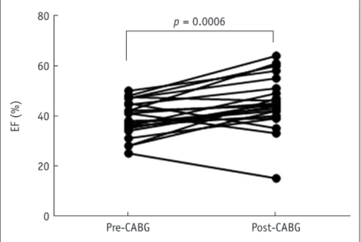
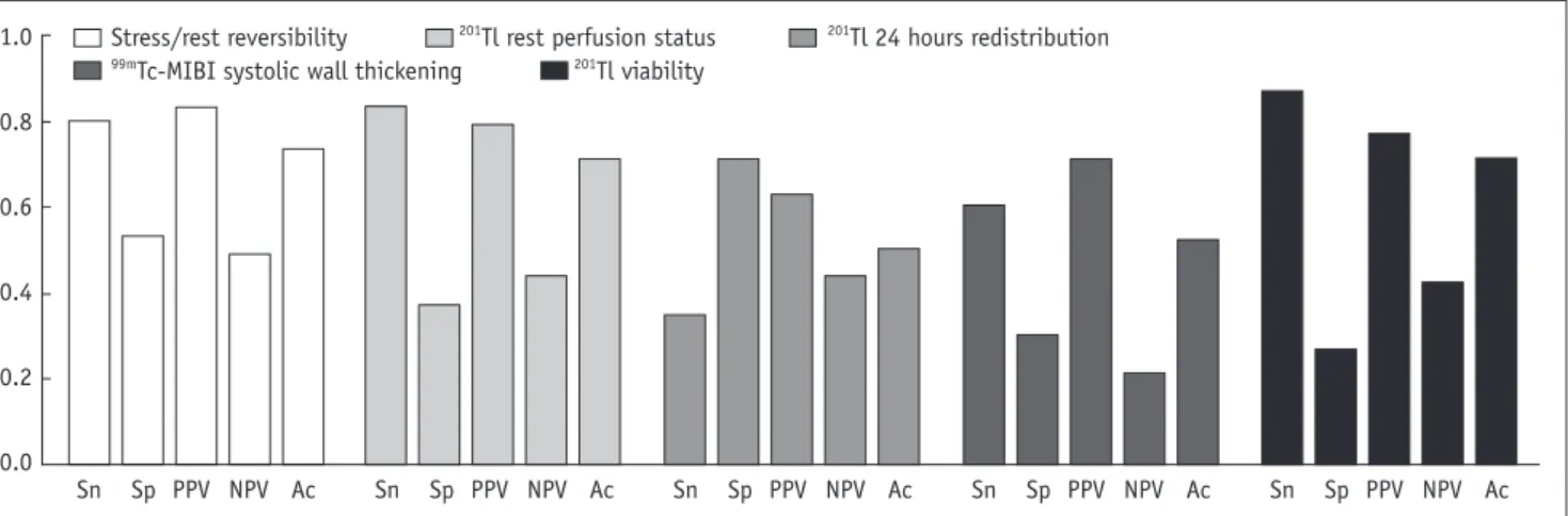
(5) Stress-Rest Reversibility as a Marker of Viable Myocardium. Tl Rest Perfusion Status Of the 115 dysfunctional segments, 90 showed preserved perfusion and 25 showed perfusion defects. The sensitivity, specificity, PPV, NPV, and accuracy were 83.5% (71 of 85), 36.7% (11 of 30), 78.9% (71 of 90), 44.0% (11 of 25), and 71.3% (82 of 115), respectively (Table 2, Fig. 4).. 201. myocardium. Viability Prediction of Markers Stress/Rest Reversibility Among the 115 dysfunctional myocardial segments, 85 turned out to be viable and 30 to be non-viable. The sensitivity, specificity, positive predictive value (PPV), negative predictive value (NPV), and accuracy were 80.0% (68 of 85), 53.3% (16 of 30), 82.9% (68 of 82), 48.5% (16 of 33), and 73.0% (84 of 115), respectively (Table 2, Fig. 4).. 80. p = 0.0006. 60. Tl 24 Hours Redistribution Of the 25 myocardial segments on which 201Tl rest perfusion status was defined as severe defect (grade 3), only 18 segments underwent 201Tl 24 hours redistribution study and of them were 8 viable and 10 non-viable; 6 redistribution-positive and 12 redistribution-negative. The sensitivity, specificity, PPV, NPV, and accuracy were 37.5% (3 of 8), 70.0% (7 of 10), 50.0% (3 of 6), 58.3% (7 of 12), and 55.6% (10 of 18), respectively (Table 2, Fig. 4). 201. Tc-MIBI Systolic Wall Thickening Of the dysfunctional 115 segments, 72 showed a good and 43 a poor myocardial wall thickening. The sensitivity, specificity, PPV, NPV, and accuracy were 60.0% (51 of 85), 30.0% (9 of 30), 70.8% (51 of 72), 20.9% (9 of 43), and 52.2% (60 of 115), respectively (Table 2, Fig. 4).. EF (%). 99m. 40. 20. 0. Pre-CABG. Post-CABG. Fig. 3. LVEF change after CABG. Gated LVEF increased significantly (p < 0.001) after CABG (45.5 ± 12.3%) in 22 patients with pre-CABG LVEFs lower than 50% (37.8 ± 9.0%). CABG = coronary artery bypass graft, LVEF = left ventricular ejection fraction. Viability Prediction as a Whole 201 Tl 24 hours redistribution was excepted because of the small number of its contributing segments. Therefore 115 myocardial segments could be evaluated using 3 viability. Table 2. Prediction of Viable Myocardium Using Individual Viability Markers Dysfunctional segments before CABG After exclusion of dysfunctional segments with normal perfusion at stress and rest (n = 27) Viability markers. 115. 115. 201. 115. 201. Tl 24 hours redistribution. 99m. Tc-MIBI systolic wall thickening. Non-Viable. 85. 30. 68 17 85 71 14 85 3 5 8 51 34 85. 14 16 30 19 11 30 3 7 10 21 9 30. 142. Stress/rest reversibility. Tl rest perfusion. Viable. 18*. 115. Positive Negative Total Positive Negative Total Positive Negative Total Positive Negative Total. Note.— *201Tl 24 hours redistribution was evaluated in only 18 myocardial segments that had rest perfusion defect. CABG = coronary artery bypass graft, 99mTc-MIBI = 99mTc-methoxyisobutylisonitrile. kjronline.org. Korean J Radiol 15(2), Mar/Apr 2014. 281.
(6) Lee et al.. 1.0. 201 Tl rest perfusion status Stress/rest reversibility 99m 201 Tc-MIBI systolic wall thickening Tl viability. 201. Tl 24 hours redistribution. 0.8 0.6 0.4 0.2 0.0. Sn. Sp PPV NPV Ac. Sn. Sp PPV NPV Ac. Sn. Sp PPV NPV Ac. Sn. Sp PPV NPV Ac. Sn. Sp PPV NPV Ac. Fig. 4. Diagnostic performances of individual viability markers for wall motion improvement. Ac = accuracy, NPV = negative predictive value, PPV = positive predictive value, Sn = sensitivity, Sp = specificity, 99mTc-MIBI = 99mTc-methoxyisobutylisonitrile. Table 3. Results of Logistic Regression Analysis Variable Stress/rest reversibility Tl rest perfusion 201 Tl 24 hours redistribution 99m Tc-MIBI systolic wall thickening 201 Tl viability 201. Univariate Logistic Regression Analysis Odds Ratio P 4.5714 < 0.001 2.9361 0.024 1.3158 0.659 0.6429 0.332 2.4463 0.088. Multiple Logistic Regression Analysis Adjusted Odds Ratio P 4.5714 < 0.001 Not included*. Not included*. Note.— *Those 2 variables were included in multiple regression analysis at first but excluded during stepwise modeling. 99mTc-MIBI = 99m Tc-methoxyisobutylisonitrile. markers (stress/rest reversibility, 201Tl rest perfusion status, and 99mTc-MIBI systolic wall thickening). When any 1 of 3 viability markers indicated myocardial viability, viability was considered positive and when all markers indicated nonviability, viability was considered negative. The sensitivity, specificity, PPV, NPV, and accuracy were 90.6% (77 of 85), 20.0% (6 of 30), 76.2% (77 of 101), 42.9% (6 of 14), and 72.2% (83 of 115), respectively. Tl Viability 201 Tl viability was defined with either normal-tomoderate degree rest perfusion decrease (grade 0–2) or severe perfusion defect (grade 3) accompanied by delayed redistribution. Of the 115 dysfunctional segments, 96 were positive and 19 negative for 201Tl viability. The sensitivity, specificity, PPV, NPV, and accuracy were 87.1% (74 of 85), 26.7% (8 of 30), 77.1% (74 of 96), 42.1% (8 of 19), and 71.3% (82 of 115), respectively. 201. Logistic Regression Analysis Using univariate analysis, stress/rest reversibility (p < 0.001) and 201Tl rest perfusion status (p = 0.024) showed significant predictability, whereas 201Tl 24 hours 282. redistribution (p = 0.738), 99mTc-MIBI systolic wall thickening (p = 0.332), and 201Tl viability (p = 0.088) were no significant predictors of wall motion improvement. However, in multiple logistic regression analysis, only stress/rest reversibility continued to show significant predictability for wall motion improvement (p < 0.001) (Table 3). Subgroup Analysis We evaluated the predictability of wall motion improvement in 2 subgroups. First, 39 myocardial segments with normal 201Tl rest perfusion were excluded because those segments may indicate stunned myocardium that would have improved wall motion even without revascularization. After exclusion of those 39 segments, the multiple logistic regression analysis in the remaining 76 segments revealed that only stress/rest reversibility was an important predictor of wall motion improvement (odds ratio = 4.0909, p = 0.022). Second, on the 22 patients with a pre-CABG LVEF lower than 50% (mean ± standard = 37.8 ± 9.0%), the stress/rest reversibility was again the most predictable marker of wall motion improvement (odds ratio = 3.8503, p = 0.005). Korean J Radiol 15(2), Mar/Apr 2014. kjronline.org.
(7) Stress-Rest Reversibility as a Marker of Viable Myocardium. DISCUSSION It is well known that reversible perfusion defects have ischemic viable myocardium in 201Tl scintigraphy (15). Myocardium with reversible perfusion defects has been considered viable. It has not been considered because there was stress-induced ischemia, but because there was some degree of reuptake or redistribution of 201Tl. With regard to the detection of viable myocardium, the reversibility from the stress defect was considered to be equivalent to the reversibility from the rest defect. Therefore stress image acquisition was not always required for a viability assessment; the 201Tl rest-redistribution study was considered sufficient for detection of viable myocardium (16, 17). It is noteworthy that the recent paradigm shift regarding the concept of hibernating myocardium puts tremendous emphasis on impaired CFR as an early manifestation of dysfunctional myocardium (18, 19). The chronic stunned state, preceding the hibernating state, presents features of abnormal wall motion and impaired CFR, while the rest perfusion is preserved (18). Myocardial hibernation occurs as a result of adaption to impaired CFR and repetitive stunning, rather than to a myocardial hypoperfusion (18). Moreover, the progression from chronic stunning to hibernation comes up with myocardial apoptosis (20), suggesting the dysfunctional myocardium in the chronic stunned state is more likely to improve wall motion after revascularization than that in a hibernating state under the influence of apoptosis. Therefore, if a certain viability marker is more inclined to detect chronic stunned myocardium, the marker may appear more competent than any other marker for viability detection. In the current study, stress/reversibility (Fig. 2A) was the single most important predictor of viable myocardium with the greatest accuracy, whereas 201Tl rest perfusion status (Fig. 2B), 201Tl 24 hours redistribution (Fig. 2C), and 99m Tc-MIBI systolic wall thickening (Fig. 2D) did not show significant predictability in the multiple logistic regression analyses. Results of the current study can be explained in several ways. First, stress perfusion has its own utility for the viability assessment by evaluating CFR and impaired CFR may indeed reflect the necessity of revascularization of ischemic viable myocardium. Not only in chronic stunned myocardium, but also in hibernating myocardium, the impaired CFR must be playing an important role in viability detection where the stress/rest reversibility (impaired CFR) proved to be the most robust detector of viable myocardium kjronline.org. Korean J Radiol 15(2), Mar/Apr 2014. in the subgroup analysis, excluding stunned myocardium. Of course, a rest study is essential in order to evaluate the CFR (here, relative CFR using myocardial SPECT rather than absolute CFR using myocardial PET), but it was not the preserved rest perfusion but the impaired CFR that required revascularization surgery for wall-motion improvement. Second, from a practical point of view to identify ischemic dysfunctional myocardium, detecting the deterioration of myocardial function may be a better method than detecting the improvement of myocardial function. For example, the residual viable myocytes in the infarct zone, hardly identified by other routine tests, have been found to trigger ventricular arrhythmia with exercise-induced ischemia. It means that functional deterioration (in this case aberrant electrical conductance) could indicate the presence of viable myocytes in the infarct zone (21). During dobutamine echocardiography, a continuous worsening of wall-motion response was a better predictor of viable myocardium than a sustained improvement response. It means that functional deterioration (wall motion abnormality) is more likely to indicate viable myocardium than wall motion improvement during dobutamine administration (22). Improvement of myocardial function during stress may take place in either normal myocardium or ischemic viable myocardium or in a mixture of both. But a deterioration of myocardial function on the usual diagnostic tests may only occur in the ischemic viable myocardium. Taken this findings together the authors believe that the deterioration of regional myocardial perfusion during stress may indicate the presence of ischemic viable myocardium. Third, individual viability markers may represent different phases during the progression of myocardial dysfunction and accordingly the time to full recovery after revascularization may differ. Dobutamine stress echocardiography has been reported to detect recovering dysfunctional myocardium just a few weeks after CABG (16). The dysfunctional myocardium with stress/rest reversibility mainly presented wall-motion improvement 3 months post-CABG (23). It took more than 6 months or up to 1 year post-revascularization for the 201 Tl uptake-positive dysfunctional myocardium to recover (16). In this regard, 201Tl uptake might have been a more significant marker of viable myocardium if we had evaluated post-CABG wall motion at periods later than 3 months postCABG. Further studies are needed to investigate the issue of a delayed functional restoration of the viable myocardium. As for 99mTc-MIBI systolic wall thickening, the low sensitivity (66%) and NPV (20.5%) were not inconsistent 283.
(8) Lee et al.. with a previous study (8). As a matter of fact, in contrast of large amounts of data regarding systolic wall thickening on echocardiography as a predictor of wall motion improvement after revascularization (24), 99mTc-MIBI systolic wall thickening has not been fully evaluated so far yet (8). Furthermore, the value of 201Tl systolic wall thickening as a marker of viable myocardium has not been thoroughly evaluated (25). We think further studies are required for gated myocardial parameters to be used as viability markers. The results of recent randomized clinical trials regarding the efficacy of revascularization over viable myocardium triggered a debate regarding the utility of viability tests (17, 26). A number of viability studies have been reported with variable degrees of success. However, the critical question regarding the most robust marker of viable myocardium was not an issue until the efficacy of revascularization was challenged by randomized clinical trials (19). In this regard, the results of the current study may lead not only nuclear medicine physicians to a glimpse of the viable myocardium, but also the results may lead back to the basic question: what is viable myocardium (2)? Limitation The most critical weak point of the current study is the use of post-stress wall motion for the evaluation of the wall motion change. Myocardial stunning may have happened in some myocardial segments during the poststress gate image acquisition. Those myocardial segments have potential of wall motion improvement even without revascularization. However, 201Tl rest SPECT seems not to be the better option than 99mTc-MIBI post-stress SPECT for the evaluation of myocardial wall motion. This issue needs further investigation. Another weak point of the present study is the fact that only wall motion improvement was assessed as a short-term outcome. Viability studies usually require a long-term outcome as final end-point (27). Furthermore, how much viable myocardium was required for long-term improvement was another critical question which could not be answered appropriately (28). Lastly, the effects of intense cardioprotective medication were not evaluated in the current study (17). Conclusion The stress/rest reversibility (in other words, impaired CFR) during dual-isotope gated myocardial perfusion SPECT was the most important marker for the prediction of wall motion improvement in 3 months post-CABG. 284. REFERENCES. 1. Rahimtoola SH. Coronary bypass surgery for chronic angina--1981. A perspective. Circulation 1982;65:225-241 2. Iskandrian AS. Myocardial viability: unresolved issues. J Nucl Med 1996;37:794-797 3. Ragosta M, Beller GA, Watson DD, Kaul S, Gimple LW. Quantitative planar rest-redistribution 201Tl imaging in detection of myocardial viability and prediction of improvement in left ventricular function after coronary bypass surgery in patients with severely depressed left ventricular function. Circulation 1993;87:1630-1641 4. Taki J, Nakajima K, Bunko H, Kawasuji M, Tonami N, Hisada K. Twenty-four-hour quantitative thallium imaging for predicting beneficial revascularization. Eur J Nucl Med 1994;21:12121217 5. Zimmermann R, Mall G, Rauch B, Zimmer G, Gabel M, Zehelein J, et al. Residual 201Tl activity in irreversible defects as a marker of myocardial viability. Clinicopathological study. Circulation 1995;91:1016-1021 6. Dilsizian V, Rocco TP, Freedman NM, Leon MB, Bonow RO. Enhanced detection of ischemic but viable myocardium by the reinjection of thallium after stress-redistribution imaging. N Engl J Med 1990;323:141-146 7. Ohtani H, Tamaki N, Yonekura Y, Mohiuddin IH, Hirata K, Ban T, et al. Value of thallium-201 reinjection after delayed SPECT imaging for predicting reversible ischemia after coronary artery bypass grafting. Am J Cardiol 1990;66:394-399 8. Snapper HJ, Shea NL, Konstam MA, Oates E, Udelson JE. Combined analysis of resting regional wall thickening and stress perfusion with electrocardiographic-gated technetium 99m-labeled sestamibi single-photon emission computed tomography: prediction of stress defect reversibility. J Nucl Cardiol 1997;4(1 Pt 1):3-10 9. Berman DS, Kiat H, Friedman JD, Wang FP, van Train K, Matzer L, et al. Separate acquisition rest thallium-201/stress technetium-99m sestamibi dual-isotope myocardial perfusion single-photon emission computed tomography: a clinical validation study. J Am Coll Cardiol 1993;22:1455-1464 10. Chua T, Kiat H, Germano G, Maurer G, van Train K, Friedman J, et al. Gated technetium-99m sestamibi for simultaneous assessment of stress myocardial perfusion, postexercise regional ventricular function and myocardial viability. Correlation with echocardiography and rest thallium-201 scintigraphy. J Am Coll Cardiol 1994;23:1107-1114 11. Cho SG, Kim JH, Cho JY, Kim HS, Bom HS. Myocardial blood flow and flow reserve in proximal and mid-to-distal lesions of left anterior descending artery measured by N-13 ammonia PET/CT. Nucl Med Mol Imaging 2013;47:158-165 12. Matzer L, Kiat H, Wang FP, Van Train K, Germano G, Friedman J, et al. Pharmacologic stress dual-isotope myocardial perfusion single-photon emission computed tomography. Am Heart J 1994;128(6 Pt 1):1067-1076 13. Mazzanti M, Germano G, Kiat H, Kavanagh PB, Alexanderson. Korean J Radiol 15(2), Mar/Apr 2014. kjronline.org.
(9) Stress-Rest Reversibility as a Marker of Viable Myocardium. E, Friedman JD, et al. Identification of severe and extensive coronary artery disease by automatic measurement of transient ischemic dilation of the left ventricle in dual-isotope myocardial perfusion SPECT. J Am Coll Cardiol 1996;27:16121620 14. Hachamovitch R, Kang X, Amanullah AM, Abidov A, Hayes SW, Friedman JD, et al. Prognostic implications of myocardial perfusion single-photon emission computed tomography in the elderly. Circulation 2009;120:2197-2206 15. Rozanski A, Berman DS, Gray R, Levy R, Raymond M, Maddahi J, et al. Use of thallium-201 redistribution scintigraphy in the preoperative differentiation of reversible and nonreversible myocardial asynergy. Circulation 1981;64:936-944 16. Alfieri O, La Canna G, Giubbini R, Pardini A, Zogno M, Fucci C. Recovery of myocardial function. The ultimate target of coronary revascularization. Eur J Cardiothorac Surg 1993;7:325-330; discussion 330 17. Bonow RO, Maurer G, Lee KL, Holly TA, Binkley PF, DesvigneNickens P, et al. Myocardial viability and survival in ischemic left ventricular dysfunction. N Engl J Med 2011;364:16171625 18. Fallavollita JA, Canty JM Jr. Differential 18F-2-deoxyglucose uptake in viable dysfunctional myocardium with normal resting perfusion: evidence for chronic stunning in pigs. Circulation 1999;99:2798-2805 19. Shah BN, Khattar RS, Senior R. The hibernating myocardium: current concepts, diagnostic dilemmas, and clinical challenges in the post-STICH era. Eur Heart J 2013;34:1323-1336 20. Lim H, Fallavollita JA, Hard R, Kerr CW, Canty JM Jr. Profound apoptosis-mediated regional myocyte loss and compensatory hypertrophy in pigs with hibernating myocardium. Circulation 1999;100:2380-2386. kjronline.org. Korean J Radiol 15(2), Mar/Apr 2014. 21. Margonato A, Mailhac A, Bonetti F, Vicedomini G, Fragasso G, Landoni C, et al. Exercise-induced ischemic arrhythmias in patients with previous myocardial infarction: role of perfusion and tissue viability. J Am Coll Cardiol 1996;27:593-598 22. Afridi I, Kleiman NS, Raizner AE, Zoghbi WA. Dobutamine echocardiography in myocardial hibernation. Optimal dose and accuracy in predicting recovery of ventricular function after coronary angioplasty. Circulation 1995;91:663-670 23. Paeng JC, Lee DS, Kang WJ, Lee BI, Kim KB, Chung JK, et al. Time course of functional recovery after coronary artery bypass grafting surgery according to the preoperative reversibility of perfusion impairment on myocardial SPECT. Eur J Nucl Med Mol Imaging 2005;32:70-74 24. Smart SC. The clinical utility of echocardiography in the assessment of myocardial viability. J Nucl Med 1994;35(4 Suppl):49S-58S 25. Lee WW, Park EK, Eo JS, Lee SW, Kim CH, So Y, et al. Evaluation of immediate post-stress wall motion on adenosine stress/rest thallium-201 gated myocardial SPECT. Int J Cardiovasc Imaging 2006;22:213-222 26. Beanlands RS, Nichol G, Huszti E, Humen D, Racine N, Freeman M, et al. F-18-fluorodeoxyglucose positron emission tomography imaging-assisted management of patients with severe left ventricular dysfunction and suspected coronary disease: a randomized, controlled trial (PARR-2). J Am Coll Cardiol 2007;50:2002-2012 27. Velazquez EJ, Lee KL, Deja MA, Jain A, Sopko G, Marchenko A, et al. Coronary-artery bypass surgery in patients with left ventricular dysfunction. N Engl J Med 2011;364:1607-1616 28. Schinkel AF, Poldermans D, Elhendy A, Bax JJ. Assessment of myocardial viability in patients with heart failure. J Nucl Med 2007;48:1135-1146. 285.
(10)
수치
관련 문서