INTRODUCTION
Esophageal cancer (EC) is one of the most frequent malignant Received: April 6, 2017 Revised: July 25, 2017
Accepted: July 27, 2017
Corresponding author: Dr. Zeqing Song, Department of Internal Medicine, Guang- dong Medical University Affiliated Longhua Central Hospital, No.187 Guan lan Avenue, Shenzhen, Guangdong 518110, China.
Tel: 86-0755-28019120, Fax: 86-0755-28015466, E-mail: [email protected]
*Xiaotian Gao and Zhanqiang Xie contributed equally to this work.
•The authors have no financial conflicts of interest.
© Copyright: Yonsei University College of Medicine 2017
This is an Open Access article distributed under the terms of the Creative Com- mons Attribution Non-Commercial License (http://creativecommons.org/licenses/
by-nc/4.0) which permits unrestricted non-commercial use, distribution, and repro- duction in any medium, provided the original work is properly cited.
Overexpression of miR-191 Predicts Poor Prognosis and Promotes Proliferation and Invasion
in Esophageal Squamous Cell Carcinoma
Xiaotian Gao1*, Zhanqiang Xie2*, Zhigang Wang2, Keluo Cheng2, Ke Liang2, and Zeqing Song3
1Department of Cardiac Surgery, The First Affiliated Hospital of Anhui Medical University, Hefei;
2Department of Cardiothoracic Surgery, Affiliated Hospital of Guangdong Medical University, Zhanjiang;
3Department of Internal Medicine, Guangdong Medical University Affiliated Longhua Central Hospital, Shenzhen, China.
Purpose: Accumulating evidence has shown that dysregulation of microRNA-191 (miR-191) is closely associated with tumorigene- sis and progression in a wide range of cancers. This study aimed to explore the potential role of miR-191 in esophageal squamous cell carcinoma (ESCC).
Materials and Methods: miR-191 expression was assessed in 93 ESCC tissue specimens by real-time polymerase chain reaction, and survival analysis was performed via Kaplan-Meier and Cox regression analyses. 3-(4,5-dimethyl-2-thiazolyl)-2,5-diphenyl- 2-H-tetrazolium bromide, plate colony-forming, BrdU, and Transwell assays were conducted to observe the effect of miR-191 on ESCC proliferation and invasion. Luciferase reporter and western blot assays were taken to identify target genes of miR-191.
Results: miR-191 was overexpressed in 93 cases of ESCC, compared with adjacent normal tissues, and miR-191 expression was significantly related to differentiation, depth of invasion, TNM stage, lymph node metastasis, and distant metastasis of tumor. Ka- plan-Meier and Cox regression analyses demonstrated that overexpression of miR-191 was an independent and significant pre- dictor of ESCC prognosis. Both gain-of-function and loss-of-function experiments showed that miR-191 promoted ESCC cell proliferation and invasion activities in vitro. Early growth response 1 (EGR1), a tumor suppressor, was predicted as a direct target of miR-191. Luciferase reporter and western blot assays proved that miR-191 reduced EGR1 expression by directly binding its 3' untranslated region. Moreover, EGR1 knockdown by siRNA enhanced ESCC cell growth and invasion.
Conclusion: Our findings provide specific biological roles of miR-191 in ESCC survival and progression. Targeting the novel miR- 191/EGR1 axis represents a potential new therapeutic way to block ESCC development.
Key Words: miR-191, esophageal squamous cell carcinoma, early growth response 1, prognosis, proliferation, invasion
pISSN: 0513-5796 · eISSN: 1976-2437 Yonsei Med J 2017 Nov;58(6):1101-1110
https://doi.org/10.3349/ymj.2017.58.6.1101
neoplasms worldwide and ranks as the sixth-leading cause of cancer-related death.1 EC consists of two main subtypes, name- ly esophageal adenocarcinoma, which is frequently occurring in developed countries, such as North America and Europe, and esophageal squamous cell carcinoma (ESCC), which is more common in developing countries, such as East Asian countries.2,3 ESCC remains the main histological subtype, ac- counting for more than 90% of all ECs, and the 5-year survival rate of patients with ESCC is less than 10%.2,3 Therefore, it is observably necessary to search and explore novel molecular targets for ESCC therapy.
Recently, microRNAs (miRNAs) have attracted more and more attention in various biological processes, including cell proliferation, migration, differentiation, and apoptosis.4,5 It is not surprising that miRNA dysregulation correlates with tu-
morigenesis in many cancers including ESCC.6 Many studies reported that microRNA-191 (miR-191) can act as a multifunc- tional miRNA, with important roles in diabetes-type 2, Crohn’s, pulmonary hypertension, Alzheimer’s, and neoplastic diseas- es.7-11 It has been reported that miR-191 expression is dysregu- lated in several cancers and correlated with malignant clinical features and tumor prognosis.12-17 On the one hand, miR-191 is overexpressed in several cancers, including breast cancer, co- lon cancer, and lung cancer, and plays an oncogenic role.12-14,18 On the other hand, miR-191 has been found to be down-regu- lated in several other cancer types, including retinoblastoma, thyroid follicular tumor, and melanoma, suggesting its tumor suppressive role.15-17 However, its clinical significance in human ESCC remains unclear.
The aims of the present study were to evaluate the relation- ships between miR-191 expression level and the clinicopatho- logical characteristics and outcomes of ESCC patients and to explore the functional role of miR-191 in ESCC cell lines. We found that miR-191 overexpression independently affects prog- nosis in ESCC. miR-191 promotes ESCC cell proliferation and invasion in vitro by directly targeting early growth response 1 (EGR1).
MATERIALS AND METHODS
Patients and specimens
Specimens of 93 ESCCs and 58 adjacent normal tissues were obtained from 93 patients who underwent surgical treatment of ESCC at Affiliated Hospital of Guangdong Medical Univer- sity between January 2010 and December 2013. All the patients were diagnosed by pathological examination, and none of the patients had received preoperative chemotherapy or radiother- apy. Patient data, including kinds of clinicopathological fea- tures, was collected, and follow-up information after surgery was acquired by telephone interview. The study protocol was approved by the Ethics Committee of Guangdong Medical Uni- versity, and all the patients provided written informed consent (Approval no. GMU2009035).
RNA isolation and quantitative real-time polymerase chain reaction (RT-PCR)
Total RNA from all tissues and cells were isolated using Trizol reagent (Invitrogen, Carlsbad, NM, USA). RT-PCR was carried out applying SYBR Premix Ex Taq II (TaKaRa, Dalian, China) and detected in a LightCycler 480 system (Roche, Basel, Swit- zerland) according the manufacturers’ instructions. All miR- NA primers were obtained from RiBoBio Co. (Guangzhou, Chi- na). EGR1 primer (TaKaRa) is shown in Supplementary Table 1 (only online). U6 or glyceraldehyde-3-phosphate dehydroge- nase was used as an internal control, and the fold change was calculated by 2-ΔΔCt.
Cell lines and transfection
ESCC cell lines EC9706 and TE-1 were purchased from the Chi- nese Academy of Medical Science (Beijing, China) and main- tained in RPMI-1640 medium supplemented with 10% fetal bovine serum (FBS), 100 U/mL penicillin, and 100 μg/mL streptomycin at 37°C in a humidified incubator with 5% CO2. miR-191 mimic, mimic control, miR-191 inhibitor, and inhibi- tor control were purchased from RiBoBio. EGR1 siRNA was obtained from Santa Cruz Biotechnology Inc. (sc-29303; Santa Cruz, Dallas, TX, USA). Cell transfection was performed ap- plying Lipofectamine 2000 (Invitrogen).
MTT cell proliferation assay
Cell proliferation was measured by the 3-(4,5-dimethyl- 2-thiazolyl)-2,5-diphenyl-2-H-tetrazolium bromide (MTT) as- say. Briefly, the cells (1×10³ cells/well) were seeded into each well of seven 96 well plates. At a different time point after trans- fection, 20 μL of MTT (5 mg/mL, Sigma, MO, USA) was added.
Then, the reaction density was read at 490 nm on a Varioskan Flash Multimode Reader (Thermo Fisher, Waltham, MA, USA).
Plate colony-forming assay
Log phase cells were trypsinized into single cell suspensions and plated in 10-cm plates at indicated cell numbers. The colo- nies were stained with Giemsa, and the total number of colo- nies was counted.
BrdU assay
The BrdU assay was performed using a BrdU Cell Proliferation Chemiluminescent Assay Kit (Cell Signaling Technology Inc., Denver, CO, USA) according to the manufacturer’s instruc- tions.
Transwell assay
Migration and invasion was detected by Transwell assay. Brief- ly, 1×10⁵ transfected cells were seeded in FBS-free media into the upper chamber of each transwell, which was pre-coated with Matrigel. Medium with 20% FBS was placed in the lower chamber. 24 hours later, non-invasive cells in the upper cham- ber were removed with a cotton swab, while the cells on the lower surface were fixed and stained with 0.1% crystal violet and photographed. Pictures of three random fields from trip- licate wells were recorded, and the number of cells was counted.
For the migration assays, all procedures were the same as in in- vasion assays, except each chamber without Matrigel coating.
Luciferase reporter assays
Wild-type 3’UTR of EGR1 and mutant constructs, which were produced via mutations in the binding site of miR-191 in EGR1’s 3’UTR were cloned downstream of the Luciferase gene in the psiCHECK-2 Luciferase vector. Luciferase reporter vectors and miR-191 mimic were transfected into HEK293T and EC9706 cells with Lipofectamine 2000, and Luciferase activity
6 5 4 3 2 1
0 Adjacent non-tumor
***
miR-191
ESCC Relative microRNA expression (normalized as to U6)
5
4
3
2
1
0 Stage I–II ESCC
***
miR-191
Stage III–IV ESCC Relative microRNA expression (normalized as to U6)
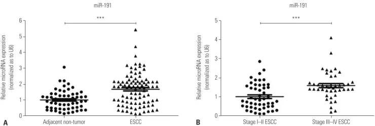
Fig. 1. miR-191 is overexpressed in ESCC tissues. (A) The relative expression levels of miR-191 in adjacent non-cancerous esophageal and ESCC samples. (B) The relative expression levels of miR-191 in Stage I–II and III–IV ESCC tissues. ***p<0.001. miRNA, microRNA; miR-191, microRNA-191;
ESCC, esophageal squamous cell carcinoma.
A B
Table 1. Associations of miR-191 Expression in ESCC Tissues with De- mographic and Clinicopathologic Characteristics
Category n=93
miR-191 expression
p value*
Low (n=41)
High (n=52)
Gender 0.976
Male 77 34 43
Female 16 7 9
Age 0.144
≤65 51 19 32
>65 42 22 20
Tumor size, cm 0.697
<5 52 22 30
≥5 41 19 22
Differentiation 0.037
Well and moderate 57 30 27
Poorly and not 36 11 25
Depth of invasion 0.010
T1–T2 24 16 8
T3–T4 69 25 44
Stages <0.001
I–II 50 32 18
III–IV 43 9 34
Lymph node metastases <0.001
0 45 28 17
≥1 48 13 35
Metastases to other organs 0.025
Present 6 0 6
Absent 87 41 46
miR-191, microRNA-191; ESCC, esophageal squamous cell carcinoma.
*p values were calculated through the χ2 test to analyze the relationship be- tween miR-191 expression and clinicopathologic characteristics.
was measured by the Dual-Luciferase Reporter Assay System (Promega, Madison, WI, USA).
Western blot
Whole-cell lysates were obtained using RIPA buffer (NobleRy- der, Beijing, China) according to the manufacturer’s instruc- tions. Western blot analysis was conducted as shown previous- ly.19 The primary antibodies used were anti-EGR1 (Abcam, Cam- bridge, UK) and β-actin (Sigma).
Statistical analysis
All statistical analyses were conducted using SPSS software (version 21.0, IBM Corp., Armonk, NY, USA). χ2 test was used to analyze relationships between miR-191 expression levels and clinicopathological factors. Overall survival curves were analyzed with the Kaplan-Meier method and compared th- rough the log-rank test. On the basis of Cox proportional haz- ards model, univariate and multivariable survival analyses were conducted. p<0.05 was regarded as statistically significant.
RESULTS
miR-191 is markedly up-regulated in ESCC tissues and correlated with malignant clinicopathological features
To classify the clinical significance of miR-191 in ESCC, we in- vestigated its expression in 93 cases of ESCC tissues with RT- PCR. The expression level of miR-191 was much higher in ESCC tissues, compared with adjacent normal samples (Fig.
1A). In addition, miR-191 expression in Stage I–II were lower than that in Stage III–IV (Fig. 1B). Next, we investigated the re- lationships between miR-191 expression and clinicopatho- logical factors of ESCC patients. According to the mean level of miR-191 expression, ESCC patients were divided into two groups, with 41 in the low-expression group and 52 in the high-
expression group. As shown in Table 1, miR-191 expression was significantly correlated with ESCC differentiation, depth of invasion, TNM stage, lymph node metastasis, and distant
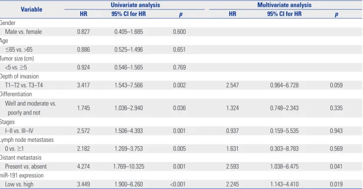
Table 2. Univariate Analysis and Multivariate Analysis of Correlations among Clinicopathological Parameters and Survival Time of Patients with Gas- tric Cancer
Variable Univariate analysis Multivariate analysis
HR 95% CI for HR p HR 95% CI for HR p
Gender
Male vs. female 0.827 0.405–1.685 0.600
Age
≤65 vs. >65 0.886 0.525–1.496 0.651
Tumor size (cm)
<5 vs. ≥5 0.924 0.546–1.565 0.769
Depth of invasion
T1–T2 vs. T3–T4 3.417 1.543–7.566 0.002 2.547 0.964–6.728 0.059
Differentiation
Well and moderate vs.
poorly and not 1.745 1.036–2.940 0.036 1.324 0.748–2.343 0.335
Stages
I–II vs. III–IV 2.572 1.506–4.393 0.001 0.937 0.159–5.535 0.943
Lymph node metastases
0 vs. ≥1 2.182 1.269–3.753 0.005 1.631 0.303–8.783 0.569
Distant metastasis
Present vs. absent 4.274 1.769–10.325 0.001 2.593 1.038–6.475 0.041
miR-191 expression
Low vs. high 3.449 1.900–6.260 <0.001 2.245 1.143–4.410 0.019
miR-191, microRNA-191; CI, confidence interval; HR, hazard ratio.
Fig. 2. Kaplan-Meier survival curves of ESCC patients with different level of miR-191 expression stratified by the TNM stage. (A) Association of miR-191 expression with overall survival (cumulative survival) in all stages. (B) Association of miR-191 expression with overall survival in Stage I–II. (C) Association of miR-191 expression with overall survival in Stage III–IV. ESCC, esophageal squamous cell carcinoma; miR-191, microRNA-191.
100 80 60 40 20 0
Survival time after operation (months) miR-191 low expression
miR-191 high expression Log-rank test: p<0.001
Stage I–IV
Cumulative survival
0 20 40 60 80
A
100 80 60 40 20 0
Survival time after operation (months) miR-191 low expression
miR-191 high expression
Log-rank test: p<0.05 Stage I–II
Cumulative survival
0 20 40 60 80
B
100 80 60 40 20 0
Survival time after operation (months) miR-191 low expression
miR-191 high expression Log-rank test: p<0.05
Stage III–IV
Cumulative survival
0 20 40 60
C metastasis, but not sex, age, and tumor size.
miR-191 is an independent predictor of ESCC prognosis In order to explore the prognostic value of miR-191 in ESCC, Kaplan-Meier analysis and log-rank test were performed to observe relationships with overall survival. Kaplan-Meier sur- vival curves indicated that higher miR-191 expression brought a shorter survival (p<0.001) (Fig. 2A). Higher expression of miR- 191 led to a poorer prognosis, compared with low miR-191, with the unadjusted hazard ratio (HR) being 3.449 [95% confi- dence interval (CI): 1.900–6.260, p<0.001] (Table 2). Further- more, the difference of overall survival between the two groups seemed to be more distinct in Stage III–IV than Stage I–II (Fig.
2B and C). The factors found to be statistically important in univariate analysis, including depth of invasion, TNM stage, differentiation, lymph node metastasis, distant metastasis, and
miR-191 expression, were selected to perform multivariate analysis. Therein, distant metastasis and miR-191 expression were independent prognostic factors for ESCC (Table 2). Taken together, these findings suggested that miR-191 expression is an independent and significant predictor of ESCC prognosis.
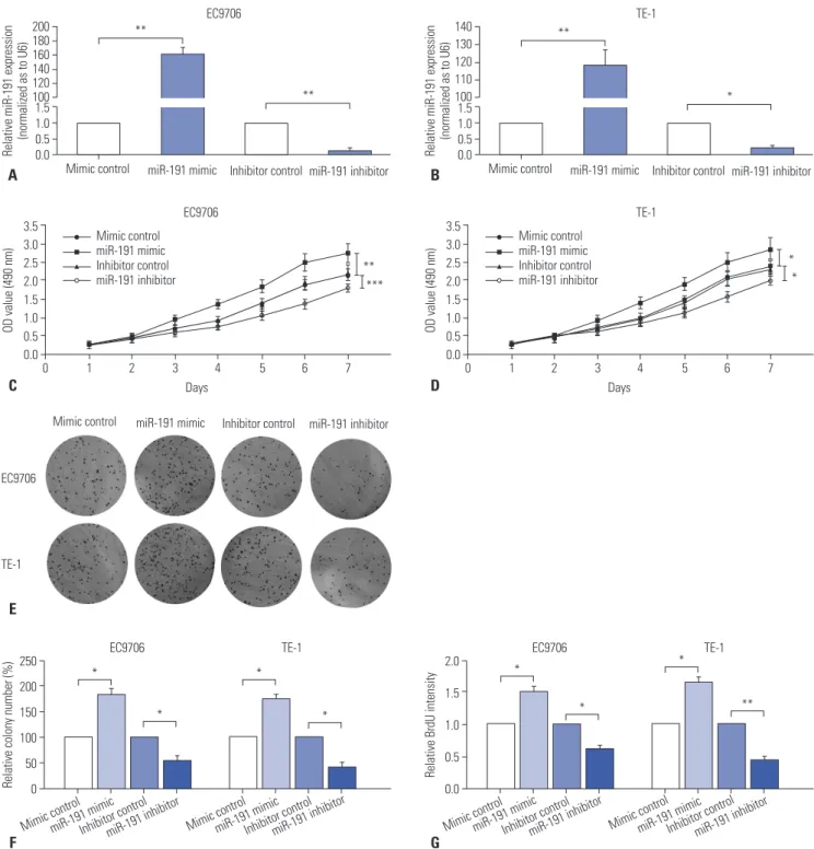
miR-191 promotes ESCC cell proliferation and invasion in vitro
To further characterize the role of miR-191 in ESCC tumori- genesis, miR-191 mimic and inhibitor were transfected into EC9706 or TE-1 cells, respectively, to conduct gain-of-function and loss-of-function assays. RT-PCR results displayed that miR-191 mimic dramatically up-regulated miR-191 expression in EC9706 and TE-1 cells, compared with mimic control, while miR-191 inhibitor significantly reduced miR-191 expression (Fig. 3A and B). MTT assays showed that up-regulation of miR-
191 significantly increased EC9706 and TE-1 cell proliferation, while down-regulation of miR-191 decreased cell growth (Fig.
3C and D). A significantly reduced number of cell colonies were observed in plates where EC9706 or TE-1 cells with down-
regulation of miR-191 were seeded, and ESCC cells with miR- 191 mimic transfection exhibited stronger capacity to form colonies (Fig. 3E and F). As DNA replication is a key step dur- ing cell mitosis, we also used BrdU to evaluate cell proliferation.
A 1.5 1.0 0.5 0.0 200180 160140 120100
EC9706
Relative miR-191 expression (normalized as to U6)
Mimic control miR-191 mimic Inhibitor control miR-191 inhibitor
**
**
F 250 200 150 100 50 0
EC9706 TE-1
Relative colony number (%)
Mimic controlmiR-191 mimicInhibitor controlmiR-191 inhibitor Mimic controlmiR-191 mimicInhibitor controlmiR-191 inhibitor
*
*
*
*
G 2.0 1.5 1.0 0.5 0.0
EC9706 TE-1
Relative BrdU intensity
Mimic controlmiR-191 mimicInhibitor controlmiR-191 inhibitor Mimic controlmiR-191 mimicInhibitor controlmiR-191 inhibitor
*
*
*
**
B 1.5 1.0 0.5 0.0 140 130 120 110 100
TE-1
Relative miR-191 expression (normalized as to U6)
Mimic control miR-191 mimic Inhibitor control miR-191 inhibitor
**
*
3.5 3.0 2.5 2.0 1.5 1.0 0.5 0.0
Days EC9706
OD value (490 nm)
0 1 2 3 4 5 6 7
C
Mimic control miR-191 mimic Inhibitor control miR-191 inhibitor
Mimic control miR-191 mimic Inhibitor control miR-191 inhibitor
**
***
3.5 3.0 2.5 2.0 1.5 1.0 0.5 0.0
Days
OD value (490 nm)
0 1 2 3 4 5 6 7
D
*
*
Mimic control miR-191 mimic Inhibitor control miR-191 inhibitor
EC9706
TE-1
E
Fig. 3. miR-191 promotes ESCC cell proliferation and invasion in vitro. (A) RT-PCR analysis of miR-191 expression in EC9706 cells transfected with miR- 191 mimic or inhibitor and corresponding control. (B) RT-PCR analysis of miR-191 expression in TE-1 cells transfected with miR-191 mimic or inhibitor and corresponding control. (C) MTT assay of EC9706 cells transfected with miR-191 mimic or inhibitor and corresponding control. (D) MTT assay of TE-1 cells transfected with miR-191 mimic or inhibitor and corresponding control. (E) The colony formation assays in EC9706 and TE-1 cells transfect- ed with miR-191 mimic or inhibitor and corresponding control. Colonies were evaluated and values were reported as the ratio. (G) The BrdU assays from EC9706 and TE-1 cells transfected with miR-191 mimic or inhibitor and corresponding control. *p<0.05, **p<0.01, ***p<0.001 miR-191, microR- NA-191; ESCC, esophageal squamous cell carcinoma; RT-PCR, real-time polymerase chain reaction; MTT, 3-(4,5-dimethyl-2-thiazolyl)-2,5-diphenyl- 2-H-tetrazolium bromide; OD, optical density.
TE-1
By doing so, we noted that ectopic miR-191 increased BrdU intensity, while miR-191 suppression decreased BrdU intensi- ty in ESCC cells (Fig. 3G). Next, the function of miR-191 in ESCC cell migration and invasion was observed. We found that up- regulation of miR-191 in EC9706 and TE-1 cells significantly promoted the migration of cells in Transwell assays without Matrigel and increased the invasion in Transwell assays with Matrigel (Fig. 3H). In contrast, down-regulation of miR-191 reduced ESCC cell migration and invasion (Fig. 3I). Collec- tively, these observations suggested that miR-191 acts as an oncogene in ESCC progression.
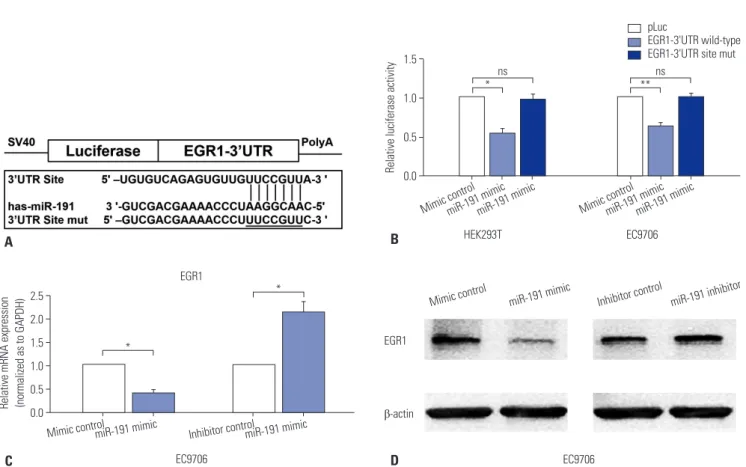
miR-191 regulates EGR1 expression by directly targeting its 3’UTR
We next investigated the specific mechanism of miR-191 in ESCC proliferation and metastasis, and bioinformatic methods were performed to identify the downstream genes of miR-191.
According to the results from three different bioinformatic programs (miRanda, Memorial Sloan-Kettering Cancer Center, New York, NY, USA; Targetscan, Whitehead Institute for Bio- medical Research, Cambridge, MA, USA; and Pictar, Rajewsky Lab, New York, NY, USA), EGR1, a tumor suppressor, was pre- dicted as a potential target gene of miR-191 (Fig. 4A). In order to validate whether miR-191 directly bound 3’UTR of EGR1 to suppress its expression, reporter vectors containing wildtype
or mutant EGR1 3’UTR binding sites were transfected into HEK293T and EC9706 cells, respectively. Luciferase reporter assays indicated that miR-191 decreased the Luciferase activi- ty in the vector containing wild-type EGR1 3’UTR, but not the one containing mutant site, suggesting a direct interaction of miR-191 and EGR1 3’UTR (Fig. 4B). In addition, both RT-PCR and western blot assays showed that overexpression of miR- 191 reduced EGR1 expression in EC9706 cells, while down- regulation of miR-191 increased EGR1 expression (Fig. 4C and D). Taken together, these results indicated that miR-191 might suppress EGRI expression by directly targeting its 3’UTR to enhance ESCC progression.
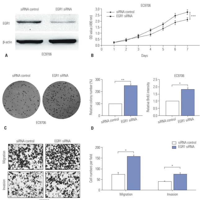
Down-regulation of EGR1 expression promotes ESCC cell growth and invasion
In order to confirm whether miR-191 promotes ESCC progres- sion via EGR1, we next examined the exact function of EGR1 during ESCC cell proliferation and invasion. EGR1 siRNA was transfected into EC9706 cells, and western blot assay con- firmed that it could significantly reduce EGR1 expression (Fig.
5A). All of the MTT, plate clone formation, and BrdU assays indicated that down-regulation of EGR1 accelerated EC9706 cell proliferation and growth (Fig. 5B, C, and D). Moreover, Transwell assay showed that EGR1 knockdown increased cell migration and invasion in vitro (Fig. 5E). Collectively, these
150
100
50
0
Cell numbers per field
Migration Invasion Migration Invasion EC9706
**
** **
**
Mimic control miR-191 mimic Inhibitor control miR-191 inhibitor
Mimic control miR-191 mimic Inhibitor control miR-191 inhibitor
MigrationInvasion
EC9706 EC9706
Mimic control miR-191 mimic Inhibitor control miR-191 inhibitor
H
150
100
50
0
Cell numbers per field
Migration Invasion Migration Invasion TE-1
**
* *
*
MigrationInvasion
TE-1 TE-1
Mimic control miR-191 mimic Inhibitor control miR-191 inhibitor
I
Fig. 3. miR-191 promotes ESCC cell proliferation and invasion in vitro. (H) Transwell assays of EC9706 cells transfected with miR-191 mimic or inhibitor and corresponding control. Left panel: representative images. Right panel: quantification of 10 randomly selected fields. (I) Transwell assays of TE-1 cells transfected with miR-191 mimic or inhibitor and corresponding control. *p<0.05, **p<0.01. miR-191, microRNA-191; ESCC, esophageal squamous cell carcinoma.
B 1.5
1.0
0.5
0.0
HEK293T EC9706
Relative luciferase activity
Mimic controlmiR-191 mimicmiR-191 mimic Mimic controlmiR-191 mimicmiR-191 mimic
ns ns
* **
pLucEGR1-3'UTR wild-type EGR1-3'UTR site mut
C 2.5 2.0 1.5 1.0 0.5 0.0
EC9706 EGR1
Relative mRNA expression (normalized as to GAPDH)
Mimic controlmiR-191 mimic Inhibitor controlmiR-191 mimic
*
* Mimic control miR-191 mimic Inhibitor control miR-191 inhibitor
EC9706 β-actin
EGR1
D
Fig. 4. miR-191 down-regulates EGR1 expression by directly targeting its 3’UTR. (A) Diagram of EGR1 3’UTR-containing reporter construct. Mutations were generated at the predicted miR-191 binding site located in the EGR1 3’UTR. (B) Relative Luciferase activity after the wild type or mutant reporter plasmids were co-transfected with miR-191 mimic or mimic control in HEK293T and EC9706 cells. (C) RT-PCR analysis of EGR1 mRNA expression in EC9706 cells transfected with miR-191 mimic or inhibitor and corresponding control. (D) Western blot analysis of EGR1 protein expression in EC9706 cells transfected with miR-191 mimic or inhibitor and corresponding control. *p<0.05, **p<0.01. ns, no significance; EGR1, early growth response 1;
miR-191, microRNA-191; mRNA, messenger RNA.
A
observations indicated that down-regulation of EGR1 promotes ESCC proliferation and invasion.
DISCUSSION
Accumulating evidence has shown that miRNAs can act either as oncogenes or as tumor suppressors in ESCC and that mea- surement of miRNA expression in malignancies may have di- agnostic and prognostic implications.20 Their very small size, in principle, makes them less prone to degradation processes, unlike messenger RNAs, which were previously proposed as molecular markers. The dysregulated expression of several miRNAs, including miR-21, miR-126, miR-138, miR-183, miR- 200b, miR-375, and miR-508, has been associated with ESCC in recent literature.20-27 However, there is still a dearth of molec- ular biomarkers suitable for clinical application in this disease.
MiR-191, together with miR-425, belongs to the miR-191/425 cluster, which is highly conserved in several metazoan species (miR-425 in 26 species, miR-191 in 30 species), suggesting it to be an important player in higher eukaryotes.7 The aberrant expression of miR-191 has been observed in more than 20 dif- ferent cancers. However, it remains controversial as to whether
miR-191 acts as an oncogene or tumor suppressor in carcino- genesis. MiR-191 functions as an oncomiRNA in hepatocellu- lar carcinoma, and its up-regulation enhanced cell growth and reduced apoptosis.28 It was also reported that miR-191 acts as an estrogen inducible oncogene and promotes breast cancer cell proliferation and invasion via targeting special AT-rich se- quence-binding protein 1 (SATB1).12 Zhang, et al.13 showed that miR-191 promotes colorectal cancer carcinogenesis via target- ing CCAAT/enhancer-binding protein β (C/EBPβ) and adjust- ing related signaling pathways. On the contrary, Di Leva, et al.29 showed that the overexpression of miR-191 suppresses cell growth and blocks carcinogenesis in aggressive breast cancer cells. Furthermore, miR-191 decreases cell proliferation and invasion by binding 3’UTR of CDK6 in thyroid cancer.16 How- ever, to our knowledge, the clinical significance of miR-191 in human ESCC remains unclear.
Herein, we revealed that miR-191 was frequently overex- pressed in ESCC tissues and significantly related to advanced clinical stage, metastasis, and poor survival rate of ESCC. More- over, multivariate Cox regression analysis indicated that up- regulation of miR-191 was an independent prognostic factor for poor survival of patients with ESCC. Previous studies have demonstrated that measurement of miR-191 expression level
may represent as a novel approach of diagnosis and prognosis in cancers.20 It was reported that miR-191 expression is a novel independent prognostic factor of patients with colorectal can- cer.30 Peng, et al.18 revealed the oncogenic role of miR-191 in gastric carcinogenesis and indicated the potential use of se- rum miR-191 as a novel and stable biomarker for diagnosis. The present study further confirmed that miR-191 could be a novel potential prognosis biomarker in ESCC.
In the present study, we also showed that miR-191 could in- crease cell proliferation and invasion, implying that miR-191 is an oncomiRNA in ESCC. Further mechanism investigation
demonstrated that miR-191 might reduce EGR1 expression by binding its 3’UTR, to be involved in ESCC progression. As a transcription factor, EGR1 is involved in tumorigenesis and development of many cancers, and plays a tumor suppressive role.31-35 It was demonstrated that decreased EGR1 expression is an important predictor of poor over survival in non-small cell lung cancer.31 Clinically, loss of EGR1 results in enhancive tumor transformation, followed by patient morbidity and mor- tality.31-33 Moreover, EGR1 is down-regulated in ESCC tissues and plays a significant role in ESCC prognosis.34 Dong, et al.35 reported that EGR1 regulates cisplatin-induced apoptosis in 3.0
2.5 2.0 1.5 1.0 0.5 0.0
Days EC9706
OD value (490 nm)
0 1 2 3 4 5 6 7
B
siRNA control
EGR1 siRNA ***
EC9706 β-actin
EGR1
A
siRNA control EGR1 siRNA
EC9706 C
siRNA control EGR1 siRNA
D 300
200
100
Relative colony number (%) 0
siRNA control EGR1 siRNA
** 2.5
2.0 1.5 1.0 0.5 0.0
EC9706
Relative BrdU intensity
siRNA control EGR1 siRNA
*
EC9706
MigrationInvasion
siRNA control EGR1 siRNA
E
200 150
100 50
0
Cell numbers per field
Migration Invasion
*
*
siRNA control EGR1 siRNA
Fig. 5. Down-regulation of EGR1 promotes ESCC cell proliferation and invasion. (A) Western blot analysis of EGR1 expression in EC9706 cells trans- fected with EGR1 siRNA or corresponding control. β-actin was used as an internal control. (B) MTT assay of EC9706 cells transfected with EGR1 siR- NA or control. (C) The colony formation assays in EC9706 transfected with EGR1 siRNA or control. Colonies were evaluated and values were reported as the ratio. (D) The BrdU assays from EC9706 transfected with EGR1 siRNA or control. (E) Transwell assays of EC9706 cells transfected with EGR1 siRNA or control. Left panel: representative images. Right panel: quantification of 10 randomly selected fields. *p<0.05, **p<0.01, ***p<0.001. EGR1, early growth response 1; ESCC, esophageal squamous cell carcinoma; MTT, 3-(4,5-dimethyl-2-thiazolyl)-2,5-diphenyl-2-H-tetrazolium bromide; OD, optical density.
ESCC and activates apoptosis related pathways. On the basis of these findings, this study further confirmed that EGR1 down- regulation promotes ESCC cell growth and invasion, provid- ing new insight of EGR1 function in ESCC development and progression.
Several limitations need to be addressed in our study. Firstly, the clinical sample size for evaluating prognostic value of miR- 191 was slightly inadequate. Secondly, due to the limited in- formation about disease-free survival, we were unable to clas- sify the role of miR-191 in disease-free survival prediction. Th- irdly, all available function studies of miR-191 and EGR1 were conducted in vitro, and further in vivo research remains to be studied.
In summary, overexpression of miR-191 predicted poor prog- nosis in patients with ESCC and miR-191 promoted ESCC cell proliferation and invasion by targeting EGR1. The results from this study suggest that miR-191 may be an important molecu- lar marker for the prognosis of ESCC. However, more rigorous clinical and basic studies are needed to confirm the present results.
ACKNOWLEDGEMENTS
We thank all the people and patients who participated in this study.
This work was conducted in Guangdong Medical University between January 2010 and December 2016.
REFERENCES
1. Siegel RL, Miller KD, Jemal A. Cancer Statistics, 2017. CA Cancer J Clin 2017;67:7-30.
2. Enzinger PC, Mayer RJ. Esophageal cancer. N Engl J Med 2003;
349:2241-52.
3. Pennathur A, Gibson MK, Jobe BA, Luketich JD. Oesophageal car- cinoma. Lancet 2013;381:400-12.
4. Yates LA, Norbury CJ, Gilbert RJ. The long and short of microRNA.
Cell 2013;153:516-9.
5. Bartel DP. MicroRNAs: target recognition and regulatory functions.
Cell 2009;136:215-33.
6. Esquela-Kerscher A, Slack FJ. Oncomirs - microRNAs with a role in cancer. Nat Rev Cancer 2006;6:259-69.
7. Nagpal N, Kulshreshtha R. miR-191: an emerging player in disease biology. Front Genet 2014;5:99.
8. Dangwal S, Stratmann B, Bang C, Lorenzen JM, Kumarswamy R, Fiedler J, et al. Impairment of wound healing in patients with type 2 diabetes mellitus influences circulating microRNA patterns via inflammatory cytokines. Arterioscler Thromb Vasc Biol 2015;35:
1480-8.
9. Tan L, Yu JT, Tan MS, Liu QY, Wang HF, Zhang W, et al. Genome- wide serum microRNA expression profiling identifies serum bio- markers for Alzheimer’s disease. J Alzheimers Dis 2014;40:1017-27.
10. Wei C, Henderson H, Spradley C, Li L, Kim IK, Kumar S, et al. Cir- culating miRNAs as potential marker for pulmonary hyperten- sion. PLoS One 2013 May 23 [Epub]. https://doi.org/10.1371/
journal.pone.0064396.
11. Paraskevi A, Theodoropoulos G, Papaconstantinou I, Mantzaris
G, Nikiteas N, Gazouli M. Circulating MicroRNA in inflammatory bowel disease. J Crohns Colitis 2012;6:900-4.
12. Nagpal N, Ahmad HM, Molparia B, Kulshreshtha R. MicroRNA-191, an estrogen-responsive microRNA, functions as an oncogenic reg- ulator in human breast cancer. Carcinogenesis 2013;34:1889-99.
13. Zhang XF, Li KK, Gao L, Li SZ, Chen K, Zhang JB, et al. miR-191 promotes tumorigenesis of human colorectal cancer through tar- geting C/EBPβ. Oncotarget 2015;6:4144-58.
14. Xu W, Ji J, Xu Y, Liu Y, Shi L, Liu Y, et al. MicroRNA-191, by pro- moting the EMT and increasing CSC-like properties, is involved in neoplastic and metastatic properties of transformed human bronchial epithelial cells. Mol Carcinog 2015;54 Suppl 1:E148-61.
15. McEvoy J, Ulyanov A, Brennan R, Wu G, Pounds S, Zhang J, et al.
Analysis of MDM2 and MDM4 single nucleotide polymorphisms, mRNA splicing and protein expression in retinoblastoma. PLoS One 2012;7:e42739.
16. Colamaio M, Borbone E, Russo L, Bianco M, Federico A, Califano D, et al. miR-191 down-regulation plays a role in thyroid follicular tumors through CDK6 targeting. J Clin Endocrinol Metab 2011;96:
E1915-24.
17. Xiao D, Barry S, Kmetz D, Egger M, Pan J, Rai SN, et al. Melanoma cell-derived exosomes promote epithelial-mesenchymal transi- tion in primary melanocytes through paracrine/autocrine signal- ing in the tumor microenvironment. Cancer Lett 2016;376:318-27.
18. Peng WZ, Ma R, Wang F, Yu J, Liu ZB. Role of miR-191/425 cluster in tumorigenesis and diagnosis of gastric cancer. Int J Mol Sci 2014;15:4031-48.
19. Yuan L, Zhang Y, Xia J, Liu B, Zhang Q, Liu J, et al. Resveratrol in- duces cell cycle arrest via a p53-independent pathway in A549 cells. Mol Med Rep 2015;11:2459-64.
20. Harada K, Baba Y, Ishimoto T, Shigaki H, Kosumi K, Yoshida N, et al. The role of microRNA in esophageal squamous cell carcinoma.
J Gastroenterol 2016;51:520-30.
21. Komatsu S, Ichikawa D, Kawaguchi T, Miyamae M, Okajima W, Ohashi T, et al. Circulating miR-21 as an independent predictive biomarker for chemoresistance in esophageal squamous cell car- cinoma. Am J Cancer Res 2016;6:1511-23.
22. Liu R, Gu J, Jiang P, Zheng Y, Liu X, Jiang X, et al. DNMT1-microR- NA126 epigenetic circuit contributes to esophageal squamous cell carcinoma growth via ADAM9-EGFR-AKT signaling. Clin Can- cer Res 2015;21:854-63.
23. Gong H, Song L, Lin C, Liu A, Lin X, Wu J, et al. Downregulation of miR-138 sustains NF-κB activation and promotes lipid raft forma- tion in esophageal squamous cell carcinoma. Clin Cancer Res 2013;19:1083-93.
24. Yang M, Liu R, Li X, Liao J, Pu Y, Pan E, et al. miRNA-183 suppresses apoptosis and promotes proliferation in esophageal cancer by targeting PDCD4. Mol Cells 2014;37:873-80.
25. Zhang HF, Alshareef A, Wu C, Jiao JW, Sorensen PH, Lai R, et al.
miR-200b induces cell cycle arrest and represses cell growth in esophageal squamous cell carcinoma. Carcinogenesis 2016;37:
858-69.
26. Osako Y, Seki N, Kita Y, Yonemori K, Koshizuka K, Kurozumi A, et al. Regulation of MMP13 by antitumor microRNA-375 markedly inhibits cancer cell migration and invasion in esophageal squa- mous cell carcinoma. Int J Oncol 2016;49:2255-64.
27. Lin C, Liu A, Zhu J, Zhang X, Wu G, Ren P, et al. miR-508 sustains phosphoinositide signalling and promotes aggressive phenotype of oesophageal squamous cell carcinoma. Nat Commun 2014;5:
4620.
28. Elyakim E, Sitbon E, Faerman A, Tabak S, Montia E, Belanis L, et al. hsa-miR-191 is a candidate oncogene target for hepatocellular carcinoma therapy. Cancer Res 2010;70:8077-87.
29. Di Leva G, Piovan C, Gasparini P, Ngankeu A, Taccioli C, Briskin D, et al. Estrogen mediated-activation of miR-191/425 cluster mod- ulates tumorigenicity of breast cancer cells depending on estro- gen receptor status. PLoS Genet 2013;9:e1003311.
30. Qin S, Zhu Y, Ai F, Li Y, Bai B, Yao W, et al. MicroRNA-191 correlates with poor prognosis of colorectal carcinoma and plays multiple roles by targeting tissue inhibitor of metalloprotease 3. Neoplas- ma 2014;61:27-34.
31. Ferraro B, Bepler G, Sharma S, Cantor A, Haura EB. EGR1 predicts PTEN and survival in patients with non-small-cell lung cancer. J Clin Oncol 2005;23:1921-6.
32. Sarver AL, Li L, Subramanian S. MicroRNA miR-183 functions as an oncogene by targeting the transcription factor EGR1 and pro-
moting tumor cell migration. Cancer Res 2010;70:9570-80.
33. Liu C, Adamson E, Mercola D. Transcription factor EGR-1 sup- presses the growth and transformation of human HT-1080 fibro- sarcoma cells by induction of transforming growth factor beta 1.
Proc Natl Acad Sci U S A 1996;93:11831-6.
34. Wu MY, Wu XY, Li QS, Zheng RM. Expression of Egr-1 gene and its correlation with the oncogene proteins in non-irradiated and irradiated esophageal squamous cell carcinoma. Dis Esophagus 2006;19:267-72.
35. Dong Q, Zhang J, Hendricks DT, Zhao X. GROβ and its downstream effector EGR1 regulate cisplatin-induced apoptosis in WHCO1 cells. Oncol Rep 2011;25:1031-7.