Cerebral Oxygenation during Laparoscopic Surgery:
Jugular Bulb versus Regional Cerebral Oxygen Saturation
Seung Ho Choi,1 Soo Hwan Kim,2 Sung Jin Lee,1 Sa Rah Soh,1 and Young Jun Oh1
1Department of Anesthesiology and Pain Medicine, Anesthesia and Pain Research Institute, Yonsei University College of Medicine, Seoul;
2Department of Anesthesiology and Pain Medicine, Kangnam Sacred Heart Hospital, Hallym University College of Medicine, Seoul, Korea.
Received: December 21, 2011 Revised: February 6, 2012 Accepted: February 24, 2012
Corresponding author: Dr. Young Jun Oh, Department of Anesthesiology and Pain Medicine, Anesthesia and Pain Research Institute,
Yonsei University College of Medicine, 50 Yonsei-ro, Seodaemun-gu, Seoul 120-752, Korea.
Tel: 82-2-2228-2423, Fax: 82-2-312-7185 E-mail: [email protected]
∙ The authors have no financial conflicts of interest.
© Copyright:
Yonsei University College of Medicine 2013 This is an Open Access article distributed under the terms of the Creative Commons Attribution Non- Commercial License (http://creativecommons.org/
licenses/by-nc/3.0) which permits unrestricted non- commercial use, distribution, and reproduction in any medium, provided the original work is properly cited.
Purpose: We hypothesized that regional cerebral oxygen saturation (rSO2) could replace jugular bulb oxygen saturation (SjvO2) in the steep Trendelenburg position under pneumoperitoneum. Therefore, we evaluated the relationship between SjvO2 and rSO2 during laparoscopic surgery. Materials and Methods: After induction of anesthesia, mechanical ventilation was controlled to increase PaCO2 from 35 to 45 mm Hg in the supine position, and the changes in SjvO2 and rSO2 were measured.
Then, after establishment of pneumoperitoneum and Trendelenburg position, ven- tilation was controlled to maintain a PaCO2 at 35 mm Hg and the CO2 step and measurements were repeated. The changes in SjvO2 (rSO2) -CO2 reactivity were compared in the supine position and Trendelenburg-pneumoperitoneum condition, respectively. Results: There was little correlation between SjvO2 and rSO2 in the supine position (concordance correlation coefficient=0.2819). Bland-Altman plots showed a mean bias of 8.4% with a limit of agreement of 21.6% and -4.7%. SjvO2 and rSO2 were not correlated during Trendelenburg-pneumoperitoneum condition (concordance correlation coefficient=0.3657). Bland-Altman plots showed a mean bias of 10.6% with a limit of agreement of 23.6% and -2.4%. The SjvO2-CO2 reac- tivity was higher than rSO2-CO2 reactivity in the supine position and Trendelen- burg-pneumoperitoneum condition, respectively (0.9±1.1 vs. 0.4±1.2% mm Hg-1, p=0.04; 1.7±1.3 vs. 0.5±1.1% mm Hg-1, p<0.001). Conclusion: There is little cor- relation between SjvO2 and rSO2 in the supine position and Trendelenburg-pneu- moperitoneum condition during laparoscopic surgery.
Key Words: Cerebral oxygenation, jugular bulb oxygen saturation, laparoscopy, pneumoperitoneum
INTRODUCTION
Lower abdominal laparoscopic surgery often requires the patient to be placed in a steep Trendelenburg position in order to secure a clear surgical field.1 However, when this position is combined with CO2 pneumoperitoneum, the risk of potential changes in cerebral hemodynamics such as an increase in cerebral blood flow (CBF) is increased.2,3
Jugular bulb oxygen saturation (SjvO2) reflects the relationship between global
For SjvO2 measurement, a 4-F dual oximeter catheterTM (Edwards Lifesciences, Irvine, CA, USA) was inserted into the left internal jugular vein according to the modified Seld- inger technique. When resistance was sensed during ad- vancement in the cephalad direction, the catheter was with- drawn about 1-2 mm and the position of the jugular bulb catheter tip was immediately confirmed radiographically.
The ideal catheter tip position is cranial to the line extend- ing from the atlanto-occipital joint space, and caudal to the lower margin of the orbit. Once correct position was con- firmed, the catheter was connected to the monitor (CCOm- bo/SvO2 Model 744HF75TM, Baxter Healthcare Corpora- tion, Irvine, CA, USA) for continuous SjvO2 monitoring and in vivo calibration was done by drawing a blood sample from the catheter. For rSO2 measurement, sensors for cere- bral oximetry were placed bilaterally at least 2 cm above the eyebrow on both sides of the forehead. The rSO2 value was continuously monitored using near-infrared spectroscopy (INVOS 5100TM, Somanetics Corp., Troy, MI, USA). Body temperature was maintained at 36.0-37.0°C by applying a forced-air warming system (Bair-HuggerTM, Augustine- Medical, Eden Prairie, MN, USA) as needed.
Conduct of the study and measurements
After induction of anesthesia, blood gases, SjvO2, rSO2, mean arterial pressure, heart rate and BIS were all mea- sured in the supine position with PaCO2 maintained at 35 mm Hg for 10 min (T1). Mechanical ventilation was then adjusted to increase PaCO2 to 45 mm Hg for 10 min, and all measurements were repeated (T2). After the patient was placed in a 30° Trendeleburg position and CO2 pneumo- peritoneum was established (intra-abdominal pressure <18 mm Hg), ventilation was controlled to maintain PaCO2 at 35 mm Hg for 10 min and all measurements were repeated (T3). Ventilation was adjusted once more to increase and maintain PaCO2 at 45 mm Hg for 10 min, and all measure- ments were repeated (T4).
At each 30 second point in time, the rSO2 values from both sides recorded during blood sampling were averaged for the same time at which the blood sample for the SjvO2 measurement was drawn.
Statistical analysis
The data are presented as means (SD) or range. We used a concordance correlation coefficient (CCC) to evaluate the agreement between SjvO2 and rSO2.12 The CCC covers components of both precision (degree of variation) and ac- cerebral oxygen supply and demand. Provided that the cere-
bral metabolic rate is constant, SjvO2 is a useful indicator of CBF.4,5 However, jugular bulb catheterization is an invasive procedure and has inherent potential complications such as bleeding and nerve damage. Near-infrared spectroscopy is a monitoring device for non-invasive assessment of regional cerebral oxygen saturation (rSO2).4,6 It is widely used in pa- tients undergoing various procedures because real-time in- formation is provided non-invasively.7,8 Previous studies evaluated the agreement between SjvO2 and rSO2 with con- tradictory results in various situations.9-11 To our knowledge, the relationship between SjvO2 and rSO2 in the supine and Trendelenburg-pneumoperitoneum condition has not been investigated. In this study, we hypothesized that rSO2 could reflect SjvO2 in the steep Trendelenburg position under pneumoperitoneum. Therefore, we evaluated the relation- ship between SjvO2 and rSO2 during laparoscopic surgery.
MATERIALS AND METHODS
After Institutional Review Board approval and the acquisi- tion of written informed consent, 35 consecutive male pa- tients scheduled for robot-assisted laparoscopic radical prostatectomy were enrolled in this study. Patients with neurological diseases, a history of carotid artery stenosis or transient ischemic attack were excluded.
Conduct of anesthesia and monitoring
No premedication was given. Continuous electrocardiogra- phy and pulse oximetry monitoring was done upon arrival at the operating room. General anesthesia was induced accord- ing to a standardized regimen of intravenous propofol 1.5 mg·kg-1, remifentanil 1 μg·kg-1 and rocuronium 0.6 mg·kg-1. After endotracheal intubation, the lungs were ventilated with 50% oxygen. Anesthesia was maintained with 1 minimum alveolar concentration end-tidal concentration of sevoflu- rane and remifentanil infusion of 0.1-0.2 μg·kg-1·min-1. A 20-G catheter was inserted in the radial artery for arterial blood pressure monitoring and arterial blood gas analysis.
Mechanical ventilation was done with a tidal volume of 8-10 mL·kg-1 to maintain PaCO2 at 35 mm Hg, and the concordance between PaCO2 and end-tidal CO2 tension was measured. A bispectral index score (BIS) monitor (A- 2000 BIS MonitorTM, Aspect Medical System Inc., Newton, MA, USA) was monitored continuously to maintain appro- priate anesthetic depth during the procedure.
SjvO2 values ranged from 59.0 to 92.7% whereas the val- ues for rSO2 ranged from 52 to 88% during study periods.
Cerebral oxygen saturation as measured by rSO2 was about 12% lower than that measured by SjvO2. With an increase of PaCO2, SjvO2 increased significantly both in the supine and Trendelenburg position (p<0.001).
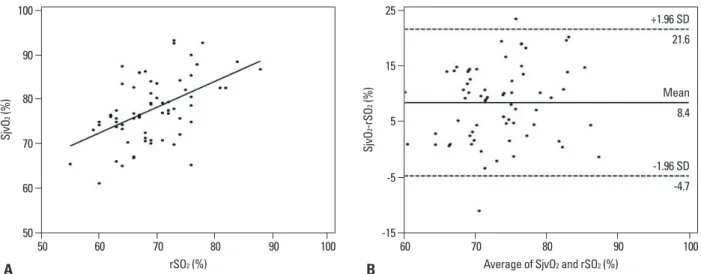
Seventy comparative measurements were performed be- tween SjvO2 and rSO2 in the supine position and Trendelen- burg-pneumoperitoneum condition, respectively. There was little correlation between SjvO2 and rSO2 in the supine po- sition (concordance correlation coefficient=0.2819) (Table 3) (Fig. 1A). Bland-Altman plots showed a mean bias of 8.4% with a limit of agreement of 21.6% and -4.7% (Fig.
1B). In addition, SjvO2 and rSO2 were not correlated during Trendelenburg-pneumoperitoneum condition (concordance correlation coefficient=0.3657) (Table 3) (Fig. 2A). Bland- Altman plots showed a mean bias of 10.6% with a limit of agreement of 23.6% and -2.4% (Fig. 2B).
The SjvO2-CO2 reactivity was higher than rSO2-CO2 reac- tivity in the supine position and Trendelenburg-pneumoperi- toneum condition, respectively (0.9±1.1 vs. 0.4±1.2%·mm curacy (degree of location or scale shift), thus providing
sound intuitive interpretations. Ranging from 0 to 1, higher values of CCC indicate more concordant data. Bland-Alt- man analysis was done to determine the magnitude of the difference between two measurements.13 Based on a previ- ous study,9 we postulated that a difference of 5% between the two parameters would be clinically acceptable under the hypothesis that the two methods are interchangeable. All data were analyzed using MedCalc software 9.3.6.0 (Med- Calc Inc., Mariakerke, Belgium).
We also compared the change in SjvO2 (or rSO2) -CO2 reactivity in the supine position and Trendelenburg-pneu- moperitoneum condition. The SjvO2 (or rSO2) -CO2 reac- tivity was defined as the % change in SjvO2 (or rSO2) by the step change induced in PaCO2. Comparison of the reac- tivity values of SjvO2 (or rSO2) -CO2 calculated in the su- pine position and Trendelenburg-pneumoperitoneum condi- tion were done using the paired t-test. Analysis of other intraoperative variables at each time period was done with repeated measures ANOVA. Data were analyzed using SPSS version 13.0 (SPSS Inc., Chicago, IL, USA). A p val- ue <0.05 was considered statistically significant.
RESULTS
Patients’ characteristics and operation data are summarized in Table 1.
Measurements of the cerebral oxygen profiles and hemo- dynamic variables during study periods are listed in Table 2.
Table 1. Patients’ Characteristics and Operation Data n=35
Age (yrs) 61.7±11.0 (43-72)
Height (cm) 166.4±4.9 (158-175)
Weight (kg) 65.9±8.3 (53-81)
Body mass index (kg·m-2) 23.7±2.5 (19.6-29.4) Duration of operation time (min) 176±22
Duration of pneumoperitoneum (min) 142±24 Values are mean±SD (range) or number of patients.
Table 2. Measurements of the Cerebral Oxygen Profiles and Hemodynamic Variables during Study Periods
T1 T2 T3 T4
SjvO2 (%) 74.2±4.9 81.0±7.5† 72.6±7.1 84.0±7.5‡
rSO2 (%) 68.1±5.3 70.3±7.1 66.1±6.1 69.3±7.7
MAP (mm Hg) 83.1±10.2 85.1±13.2 92.2±10.1* 88.8±11.2
HR (beats·min-1) 64.3±12.9 65.6±10.4 69.4±9.5 67.5±14.8
SjvO2, jugular bulb oxygen saturation; rSO2, regional cerebral oxygen saturation; MAP, mean arterial pressure; HR, heart rate.
Values are mean±SD. T1 and T2, PaCO2 of 35 and 45 mm Hg in supine position, respectively; T3 and T4, PaCO2 of 35 and 45 mm Hg in the Trendelenburg position under pneumoperitoneum, respectively.
*p<0.05 and †p<0.001 compared with the value at T1.
‡p<0.001 compared with the value at T3.
Table 3. The Relationship between Jugular Bulb Oxygen Saturation and Regional Cerebral Oxygen Saturation during Study Periods
Supine Trendelenburg-pneumoperitoneum
Concordance correlation coefficient 0.2819 0.3657
95% confidence interval 0.1527-0.4017 0.2492-0.4718
Precision (Pearson’s ρ) 0.5104 0.6977
Accuracy (Bias correction factor) 0.5523 0.5241
There are some studies investigating the correlation be- tween rSO2 and SjvO2 under specific clinical situations with contrary results. Kim, et al.14 reported good agreement be- tween rSO2 and SjvO2 measurements in healthy volunteers during isocapnic hypoxia. However, Leyvi, et al.10 demon- strated that there was only a weak correlation between rSO2 and SjvO2, and individual variation was wide during deep hy- pothermic circulatory arrest. Nagdyman, et al.9 reported that rSO2 demonstrated a substantial bias of the measurements to SjvO2 in children with congenital heart disease, which is in agreement with our results. In our study, there was poor agreement, significant bias, and imprecision between SjvO2 and rSO2 in the supine position and Trendelenburg-pneumo- peritoneum condition during laparoscopic surgery.
The disagreement between SjvO2 and rSO2 may be at- tributed to several factors. First, there is a significant differ- Hg-1, p=0.04; 1.7±1.3 vs. 0.5±1.1%·mm Hg-1, p<0.001). The
SjvO2-CO2 reactivity was higher in the Trendelenburg-pneu- moperitoneum condition compared to the supine position (1.7±1.3 vs. 0.9±1.1%·mm Hg-1, p<0.001). No adverse ef- fects related to jugular venous catheterization were observed.
DISCUSSION
Our main result is that there is little correlation between SjvO2 and rSO2 in the supine position and Trendelenburg- pneumoperitoneum condition during laparoscopic surgery.
Although episodes of clinically significant cerebral desatu- ration were not detected in this clinical setting, Bland-Alt- man analysis demonstrated that both rSO2 and SjvO2 are not interchangeable values in this study.
Fig. 1. Concordance correlation (A) and Bland-Altman analysis (B) of the measured difference between jugular bulb oxygen saturation (SjvO2) and regional cerebral oxygen saturation (rSO2) in the supine position.
Fig. 2. Concordance correlation (A) and Bland-Altman analysis (B) of the measured difference between jugular bulb oxygen saturation (SjvO2) and regional cerebral oxygen saturation (rSO2) in the Trendelenburg-pneumoperitoneum condition.
50
50
-15
-10 60
60
-5
0 70
70 80
80
5
10 90
90
15
20 100
100
25
30
SjvO2 (%)SjvO2 (%) SjvO2-rSO2 (%)SjvO2-rSO2 (%)
50
50
60
55 60
60
70
65 70
70
80
80
80
75 90
90
90
85 100
100
100
95 rSO2 (%)
rSO2 (%)
Average of SjvO2 and rSO2 (%)
Average of SjvO2 and rSO2 (%)
-4.7
-2.4 8.4
10.6 21.6
23.6 -1.96 SD
-1.96 SD Mean
Mean +1.96 SD
+1.96 SD
A
A
B
B
change of PaCO2 more accurately than rSO2 during laparo- scopic surgery. There is general agreement that rSO2 may be valuable as a trend monitor, but that it is less useful as an indicator of cerebral ischemia. Our results demonstrate that the validity of rSO2 is also questionable during Trendelen- burg-pneumoperitoneum condition.
In this study, SjvO2-CO2 reactivity was significantly high- er in the Trendelenburg-pneumoperitoneum condition com- pared to the supine position. This result means that the change of CBF according to the change of PaCO2 was greater in the Trendelenburg-pneumoperitoneum condition than in the supine position. Therefore, we suggested that it is necessary to control PaCO2 for the prevention of an in- crease of CBF during laparoscopic surgery.
There were some limitations in this study. First, we mea- sured SjvO2 unilaterally and compared it with the average value of rSO2 from both sides. Secondly, the patients of the study were all American Society of Anesthesiologists phys- ical status I or II without any cardiopulmonary diseases.
This may limit the extrapolation of our results to patients with severe cardiopulmonary compromise. Lastly, intraop- erative variables were measured at arbitrary time points without exact information on time dependent cardiopulmo- nary changes. Therefore, the optimal time points of evalua- tion when the patient is in the Trendelenburg position with CO2 pneumoperitoneum cannot be guaranteed.
In conclusion, there is little correlation between SjvO2 and rSO2 in the supine position and Trendelenburg-pneu- moperitoneum condition. Therefore, both rSO2 and SjvO2 are not interchangeable values in this condition.
ACKNOWLEDGEMENTS
This research was supported by Basic Science Research Program through the National Research Foundation of Ko- rea (NRF) funded by the Ministry of Education, Science and Technology (NRF-2010-0022999).
REFERENCES
1. Mottrie A, Van Migem P, De Naeyer G, Schatteman P, Carpentier P, Fonteyne E. Robot-assisted laparoscopic radical prostatectomy:
oncologic and functional results of 184 cases. Eur Urol 2007;52:
746-50.
2. Fujii Y, Tanaka H, Tsuruoka S, Toyooka H, Amaha K. Middle ce- rebral arterial blood flow velocity increases during laparoscopic
ence in measuring cerebral oxygen saturation between SjvO2 and rSO2. rSO2 measures cerebral oxygen saturation in a small region of the brain and may be influenced by blood distribution or signals caused by extracerebral tissues,15,16 and SjvO2 represents global cerebral oxygen saturation. Further- more, Knirsch, et al.17 demonstrated that rSO2 correlates bet- ter with central venous oxygen saturation than SjvO2. rSO2 is influenced by both cerebral and extracerebral components;
therefore, the impact of extracerebral components on the rSO2 reading should not be underestimated. In our study, ce- rebral oxygen saturation as measured by rSO2 was about 12% lower than that measured by SjvO2. Also, changes in extracerebral blood flow, variation in inter-individual ab- sorption differences and changed position of the probes over time may affect the measurements of rSO2.18,19
Body position and PaCO2 can also influence cerebral ox- ygen saturation. A previous study demonstrated that rSO2 was decreased in association with the Trendelenburg posi- tion and was further impaired by hypercapnia and pneumo- peritoneum during laparoscopic surgery.20 Another study demonstrated that rSO2 increased during Trendelenburg- pneumoperitoneum condition and PaCO2 increased in a similar manner,21 which is in accordance with our study. In this study, an increase of PaCO2 also increased SjvO2 sig- nificantly both in the supine and Trendelenburg position, and rSO2 increased slightly in this period. Therefore, it is suggested that PaCO2 should be maintained within the nor- mal range during the Trendelenburg-pneumoperitoneum position. It has also been suggested that rSO2 measure- ments can best be assessed if patient’s body position and PaCO2 are held constant.22
The rSO2 value from near-infrared spectroscopy reflects saturation in a mixture of 25% arterial, 70% venous and 5% capillary compartments. The changes in body position may alter the ratio of arterial and venous blood compart- ment in the cerebral circulation; therefore, the validity of rSO2 is questionable in this situation.
CBF-CO2 reactivity represents the ability of cerebral vas- culature to respond to changes in cerebral metabolic de- mands. In this study, the SjvO2-CO2 reactivity was higher than rSO2-CO2 reactivity in the supine position and Tren- delenburg-pneumoperitoneum condition, respectively. We previously demonstrated that CBF-CO2 reactivity measured by SjvO2 was preserved in the modest Trendelenburg posi- tion under pneumoperitoneum during sevoflurane anesthe- sia if PaCO2 was controlled.23 Therefore, it is suggested that SjvO2 may represent the change of CBF in relation to the
14. Kim MB, Ward DS, Cartwright CR, Kolano J, Chlebowski S, Henson LC. Estimation of jugular venous O2 saturation from ce- rebral oximetry or arterial O2 saturation during isocapnic hypoxia.
J Clin Monit Comput 2000;16:191-9.
15. Brown R, Wright G, Royston D. A comparison of two systems for assessing cerebral venous oxyhaemoglobin saturation during car- diopulmonary bypass in humans. Anaesthesia 1993;48:697-700.
16. Fearn SJ, Chant HJ, Picton AJ, Mortimer AJ, McCollum CN. The contribution of extracranial blood oxygenation on near-infrared spectroscopy during carotid endarterectomy. Anaesthesia 1997;52:
704-5.
17. Knirsch W, Stutz K, Kretschmar O, Tomaske M, Balmer C, Schmitz A, et al. Regional cerebral oxygenation by NIRS does not correlate with central or jugular venous oxygen saturation during interventional catheterisation in children. Acta Anaesthesiol Scand 2008;52:1370-4.
18. Dullenkopf A, Frey B, Baenziger O, Gerber A, Weiss M. Mea- surement of cerebral oxygenation state in anaesthetized children using the INVOS 5100 cerebral oximeter. Paediatr Anaesth 2003;13:384-91.
19. Yoshitani K, Kawaguchi M, Tatsumi K, Kitaguchi K, Furuya H. A comparison of the INVOS 4100 and the NIRO 300 near-infrared spectrophotometers. Anesth Analg 2002;94:586-90.
20. Lee JR, Lee PB, Do SH, Jeon YT, Lee JM, Hwang JY, et al. The effect of gynaecological laparoscopic surgery on cerebral oxygen- ation. J Int Med Res 2006;34:531-6.
21. Park EY, Koo BN, Min KT, Nam SH. The effect of pneumoperi- toneum in the steep Trendelenburg position on cerebral oxygen- ation. Acta Anaesthesiol Scand 2009;53:895-9.
22. Pollard V, Prough DS, DeMelo AE, Deyo DJ, Uchida T, Widman R. The influence of carbon dioxide and body position on near-in- frared spectroscopic assessment of cerebral hemoglobin oxygen saturation. Anesth Analg 1996;82:278-87.
23. Choi SH, Lee SJ, Rha KH, Shin SK, Oh YJ. The effect of pneu- moperitoneum and Trendelenburg position on acute cerebral blood flow-carbon dioxide reactivity under sevoflurane anaesthesia. An- aesthesia 2008;63:1314-8.
cholecystectomy. Anesth Analg 1994;78:80-3.
3. Huettemann E, Terborg C, Sakka SG, Petrat G, Schier F, Reinhart K. Preserved CO(2) reactivity and increase in middle cerebral ar- terial blood flow velocity during laparoscopic surgery in children.
Anesth Analg 2002;94:255-8.
4. Smythe PR, Samra SK. Monitors of cerebral oxygenation. Anes- thesiol Clin North America 2002;20:293-313.
5. Macmillan CS, Andrews PJ. Cerebrovenous oxygen saturation monitoring: practical considerations and clinical relevance. Inten- sive Care Med 2000;26:1028-36.
6. Tobias JD. Cerebral oxygenation monitoring: near-infrared spec- troscopy. Expert Rev Med Devices 2006;3:235-43.
7. Casati A, Spreafico E, Putzu M, Fanelli G. New technology for noninvasive brain monitoring: continuous cerebral oximetry. Mi- nerva Anestesiol 2006;72:605-25.
8. Yao FS, Tseng CC, Ho CY, Levin SK, Illner P. Cerebral oxygen desaturation is associated with early postoperative neuropsycho- logical dysfunction in patients undergoing cardiac surgery. J Car- diothorac Vasc Anesth 2004;18:552-8.
9. Nagdyman N, Ewert P, Peters B, Miera O, Fleck T, Berger F.
Comparison of different near-infrared spectroscopic cerebral oxy- genation indices with central venous and jugular venous oxygen- ation saturation in children. Paediatr Anaesth 2008;18:160-6.
10. Leyvi G, Bello R, Wasnick JD, Plestis K. Assessment of cerebral oxygen balance during deep hypothermic circulatory arrest by continuous jugular bulb venous saturation and near-infrared spec- troscopy. J Cardiothorac Vasc Anesth 2006;20:826-33.
11. Nagdyman N, Fleck T, Schubert S, Ewert P, Peters B, Lange PE, et al. Comparison between cerebral tissue oxygenation index mea- sured by near-infrared spectroscopy and venous jugular bulb satu- ration in children. Intensive Care Med 2005;31:846-50.
12. Barnhart HX, Haber M, Song J. Overall concordance correlation coefficient for evaluating agreement among multiple observers.
Biometrics 2002;58:1020-7.
13. Bland JM, Altman DG. Statistical methods for assessing agree- ment between two methods of clinical measurement. Lancet 1986;1:307-10.