J. Biomed. Lab. Sci. 10 (2004) 65–73
Specific Gene Silencing by Single Stranded Large Circular Antisense Molecules
Jong-Gu Park1,2†
1Department of Medical Genetic Engineering, Keimyung University School of Medicine, Dongsan Medical Center, 194 Dongsandong, Joonggu, Daegu 700-712, Korea
2WelGENE Inc., 71B-4L, Hightech Sector 2, Sungseo Industrial Park 3, Dalseogu, Daegu 704-230, Korea
I report that single-stranded antisense as a part of large circular (LC-) genomic DNA of recombinant M13 phage exhibits enhanced stability, sequence specific antisense activity, and no need for target site search. A cDNA fragment (708 bp) of rat TNF-α was inserted into a phagemid vector, and TNF-α antisense molecules (TNFα-LCAS) were produced as single-stranded circular DNA. When introduced into a rat monocyte/macrophage cell line, WRT7/P2, TNFα-LCAS was able to ablate LPS-induced TNF-α mRNA to completion. The antisense effect of TNFα-LCAS was shown to be sequence-specific because expressions of three control genes (β-actin, GAPDH and IL-1β) were not significantly altered by the antisense treatment. Further, TNFα-LCAS was found to be highly efficacious as only 0.1 µg (0.24 nM) of TNFα-LCAS was sufficient to block TNF-α expression in 1×10⁵ WRT7/P2 cells. I have also observed specific antisense activity in reduction of NF-κB gene expression. The results suggest that an antisense sequence as a part of single-stranded circular genomic DNA has a specific antisense activity.
Key Words: Large circular (LC-) antisense, Bacteriophage genome, TNF-α, NF-κB, Functional genomics, Target validation
서 론
안티센스 기술 (antisense technology)은 유전자 기능분석 및 의약품 개발을 위한 표적 유전자 (target gene)의 발굴에 광범위하게 응용되고 있다. 안티센스 분자는 세포내에서 표 적 mRNA와 상보적 결합을 함으로써, 이 결합체를 인식하 여 mRNA를 분해하는 RNaseH의 작용을 유도하거나, 리보 좀 복합체의 mRNA에로의 접근을 방해함으로써 궁극적으로 표적 유전자의 발현을 억제한다1). 기존에는 주로 antisense oligonucleotide (AS-oligo) 분자에 의한 유전자 발현 억제를 통해 유전자의 기능을 분석하거나2~4), 항암제를 개발하여 왔
다5~11). 이 과정에서 AS-oligo가 가지는 문제점들이 발견되었
다. AS-oligo는 표적 서열로의 비접근성12,13), 핵산분해효소에 대한 불안정성14~16), 낮은 서열 특이성 및 체내 부작용 유발
등의 단점을 가지는 것으로 밝혀졌다. 이 중 불안정성 문제는 AS-oligo를 phosphorothioate (PS) 기 또는 methylphosphonate (MP) 기 등으로 화학적 수식을 함으로써 부분적으로 개선되 어 왔다17~19). 그러나, 서열 특이적 반응성의 결핍, RNaseH 의 작용에 대한 둔감성, 분해된 수식 뉴클레오티드가 DNA 복제 및 수복 과정에서 일으키는 돌연변이 현상 등은 개선해 야 할 문제점이다.
본 연구실은 지금까지 stem-loop 구조13), CMAS (Covalently- Closed Multiple Antisense) 구조20), RiAS (Ribbon Antisense)21) 구조를 가지는 안티센스 분자들을 개발하여 왔다. 분자내 공 유결합을 통해 환형 폐쇄 구조를 가지게 함으로써 핵산분해 효소의 작용점을 원천적으로 제거한 CMAS 및 RiAS 분자는 개선된 안정성을 보였으며, 표적 mRNA의 특이적 제거에도 효과적이었다.
한편, M13 박테리오페이지 등은 단가닥의 폐쇄적 환형 분 자 (single stranded closed circular structure)를 지놈 (genome) 으로 가져, 지금까지는 주로 돌연변이 연구 및 DNA 염기 서열 분석 등에 활용되어 왔다. 구축한 재조합 페이지미드 (recombinant phagemid)를 숙주세포에 형질전환한 후 헬퍼 박 테리오페이지 (helper bacteriophage)를 감염시키면, 배양 상청
*논 문 접 수: 2004년 4월 9일 수정재접수: 2004년 5월 6일
†별책 요청 저자: 박종구, (우) 700-712 대구광역시 중구 동산동 194, 동산의료원 내 계명대학교 의과대학 의학유전공학교실 Tel: 053-250-7854, Fax: 053-250-7854
e-mail: [email protected]
액에 존재하는 재조합 박테리오페이지 (recombinant bacterio- phage)로부터 단가닥 형태의 large circular (LC-) DNA를 정 제할 수 있다. 이때, M13 페이지미드는 LC-DNA가 특정 유 전자 유래의 안티센스 서열을 함유할 수 있게 재조합될 수 있다. 이러한 LC-antisense는 폐쇄된 환형 구조를 가져 핵산 분해효소에 대한 높은 안정성을 가질 수 있고, 제조원리상 페이지미드를 이용하므로 수천 내지 수만 개 유전자들로 구 성된 안티센스 라이브러리 (antisense library)를 쉽게 구축할 수 있을 것으로 기대하였다.
본 연구에서는 TNF-α 및 NF-κB 유전자의 안티센스 서열 을 가진 LC-antisense 분자들을 대량 제작한 후, 이들의 핵산 분해효소에 대한 안정성 및 표적 유전자에 대한 서열 특이적 안티센스 활성을 조사하였다.
재료 및 방법
1. 세포 배양
THP-1 (acute monocytic leukemia) 세포를 한국세포주은행 으로부터 분양 받았으며, WRT7/P2 (mouse monocytic cell line) 세포를 일본 쥰텐도 대학의 야기타 박사로부터 제공받았다.
세포주들을 10% FBS, 100 U/ml penicillin, 100 µg/ml strepto- mycin을 함유한 RPMI 1640 또는 EMEM 배지 (JBI, Korea) 로 37℃, 5% CO2 조건에서 배양하였다. 형질감염 (transfec- tion) 하루 전날 세포들을 신선한 배지로 교환해 주었으며, 형질감염을 위한 생존 세포수의 측정은 trypan blue (0.4%) 염 색법으로 하였다.
2. 재조합 페이지미드의 구축
표준 방법에 따라 TNF-α 및 NF-κB 유전자를 함유하는 재조합 페이지미드를 제작하였다22). 먼저, 48-웰 플레이트에 1×10⁵개의 WRT7/P2 세포를 분주하고, 다음 날 랫트 TNF-α 유전자의 발현을 유도하기 위해 최종농도 30 µg/ml의 lipo- polysaccharide (LPS, Sigma-Aldrich, USA)를 24시간 동안 처리 하였다. 세포로부터 적정량의 TRIzol 시약 (GibcoBRL, USA) 을 사용하여 total RNA를 분리하였다. 이어 한 쌍의 PCR 프 라이머 (forward: 5'-GATCGTCGACGATGAGCACAGAAAGC- ATGATCC-3', reverse: 5'-GATCGAATTCGTCACAGAGCAAT- GACTCCAAAG-3')를 사용하여 TNF-α 유전자의 전체 코드 영역을 함유하는 cDNA 단편 (708 bp)을 증폭하였다. 획득한 cDNA 단편을 SalI/EcoRI 제한효소로 분해한 후 pBS KS (-) phagemid (Stratagene, USA)에 클로닝하였다. 동일한 방법으 로, THP-1 세포로부터 한 쌍의 PCR 프라이머 (forward: 5'- GATCGTCGACGCGCCACCCGGCTTCAGAATGGC-3', reve- rse: 5'-GATCGAATTCGGTGAAGCTGCCAGTGCTATCCG-3') 를 사용하여 NF-κB 유전자의 cDNA 단편 (700 bp)을 증폭한
다음 pBS KS (-)의 EcoRV 부위에 클로닝하였다. 대조군으 로서 LC-SE (표적 유전자의 센스 서열을 함유하는 단일가닥 페이지 지놈) 및 LCSS (insert 서열을 함유하지 않은 단일가 닥 페이지 지놈) 분자들도 구축한 다음, 재조합 페이지미드 들을 대장균 숙주세포 (XL-1 Blue, Stratagene)에 calcium- chloride 법으로 형질전환하였다. cDNA 단편들의 클로닝 방 향은 염기서열 분석 및 제한효소 처리로 확인하였다.
3. LC-antisense 분자의 생산 및 정제
재조합 페이지미드를 헬퍼 박테리오페이지 M13K07을 미 리 감염해 놓은 숙주대장균에 형질전환한 후, ampicillin (최 종농도 50 µg/ml)을 함유한 LB 고체배지로 37℃에서 종야 배양하였다. 다음날, 형성된 single colony를 2× YT 액체배지 (10 g NaCl, 10 g yeast extract, 16 g bactotrypton/1000 ml)에 접 종하고, 37℃에서 16시간 동안 진탕 배양하였다. 배양액을 원 심분리한 후, 얻은 배양 상청액에 PEG/NaCl (20% PEG8000, 2.5 M NaCl) 용액을 첨가하였다. 박테리오페이지 침전물을 TE (pH 8.0) 완충용액으로 재용해한 후, phenol 추출 및 ethanol 침전을 통하여 LC-antisense 분자를 회수하였다.
이어, 잔존하는 헬퍼 박테리오페이지 및 대장균 숙주세포 유래의 지놈 DNA로부터 LC-antisense 분자를 대량으로 정 제하기 위하여 겔 여과 칼럼 크로마토그래피 (gel filtration column chromatography)를 이용하였다. 수지로는 Superfine Sephacryl S-1000 (Amersham Biosciences, Sweden)을 충진하 였고, 0.2 M NaCl을 함유한 50 mM Tris-HCl (pH 8.3) 완충용 액으로 평형화하였다. 정제 시작 부피는 겔 유효 부피의 5%
로 조정하였고, DNA 용출 (유출 속도: 0.3 ml/min)은 수지 평형화에 사용하였던 완충용액으로 하였다. 이때, 샘플들을 260/280 nm 파장으로 UV scan하면서 매 5~10분 간격으로 수집하였다. 수집한 샘플 분획 I~VII들을 ice-cold ethanol로 침전한 후, PBS (phosphate buffered saline) 용액으로 용해하 였다. 정제한 LC-antisense 분자들의 양 및 순도를 확인하기 위해 1% 아가로스 겔 전기영동을 하였다. 단가닥 LC-anti- sense 분자들의 서열 충실도를 확인하기 위해 universal primer 를 사용한 DNA 염기서열 분석을 하였다. 동일한 방법으로 대조군으로서 LC-SE 및 LC-SS 분자도 정제하였다.
4. LC-antisense 분자의 안정성 조사
LC-antisense 분자의 안정성을 확인하기 위해, 1 µg의 LC- antisense 분자를 XhoI (10 U/µg DNA), Exonuclease III (160 U/µg DNA) 및 S1 nuclease (10 U/µg DNA)로 37℃에서 3시간 동안 각각 처리하였다. 또한, 혈청 속에서의 LC-antisense 분 자의 안정성을 확인하기 위해, 1 µg의 DNA를 1:3 (w/w)의 비율로 리포좀과 복합체를 형성하고, 여기에 열처리를 하지 않은 30% 소태아혈청을 첨가한 후 37℃에서 최대 48시간까
지 다양한 시간 동안 반응하였다. 이때, 대조군으로서 리포좀 과의 복합체를 형성하지 않은 LC-antisense 분자를 동일하게 반응하였다. 반응액으로부터 페놀 추출 및 ethanol 침전을 통 해 DNA를 회수하고, 1% 아가로스 겔에 전기영동하였다.
5. LC-antisense의 형질감염 및 RT-PCR
표적 유전자의 mRNA를 제거함에 있어 LC-antisense 분 자의 활성을 조사하기 위해, LC-antisense 분자를 세포에 형 질감염한 후 RT-PCR을 하였다. 이를 위하여, 먼저 세포들 을 200 µl의 배양배지로 48-웰 플레이트에 분주하고, LC- antisense 분자와 양이온 리포좀의 일종인 Lipofectamine (In- vitrogen, USA) 시약을 표적 세포주의 종류에 따라 다양한 비 율 (w/w)로 혼합하였다. 리포좀-DNA 복합체의 혼합은 Opti- MEM (Invitrogen) 배지로 한 후 제조사의 지침에 따라 세포 에 처리하였다. 37℃에서 6시간 동안 형질감염한 후, 배지에 2× FBS 및 항생제들을 첨가하고 18시간 더 배양하였다.
한편, 랫트 TNF-α 유전자의 발현을 유도하기 위하여 TNF- α 유래 LC-antisense 분자를 형질감염 해 놓은 THP-1 세포에 lippopolysaccharide (LPS, 30 µg/ml)를 처리하였다. LC-anti- sense 분자의 활성을 비교하기 위하여, 리포좀 자체 및 con- trol DNA-리포좀 복합체도 동시에 처리하여 분석하였다.
RNA 조제는 Tri 시약 (Molecular Research Center, USA)을 사용하여 제조사의 지침에 따라 하였다. 각 웰로부터 회수한 세포들을 1 ml의 Tri 시약으로 균질화하고 chloroform 추출 및 isopropanol 침전으로 RNA를 정제하였다. 이어 Access
RT-PCR Kit (Promega, USA)을 사용하여 제조사의 지침에 따 라 RT-PCR을 하였다. 총 50 µl의 PCR 반응액에 total RNA (1 µg), 한 쌍의 프라이머 (TNF-α: forward, 5'-CATCTCCCT- CCGGAAAGGACAC-3', reverse, 5'-CGGATGAACACGCCAG- TCGC-3', NF-κB: forward, 5'-CCTGGCCGGAGCCACTAGAC- -3', reverse, 5'-CTATACTCAGATCCATCACC-3'), AMV reverse transcriptase (5 U/µl), Tfl DNA polymerase (5 U/µl), dNTP (10 mM, 1 µl) 및 MgSO4 (25 mM, 2.5 µl)를 혼합하여 조제하였다.
역전사 및 PCR 반응은 thermal cycler (MJ Research, USA)로 하였다. First strand cDNA의 합성은 48℃에서 45분 동안, 이 어지는 DNA의 증폭은 일련의 94℃/30초, 59℃/1분, 68℃/2분 반응들을 30회 반복함으로써 수행하였다. 획득한 PCR 산물 을 1% 아가로스 겔 전기영동으로 확인하였고, 증폭 DNA의 정량을 위해 AlphaImager 1220 (Alpha Innotech, USA) 기기를 사용하였다.
6. ELISA
LC-antisense 분자의 형질감염에 의한 TNF-α 단백질의 발 현 변화를 ELISA법으로 측정하였다. 이를 위하여, 세포 배 양 상청액을 50배 희석하여 anti-TNF-α 항체가 코팅된 플레 이트 (R&D System, USA)에 첨가하였다. Biotin 표식 2차 항 체를 각 웰에 다시 첨가한 후 상온에서 90분간 반응하였다.
플레이트를 3회 세정한 후 streptavidin-peroxidase를 첨가하여 45분간 반응하였다. 이어서 플레이트를 4회 세정하여 결합하 지 않은 streptavidin-peroxidase를 제거하고, 발색을 위해 ch-
Fig. 1. A schematic diagram of construction of the phage genomic LC-antisense molecule for rat TNF-α (TNFα-LCAS). Rat TNF-α was cloned into the multiple cloning site of the phagemid vector, pBS KS (-). Whereas single-stranded sense molecules were obtained by placing the sense strand of TNF-α cDNA in the reverse orientation as that of the lacZ gene, LC-antisense molecules were obtained by placing the sense strand in the same orientation to the lacZ gene. These constructs allow the rescue of either single stranded LC-antisense or sense control molecules of TNF-α when transformed into competent bacterial cells that were already superinfected with helper phage, M13KO7.
romogen을 첨가한 후 20분간 반응하였다. 흡광도는 450 nm 에서 측정하였다.
결 과
1. 재조합 M13 박테리오페이지 시스템을 이용한 LC- antisense의 구축
본 연구실은 공유결합으로 말단을 폐쇄한 환형 안티센스 분자가 표적 유전자의 발현을 저해함에 있어, 높은 안정성과
효능을 가짐을 보고한 바 있다20,21). 이러한 결과들을 기초로 M13 박테리오페이지의 단가닥 환형 지놈도 안티센스 분자 로서 유용성을 가질 것으로 기대하였다. 표적 유전자의 센스 또는 안티센스 서열을 함유하는 단일가닥의 환형 페이지 지놈을 생성하기 위해 M13 페이지미드의 F1 복제 개시점 (replication origin)을 활용하였다. 페이지 지놈의 일부로서 LC-antisense 분자를 조제하기 위하여 랫트 TNF-α cDNA를 pBS KS (-) 페이지미드에 클로닝하였다 (Fig. 1). 동일한 방 법으로 NF-κB cDNA도 클로닝하였다. Jupin 등의 방법23)에
Fig. 2. Chromatographic purification and biochemical properties of TNFα-LCAS. A. Elution profiles for large circular antisense mole- cules from gel filtration column chromatography. B. Electrophoretic patterns of fractions obtained from gel filtration column chromato- graphy. Lane 1, crude DNA; and lanes 2~8, fractions I-VII (correspond to retention time from panel A). C. Characterization of TNFα- LCAS molecules. DNA was either double-stranded (ds) phagemid or single-stranded (ss) LC-antisense: Lane M, λ-HindIII DNA size marker; lane 1, phagemid DNA containing TNF-α cDNA (TNFα-phagemid); lane 2, TNFα-LCAS; lane 3, TNFα-phagemid/XhoI; lane 4, TNFα-LCAS/XhoI; lane 5, TNFα-phagemid/S1 nuclease; lane 6, TNFα-LCAS/S1 nuclease; lane 7, TNFα-phagemid/XhoI/exonuclease III; lane 8, TNFα-LCAS/XhoI/exonuclease III. D. Stability test of LC-antisense molecules. Antisense molecules treated with sera were run on a 1% agarose gel and visualized with ethidium bromide staining. Lanes 1~8, treated with 30% fetal bovine serum for different periods of time as indicated; and lane 9, sham treated control.
A
1 2 3 4 5 6 7 8 B
M 1 2 3 4 5 6 7 8 ds ss ds ss ds ss ds ss
Control XhoI S1 XhoI/ExoIII C
1 2 3 4 5 6 7 8 9 30% FBS
1h 2h 3h 4h 6h 16h 24h 48h
Control
D
따라 헬퍼 페이지를 미리 감염해 놓은 대장균 숙주세포에 재조합 페이지미드들을 형질전환한 후 배양하였다.
2. 크로마토그래피를 이용한 LC-antisense 분자의 대량 정제
재조합 박테리오페이지 시스템으로 생산한 LC-antisense 분자는 소량의 M13 페이지 및 헬퍼 페이지 유래 지놈 DNA 와 혼재되어 분리될 수 있다. 또한, 형질감염 효율을 감소시 키거나 세포주의 성장에 영향을 주는 LPS 분자도 다량 함유 하게 된다. 따라서, 형질전환체의 배양 상청액으로부터 LC- antisense 분자를 대량 정제하기 위하여 겔 여과 칼럼 크로마 토그래피를 이용하였다. Major peak는 75~120분의 용출 시 간대에서 분리되었다 (Fig. 2A). Peak들을 시간대별로 7개 (fractions I~VII)의 분획으로 나누어 분리한 후, LC-antisense 분자의 정제 순도를 확인하기 위해 1% 아가로스 겔 전기영 동을 하였다 (Fig. 2B). M13 페이지의 wild-type 지놈 DNA 는 분획 III (75분대)에서, LC-antisense 분자는 분획 IV~VII (80~110분대)에서 분리되었다. 분획 IV~VII들을 모은 후 ethanol 침전을 하였다. LC-antisense 분자를 60%의 회수율로 정제할 수 있었으며, OD260 / 280nm 측정법을 통한 순도 분석
결과 1.8 이상의 값을 가짐을 알 수 있었다. LAL endotoxin 시험을 통하여, LPS도 대량 제거되었음을 확인하였다. 또한, 정제한 단일가닥 페이지 지놈 DNA가 실제로 표적 유전자의 안티센스 서열을 함유함을 T3 프라이머를 사용한 염기서열 분석으로 확인하였다. 본 과정을 통해 정제한 TNF-α 및 NF- κB 유전자 유래 LC-antisense 분자들을 각각 TNFα-LCAS 및 NFκB-LCAS로 명명하였다.
3. LC-antisense 분자의 구조 및 안정성
LC-antisense 분자는 그 제조원리상 단가닥의 폐쇄적 환형 구조를 가지므로 exonuclease에 대한 안정성을 가질 것으로 기대하였다. 이를 확인하기 위하여 TNFα-LCAS를 다양한 효 소 또는 소태아혈청 (fetal bovine serum)과 반응해 보았다 (Fig. 2C). TNFα-LCAS의 경우, 제한효소 XhoI과 exonuclease III를 3시간 이상 처리하여도 분해되지 않은 반면, TNF-α cDNA를 함유하는 두 가닥의 재조합 페이지미드는 XhoI의 처리로서 완전히 분해되었다. 또한 재조합 페이지미드는 XhoI과 exonuclease III를 동시에 처리하였을 때도 완전히 분 해되었다. TNFα-LCAS 분자가 단가닥의 DNA라는 사실은, 단가닥 DNA만 특이적으로 인식하는 S1 nuclease가 동 안티
Fig. 3. Antisense activity of TNFα-LCAS on TNF-α mRNA levels in WRT7/P2 cells. A. RT-PCR was performed with 2 sets of pri- mers, either TNF-α primers or β-actin primers: Lane M, λ-HindIII DNA size marker; lane 1, liposome alone; lane 2, TNFα-LCAS (1.4 nM);
lane 3, TNFα-LCSE (1.4 nM); lane 4, LCSS (1.4 nM). B. Both IL-1β and GAPDH genes were amplified by RT-PCR to examine non- specific antisense effects. The amounts of total RNA and single stranded circular molecules, including the antisense compounds were the same as those in panel A; Lane M, 100 bp DNA ladder; lane 1, liposome alone; lane 2, TNFα-LCAS (1.4 nM); lane 3, TNFα-LCSE (1.4 nM); lane 4, LCSS (1.4 nM). C. Southern hybridization of panel A. D. Dose dependent effects of TNFα-LCAS on TNF-α mRNA expre- ssion: Lane M, 100 bp DNA ladder; lane 1, liposome alone; lanes 2~5, TNFα-LCAS (0.03 nM, 0.14 nM, 0.28 nM and 0.56 nM, orderly);
lane 6, TNFα-LCSE (0.28 nM); lane 7, LCSS (0.28 nM).
Rat TNF-α
β-actin 1 2 3 4
C
Rat TNF-α β-actin M 1 2 3 4 5 6 7
D M 1 2 3 4
Rat TNF-α (752 bp) β-actin (318 bp) A
M 1 2 3 4
IL-1β (550 bp) GAPDH (330 bp) B
센스 분자를 완전히 분해함을 확인함으로써 증명하였다. 한 편, TNFα-LCAS 분자를 양이온 리포좀과 복합체를 형성한 후 30% FBS와 24시간 이상 반응하여도 대부분의 분자들은 분해되지 않았다 (Fig. 2D). 이러한 결과들은, 페이지 지놈 유래의 LC-antisense 분자가 단가닥의 환형구조를 가지며, 생 체내에 존재하는 분해효소들에 대하여 높은 안정성을 가짐 을 증명해 준다.
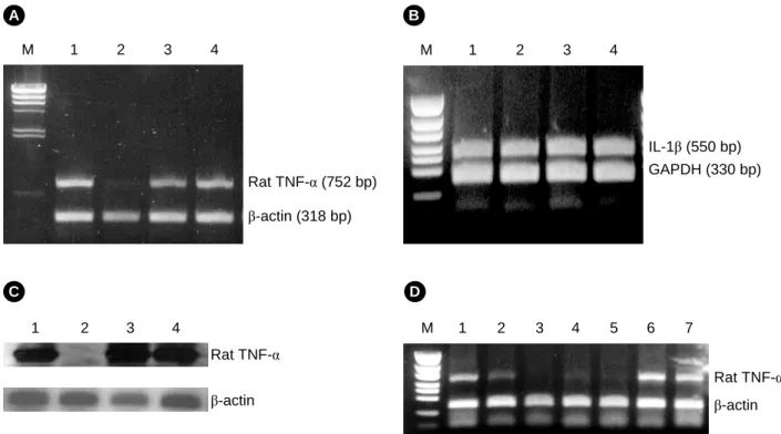
4. TNFα-LCAS에 의한 랫트 TNF-α mRNA의 특이적 제거
상기의 실험들을 통해 그 안정성을 확인한 LC-antisense 분자의 안티센스 활성을 조사하였다. TNFα-LCAS (0.5 µg, 1.4 nM)를 양이온 리포좀과 복합체를 형성하여 WRT7/P2 세포 에 처리한 후, TNF-α 유전자의 발현을 유도하기 위해 LPS를 처리하였다. 그 결과, TNFα-LCAS를 처리한 세포에서는 TNF- α mRNA의 발현 수준이 현저히 감소함을 알 수 있었다. 반 면에, TNFα-LCSE 또는 LCSS 분자의 처리는 TNF-α mRNA 의 발현에 있어 큰 변화를 일으키지 않았다 (Fig. 3A). 또한, 동일한 조건으로 TNFα-LCAS를 처리한 후 β-actin, GAPDH (glyceraldehyde 3-phosphate dehydrogenase) 및 IL-1β 유전자 들의 mRNA 수준도 확인하였으나, 이러한 유전자들의 발현 에는 큰 영향을 주지 않았다 (Fig. 3A 및 3B). 한편, 획득한 PCR 산물이 TNF-α 유전자에서 유래한 것임을 Southern blot- ting을 통하여 증명하였다 (Fig. 3C). 이때 probe로는 증폭된
DNA 단편의 일부 영역과 일치하는 서열 (5'-GATGAGAGG- GAGCCCATTTGGG-3')을 가진 것을 사용하였다. 다음으로는, WRT7/P2 세포에 다양한 양의 TNFα-LCAS를 처리하여 처리 량의 증가에 따른 활성 변화를 조사해 보았다. TNF-α 유전자 의 발현은 0.05 µg (0.03 nM)의 TNFα-LCAS를 처리하였을 때 소폭 감소되었고, 0.1 µg (0.14 nM)의 처리시 현저히 감소되기 시작하여 0.5 µg (0.28 nM)의 처리로 완전히 억제되었다 (Fig.
3D). 이러한 결과들은 TNFα-LCAS 분자가 서열 특이적으로, 그리고 기존의 안티센스 분자들에 비해 소량으로도 표적 유 전자의 발현을 효율적으로 감소시킴을 보여준다.
5. NFκB-LCAS에 의한 NF-κB mRNA의 특이적 제거 LC-antisense 분자의 범용성을 확인하기 위하여, 동 안티 센스 분자가 TNF-α 이외 다른 표적 유전자의 발현도 저해할 수 있는지를 조사하였다. 이를 위하여, 다양한 양의 NFκB- LCAS 분자를 리포좀과 복합체를 형성한 다음 THP-1 세포 에 처리하였다. 0.05 µg (0.14 nM)의 NFκB-LCAS를 처리하였 을 때, NF-κB mRNA의 수준은 70% 정도 감소하였고, 0.1 µg (0.28 nM) 또는 0.2 µg (0.56 nM)을 처리하였을 때 90% 이상 감소하였다 (Fig. 4A). 반면에, NFκB-LCSE 또는 LCSS 분자 를 처리한 세포들은 NFκB mRNA의 발현에 있어 큰 변화를 나타내지 않았다. 증폭한 PCR 산물이 NFκB 유전자에서 유 래한 것임을 Southern blotting으로 확인하였다 (Fig. 4B). 이 때 probe로는 증폭된 DNA 단편의 일부 영역과 일치하는 서 열 (5'-CTTCCAGTGCCCCCTCCTCCACCGC-3')을 가진 것을 사용하였다. 이러한 결과들은 LC-antisense 분자가 다른 종류 의 표적 유전자에 대해서도 그 발현을 저해할 수 있는 범용
NF-κB (611 bp) β-actin (318 bp) M 1 2 3 4 5 6 7 8 9
A
NF-κB 1 2 3 4 5 6 7 8 9 B
Fig. 4. Effects of NFκB-LCAS on mRNA levels of NF-κB gene in THP-1 cells. Amplified PCR fragments were run on a 1%
agarose gel and visualized with ethidium bromide staining. A. RT- PCR results. Lane M, 100 bp ladder marker; lane 1, sham treated control; lanes 2~4, NFκB-LCAS (0.14 nM, 0.28 nM and 0.56 nM, orderly); lanes 5~6, NFκB-LCSE (0.28 nM and 0.56 nM);
lanes 7~8, LCSS (0.28 nM and 0.56 nM); lane 9, liposome alone.
B. Southern blotting of panel A.
Fig. 5. Effect of TNFα-LCAS on the level of TNF-α protein.
ELISA of TNF-α protein in media: WRT7/P2 cells transfected with TNFα-LCAS, TNFα-LCSE or LCSS. Each bar value repre- sents the mean ± S.D. of triplicate experiments. Statistical signi- ficance was calculated with students t-test. A value of P<0.05 was considered to be statistically significant.
성을 가진다는 사실을 증명해 준다.
6. TNFα-LCAS에 의한 TNF-α 단백질 수준의 감소 LC-antisense 분자가 표적 유전자의 mRNA를 제거할 뿐만 아니라, 궁극적으로 단백질의 수준도 감소시키는지를 확인 해 보았다. 이를 위하여, WRT7/P2 세포에 TNFα-LCAS를 처 리한 다음, 배양 상청액으로 분비된 TNF-α 단백질의 수준을 ELISA법으로 측정한 결과, 세포내 mRNA의 수준이 감소한 바와 같이, 단백질의 수준도 90% 이상 감소함을 알 수 있었 다 (Fig. 5). 반면에, TNFα-LCSE 또는 LCSS 분자 등의 대조 군 처리는 TNF-α 단백질의 수준에 큰 변화를 주지 않았다.
이러한 결과는 LC-antisense 분자에 의한 mRNA 수준의 특 이적 감소는 세포내 표적 단백질의 수준 감소로도 이어짐을 증명해 준다.
고 찰
본 연구에서는 재조합 박테리오페이지가 가지는 단가닥 지놈 DNA의 일부로서 안티센스 서열을 함유하는 LC-anti- sense 분자를 대량 정제하였으며, 그 안정성 및 활성을 조사 하였다. 본 연구실은 증가된 안정성 및 안티센스 활성을 가 지는 일련의 환형 AS-oligo 분자들을 고안하여 왔으며, 최근 에는 RiAS 분자에 대하여 보고하였다21). c-myb 유전자에 대 한 RiAS (ribbon antisense) 분자를 구축하였고 이를 암세포주 에 처리함으로써, 동 안티센스 분자가 표적 유전자의 발현을 억제하며 나아가 세포의 성장도 저해함을 확인하였다. 본 연 구에서 시험한 LC-antisense 분자 역시 환형 구조를 가지며, 개선된 안정성 및 활성을 보여 주었다.
TNF-α 유전자는 cytokine의 일종이며 류마티스 관절염, 알 레르기, 그리고 패혈증 등의 기타 면역질환들과 밀접한 관련 성을 가진다24~26). 따라서, TNF-α 유전자의 효율적 발현 조 절은 특정 면역질환들의 잠재적 치료법으로서 추구되어 왔
다27,28). 본 연구를 통하여 TNFα-LCAS가 TNF-α mRNA의
발현 저해에 유용함을 확인하였다. 또한, 동일한 접근법으로 서 NF-κB 유전자의 발현 감소도 확인함으로써, LC-antisense 분자가 모든 유전자에 대하여 범용성을 가진다는 사실도 검 정하였다.
새로운 형태의 LC-antisense 분자는 핵산분해효소들에 대해 높은 안정성을 가질 뿐만 아니라, 기존 AS-oligo 분자들의 사용량에 비해 1/10의 수준으로도 표적 유전자를 효율적으로 제거하였다. 이러한 LC-antisense 분자의 활성 증가는 다음의 두 가지 이유로 설명할 수 있다. 첫째, LC-antisense 분자는 긴 영역의 안티센스 서열을 가지므로, 표적 mRNA의 상보적 서열과 보다 안정된 결합체를 형성함으로써 보다 장시간 동 안 RNaseH 활성에 대한 기질로서 작용할 수 있다. 둘째,
mRNA는 분자내 서열간의 상보성 및 세포질내에 존재하는 RNA 결합 단백질 (RNA-binding protein)과의 상호작용으로 인하여 광범위한 2차 구조 및 3차 구조를 형성하는 경향을 가진다. 따라서, 길이가 짧은 안티센스 분자는 표적 서열에 접근하지 못할 확률이 높다. 이러한 이유 때문에, AS-oligo의 경우 표적 유전자의 효율적 발현 억제를 위해 번거로운 표적 부위 설정을 거쳐야 한다. 반면에, LC-antisense 분자는 긴 길 이의 안티센스 영역을 가지므로 특정영역이 표적 mRNA 서 열에 보다 쉽게 결합할 수 있다. 따라서, 짧은 영역의 안티 센스 서열을 가진 기존의 AS-oligo 분자보다 표적 mRNA를 보다 효율적으로 제거할 가능성이 높아질 것이다. 지금까지 긴 길이의 AS-oligo 분자는 특이성의 저하, 합성 길이의 한 계성, 그리고 비효율적 세포내 전달 등의 문제점으로 인하여 응용하기 힘든 것으로 인식되어 왔다. 그러나, 표적 유전자를 세포에 형질감염한 후 긴 길이의 antisense RNA를 발현하고, 이를 다양한 in vitro 연구에 활용하고 있음은 주지의 사실이 다29). 이러한 연구흐름은 긴 길이의 안티센스 분자가 반드시 증가된 비특이성을 가지는 것은 아님을 반증해 주는 것이다.
한편, TNFα-LCAS의 형질감염은 표적 mRNA의 발현 감소 뿐만 아니라 TNF-α 단백질의 수준에도 영향을 준다는 사실 을 확인하였다.
안티센스 분자에 의한 유전자 발현 저해는 표적 유전자의 기능 연구를 위한 효율적인 수단으로서 활용되고 있다30~32). 안티센스에 의한 유전자 기능연구는 기존의 유전자 발현 저 해법들보다 빠른 속도로 진행될 수 있다. LC-antisense 분자 는 보다 높은 안정성을 가지며 소량으로도 높은 활성을 나 타낸다. 특히, 박테리오페이지 및 페이지미드 시스템을 이용 하므로 수천 내지 수만 개의 다른 유전자에서 유래한 안티센 스 분자들을 대량으로 얻을 수 있다. 따라서, LC-antisense 분 자는 antisense library의 형태로서 쉽게 구축될 수 있어 대규 모 유전자 기능분석을 위한 이상적인 도구로서 활용할 수 있을 것이다.
Acknowledgements
This study was supported by generous grants of the CDRC of the Korean Science & Engineering Foundation (research grant R01-2000-00138) and WelGENE Inc, a biotechnology company founded by Dr. Jong-Gu Park.
참 고 문 헌
1) Dolnick BJ (1991): Antisense agents in cancer research and therapeutics. Cancer Invest, 9: 185-194.
2) Thompson CB, Challoner PB, Neiman PE and Groudine M (1985): Levels of c-myc oncogene mRNA are invariant throu-
ghout the cell cycle. Nature, 314: 363-366.
3) Anfossi GA, Gewirtz M and Calabretta B (1989): An oligomer complementary to c-myb-encoded mRNA inhibits proliferation of human myeloid leukemia cell lines. Proc Natl Acad Sci USA, 86: 3379-3383.
4) Melani C, Rivoltini L, Parmiani G, Calabretta B and Colombo MP (1991): Inhibition of proliferation by c-myb antisense oli- godeoxynucleotides in colon adenocarcinoma cell lines that express c-myb. Cancer Res, 51: 2897-2901.
5) Kamano H, Ohnishi H, Tanaka T, Ikeda K, Okabe A and Irino S (1990): Effects of the antisense v-myb expression on K562 human leukemia cell proliferation and differentiation. Leuk Res, 14: 831-839.
6) Melotti P and Calabretta B (1996): Induction of hematopoietic commitment and erythromyeloid differentiation in embryonal stem cells constitutively expressing c-myb. Blood, 87: 2221- 2234.
7) Ferrari S, Donelli A, Manfredini R, Sarti M, Roncaglia R, Tagliafico E, Rossi E, Torelli G and Torelli U (1990): Diffe- rential effects of c-myb and c-fos antisense oligodeoxynu- cleotides on granulocytic differentiation of human myeloid leukemia HL60 cells. Cell growth Differ, 1: 543-548.
8) Ratajczak MZ, Hijiya N, Catani L, DeRiel K, Luger SM, McGlave P and Gewirtz AM (1992): Acute- and chronic-phase chronic myelogenous leukemia colony forming units are hi- ghly sensitive to the growth inhibitory effects of c-myb anti- sense oligodeoxynucleotides. Blood, 79: 1956-1961.
9) Kastan MB, Stone KD and Civin CI (1989): Nuclear oncopro- tein expression as a function of lineage, differentiation stage, and proliferative status of normal human hematopoietic cells.
Blood, 74: 1517-1524.
10) Thaler DS, Liu S and Tombline G (1996): Extending the chemistry that supports genetic information transfer in vivo:
phosphorothioate DNA, phosphorothioate RNA, 2'-O-methyl RNA, and methylphosphonate DNA. Proc Natl Acad Sci USA, 93: 1352-1356.
11) Wagner RW (1994): Gene inhibition using antisense oligode- oxynucleotides. Nature, 372: 333-335.
12) Flanagan WM and Wagner RW (1997): Potent and selective gene inhibition using antisense oligodeoxynucleotides. Mol Cell Biochem, 172: 213-225.
13) Matsuda M, Park JG, Wang DC, Hunter S, Chien P and Sch- reiber AD (1996): Abrogation of the Fc gamma receptor IIA-mediated phagocytic signal by stem-loop Syk antisense oligonucleotides. Mol Biol Cell, 7: 1095-1106.
14) Akhtar S, Kole R and Juliano RL (1991): Stability of anti- sense DNA oligodeoxynucleotide analogs in cellular extracts and sera. Life Sci, 49: 1793-1801.
15) Wagner RW, Matteucci MD, Lewis JG, Gutierrez AJ, Moulds C and Froehler BC (1993): Antisense gene inhibition by oligonucleotides containing C-5 propyne pyrimidines. Science, 260: 1510-1513.
16) Gryaznov S, Skorski T, Cucco C, Nieborowska-Skorska M, Chiu CY, Lloyd D, Chen JK, Koziolkiewicz M and Calabretta B (1996): Oligonucleotide N3'→P5' phosphoramidates as antisense agents. Nucleic Acids Res, 24: 1508-1514.
17) Helene C (1991): Rational design of sequence-specific onco- gene inhibitors based on antisense and antigene oligonucleo- tides. Eur J Cancer, 27: 1466-1471.
18) Bayever EP, Iversen L, Bishop MR, Sharp JG, Tewary HK, Arneson MA, Pirruccello SJ, Ruddon RW, Kessinger A, Zon G and Armitage JO (1993): Systemic administration of a pho- sphorothioate oligonucleotide with a sequence complemen- tary to p53 for acute myelogenous leukemia and myelo- dysplastic syndrome: Initial results of a phase I trial. Anti- sense Res Dev, 3: 383-390.
19) Baker BF and Monia BP (1999): Novel mechanisms for anti- sense mediated regulation of gene expression. Biochim Bio- phys Acta, 1489: 3-18.
20) Moon IJ, Lee Y, Kwak CS, Lee JH, Choi K, Schreiber AD and Park JG (2000): Target site search and effective inhibition of leukaemic cell growth by a covalently multiple antisense oligonucleotide to c-myb. Biochem J, 346: 295-303.
21) Moon IJ, Choi K, Choi YK, Kim JE, Lee Y, Schreiber AD and Park JG (2000): Potent growth inhibition of leukemic cells by novel ribbon-type antisense oligonucleotides to c-myb. J Biol Chem, 275: 4647-4653.
22) Sambrook J and Russell DW (2001): Plasmids and their use- fulness in molecular cloning. pp.1.1-1.138. "Molecular clon- ing; A laboratory manual", 3rd Ed., Cold Spring Harbor La- boratory Press, Cold Spring Harbor, New York.
23) Jupin I and Gronenborn B (1995): Abundant, easy and repro- ducible production of single-stranded DNA from phagemids using helper phage-infected competent cells. Nucleic Acid Res, 23: 535-536.
24) Waszczykowska E, Robak R, Wozniacka A, Narbutt J, Tor- zecka JD and Sysa-Jedrzejowska A (1999): Estimation of SLE activity based on the serum level of chosen cytokines and superoxide radical generation. Mediators Inflamm, 8: 93-100.
25) Pulsatelli L, Dolzani P, Piacentini A, Silvestri T, Ruggeri R,
Gualtieri G, Meliconi R and Facchini A (1999): Chemokine production by human chondrocytes. J Rheumatol, 26: 1992- 2001.
26) Yoshino S, Sasatomi E, Mori Y and Sagai M (1999): Oral administration of lipopolysaccharide exacerbates collagen- induced arthritis in mice. J Immunol, 163: 3417-3422.
27) Maini R, St Clair EW, Breedveld F, Furst D, Kalden J, Weis- man M, Smolen J, Emory P, Harriman G, Feldmann M and Lipsky P (1999): Infliximab (chimeric anti-tumour necrosis factor alpha monoclonal antibody) versus placebo in rheuma- toid arthritis patients receiving concomitant methotrexate: a randomised phase III trial. ATTRACT study Group. Lancet, 354: 1932-1939.
28) Kim KN, Watanabe S, Ma Y, Thornton S, Giannini EH and Hirsch R (2000): Viral IL-10 and soluble TNF receptor act synergistically to inhibit collagen-induced arthritis following adenovirus-mediated gene transfer. J Immunol, 164: 1576- 1581.
29) Barnor JS, Miyano-Kurosaki N, Yamaguchi K, Kobayashi H,
Ishikawa K, Osei-Kwasi M, Ampofo WK, Ofori-Adjei D, Inagaki Y, Yamamoto N and Takaku H (2002): Down re- gulation of human immunodeficiency virus type-1 (HIV-1) expression by Vif antisense RNA expression vectors in trans- fected cells. Nucleic Acids Res Suppl, 123-124.
30) Bennett CF and Cowsert LM (1999): Antisense oligonucleo- tides as a tool for gene functionalization and target validation.
Biochim Biophys Acta, 1489: 19-30.
31) De Backer MD, Nelissen B, Logghe M, Viaene J, Loonen I, Vandoninck S, De Hoogt S, Dewaele S, Simons FA, Ver- hasselt P, Vanhoof G, Contreras R and LuytenDe WHML (2001): An antisense-based functional genomics approach for identification of genes critical for growth of Candida albicans.
Nat Biotechnol, 19: 235-241.
32) Ji Y, Zhang B, Van SF, Patrick Warren H, Woodnutt G, Burn- ham MKR and Rosenberg M (2001): Identification of critical Staphylococcal genes using conditional phenotypes generated by antisense RNA. Science, 293: 2266-2269.