Open Access
Investigating the Feasibility of Targeted Next-Generation Sequencing to Guide the Treatment of Head and Neck Squamous Cell Carcinoma
Original Article
Purpose
Head and neck squamous cell carcinoma (HNSCC) is a deadly disease in which precision medicine needs to be incorporated. We aimed to implement next-generation sequencing (NGS) in determining actionable targets to guide appropriate molecular targeted therapy in HNSCC patients.
Materials and Methods
Ninety-three tumors and matched blood samples underwent targeted sequencing of 244 genes using the Illumina HiSeq 2500 platform with an average depth of coverage of greater than 1,000. Clinicopathological data from patients were obtained from 17 centers in Korea, and were analyzed in correlation with NGS data.
Results
Ninety-two of the 93 tumors were amenable to data analysis. TP53 was the most common mutation, occurring in 47 (51%) patients, followed by CDKN2A (n=23, 25%), CCND1 (n=22, 24%), and PIK3CA (n=19, 21%). The total mutational burden was similar between human papillomavirus (HPV)–negative vs. positive tumors, although TP53, CDKN2A and CCND1 gene alterations occurred more frequently in HPV-negative tumors. HPV-positive tumors were significantly associated with immune signature-related genes compared to HPV-neg- ative tumors. Mutations of NOTCH1 (p=0.027), CDKN2A (p < 0.001), and TP53 (p=0.038) were significantly associated with poorer overall survival. FAT1 mutations were highly enriched in cisplatin responders, and potentially targetable alterations such as PIK3CA E545K and CDKN2A R58X were noted in 14 patients (15%).
Conclusion
We found several targetable genetic alterations, and our findings suggest that implemen- tation of precision medicine in HNSCC is feasible. The predictive value of each targetable alteration should be assessed in a future umbrella trial using matched molecular targeted agents.
Key words
Squamous cell carcinoma of the head and neck, Next-generation sequencing, Molecular targeted therapy, Biomarkers, Clinical trial
*A list author’s aliations appears at the end of the paper.
+ + + + + + + + + + + + + + + + + + + + + + + + + + + + + + + + + + + + + + + + + + + + + + + + + + + + + + + + + + + + + + + + + + + + + + + + + + + + + + + + + + + + + + + + + + + + + + + + + + + + + + + + + + + + + + + + + + + + + + + + + + + + + + + + + + + + + + + + + + + + + + + + + + + + + + + + + + + + + + + + + + + + + + + + + + + + + + + + + + + + + + + + + + + + + + + + + + + + + + + + + + + + + + + + + + + + + + + + + + + + + + + + + + + + + + + + + + + + + + + + + + + + + + + + + + + + + + + + + + + + + + + + + + + + + + + + + + + + + + + + + + + + + + + + + + + + + + + + + + + + + + + + + + + + + + + + + + + + + + + + + + + + + + + + + + + + + + + + + + + + + + + + + + + + + + + + + + + + + + + + + + + + + + + + + + + + + + + + + + + + + + + + + + + + + + + + + + + + + + + + + + + + + + + + + + + + + + + + + + + + + + + + + + + + + + + + + + + + + + + + + + + + + + + + + + + + + + + + + + + + + + + + + + + + + + + + + + + + + + + + + + + + + + + + + + + + + + + + + + + + + + + + + + + + + + + + + + + + + + + + + + + + + + + + + + + + + + + + + + + + + + + + + + + + + + + + + + + + + + + + + + + + + + + + + + + + + + + + + + + + + + + + + + + + + + + + + + + + + + + + + + + + + + + + + + + + + + + + + + + + + + + + + + + + + + + + + + + + + + + + + + + + + + + + + + + + + + + + + + + + + + + + + + + + + + + + + + + + + + + +
Correspondence: Hwan Jung Yun, MD Department of Internal Medicine, Chungnam National University Hospital, 282 Munhwa-ro, Jung-gu, Daejeon 35015, Korea Tel: 82-42-280-7157
Fax: 82-42-257-5753 E-mail: [email protected]
Co-correspondence: Sangwoo Kim, PhD Department of Biomedical Systems Informatics and Brain Korea 21 PLUS Project for Medical Science, Yonsei University College of Medicine, 50-1 Yonsei-ro, Seodaemun-gu, Seoul 03722, Korea Tel: 82-2-2228-0913
Fax: 82-2-2227-8129 E-mail: [email protected]
Co-correspondence: Hye Ryun Kim, MD, PhD Division of Medical Oncology, Department of Internal Medicine, Yonsei University College of Medicine, 50-1 Yonsei-ro, Seodaemun-gu, Seoul 03722, Korea
Tel: 82-2-2228-8125 Fax: 82-2-393-3652 E-mail: [email protected]
Received January 3, 2018 Accepted May 4, 2018 Published Online May 9, 2018
*Sun Min Lim, Sang Hee Cho, and
In Gyu Hwang contributed equally to this work.
Sun Min Lim, MD
1Sang Hee Cho, MD, PhD
2In Gyu Hwang, MD, PhD
3Jae Woo Choi, BS
4Hyun Chang, MD, PhD
5Myung-Ju Ahn, MD, PhD
6Keon Uk Park, MD, PhD
7Ji-Won Kim, MD, PhD
8Yoon Ho Ko, MD, PhD
9Hee Kyung Ahn, MD
10Byoung Chul Cho, MD, PhD
11Byung-Ho Nam, PhD
12Sang Hoon Chun, MD
13Ji Hyung Hong, MD, PhD
14Jung Hye Kwon, MD, PhD
15Jong Gwon Choi, MD, PhD
16Eun Joo Kang, MD, PhD
17Tak Yun, MD
18Keun-Wook Lee, MD, PhD
8Joo-Hang Kim, MD, PhD
1Jin Soo Kim, MD, PhD
19Hyun Woo Lee, MD
20Min Kyoung Kim, MD, PhD
21Dongmin Jung, MS
22Ji Eun Kim, MD, PhD
23Bhumsuk Keam, MD, PhD
24Hwan Jung Yun, MD
25Sangwoo Kim, PhD
26Hye Ryun Kim, MD, PhD
11Introduction
Head and neck squamous cell carcinoma (HNSCC) is the sixth most common malignancy worldwide, and is usually curable, if diagnosed early. Unfortunately, patients often present with advanced disease that is incurable or requires aggressive treatment, which results in functional disability, dismal prognosis and high mortality. Low survival outcomes in combination with significant toxicity of current treatment strategies emphasize the necessity for novel therapeutic modalities. Until recently, the only targeted therapy in HNSCC was cetuximab, a monoclonal antibody against epidermal growth factor receptor, which has shown a response rate of 10% to 15% in the patients with recurrent or metastatic dis- ease [1]. However, there is no validated biomarker for pre- dicting cetuximab efficacy, which dampens the precise selec- tion of patients. Antiprogrammed death 1 agents including pembrolizumab and nivolumab were recently approved for HNSCC that is refractory to platinum-based therapy. How- ever, the presence of programmed death-ligand 1 (PD-L1) on tumor cells did not satisfactorily predict response, with 22% of PD-L1 positive patients responding vs. 4% of PD-L1 negative patients responding [2]. Therefore, more effective treatment strategies for personalized treatment of HNSCC are urgently needed.
Next-generation sequencing (NGS) of tumors has greatly expanded our understanding of genetic profiles, and several studies have found novel genetic alterations in HNSCC [3-6]. However, these studies were performed retrospectively in surgical specimens without incorporated clinical data on the response to therapy. Although potentially targetable genetic alterations such as PIK3CA, epidermal growth factor receptor (EGFR), and fibroblast growth factor receptor (FGFR) mutations have been identified, functional studies to validate the roles of such mutations as biomarkers remain scarce.
Herein, we describe our implementation of a precision medicine approach in 93 patients with HNSCC. This is a fea- sibility study of “Translational biomarker-driven umbrella project for head and neck and esophageal squamous cell car- cinoma (TRIUMPH)” study by the Korean Cancer Study Group (NCT03292250) (S1 Fig.). TRIUMPH is the first, prospective, biomarker-driven umbrella trial for patients with HNSCC, consisting of multiple targeted therapies including phosphoinositide 3-kinase (PI3K) inhibitor, pan- HER inhibitor, FGFR inhibitor and CDK4/6 inhibitor.
Patients without actionable targets are to be allocated into an immunotherapy arm. Before the start of TRIUMPH study, we conducted this feasibility study in which tumors and matched blood samples were analyzed by multiplexed tar- geted NGS assays. The objective of this study is to examine
the feasibility of implementing NGS to guide treatment in HNSCC patients, and to find the associations between somatic alterations and clinical outcome.
Materials and Methods
1. Patients and data collection
Pretreatment tumor tissues (somatic) and matched normal DNA (germline) from prospectively recruited patients with HNSCC were obtained between 2016 and 2017 under the approval of Institutional Review Board of 19 institutions in Korea. HNSCC patients with initial stages 1-4 were included in this study. Clinicopathological data were collected from patient charts in accordance with an IRB-approved protocol.
Clinical information including age, sex, anatomic site of tumor, tobacco and alcohol use, clinical stage, treatment his- tory, and survival data were collected.
2. Targeted sequencing of tumors
Genomic DNA was isolated from formalin-fixed paraffin- embedded (FFPE) samples using the QIAamp DNA FFPE Tissue Kit (Qiagen, Hilden, Germany) for the targeted sequencing of 244 head and neck cancer-related genes selected based on a literature search (S2 Table). The genomic regions of the 244 genes were captured by the customized SureSelectXT Target Enrichment library generation kit (Agilent, Santa Clara, CA), and sequenced using the Illumina HiSeq 2500 platform with a depth of coverage > 1,000.
3. Variant calling and functional annotation
By default, base quality trimming for short reads from the targeted sequencing was performed using Sickle [7]. Filtered reads were mapped to the human reference genome (GRCh- 37/hg19) using Burrows-Wheeler Aligner [8]. All reads that were mapped with < 23 mapping quality were discarded.
The aligned reads (BAM file) were further processed with the Genome Analysis ToolKit v3.5, including MarkDuplicate, Local Realignment, and Base Quality Score Recalibration [9].
Initial somatic mutations candidates were called by MuTect ver. 1.17 with a default parameter [10]. Somatic insertions/
deletions (indels) were called by Varscan2 ver. 2.3.7 with
somatic p < 0.05 [11]. From the initial call set, FoxoG artefacts
were removed using the in-house Python program ver. 3.6
to discard skewed read-orientation variants [12]. The func-
tionality of final high confidence variants was annotated with
ANNOVAR software [13], including the consequences, pre-
dicted impacts and reported allele frequencies in population.
In particular, non-rare variants (minor allele frequency
> 0.05) were discarded to retain only pathogenic variants.
Finally, the clinical interpretation of targeted therapy was annotated using the CIVIC [14] and DoCM [15] databases.
Copy number alterations (CNAs) were called using CNVkit [16] for targeted deep sequencing. To reduce ambiguity from individual variations, all normal samples were pooled and used as a control. Of the initial CNA calls, genes with 4 and 0 measured copy numbers were considered amplified and deleted, respectively, to secure high confidence. To visualize the overall landscape of mutations, ‘Oncoprint’ was drawn using the R package ‘ComplexHeatmap’ of R ver. 3.4. Lol- lipop plots were drawn for frequently mutated genes using MAFtools to check the recurrence of genomic loci with vari- ants.
4. Nanostring assay
Total tumor RNA was isolated using the RNeasy kit (Qia- gen). The nCounter Analysis System (Nanostring Technolo- gies, Seattle, WA) was used to screen for the expression of 55 immune-related genes. Counts were normalized to internal controls and reference genes using the nSolver software ver.
3.0. We obtained gene expression data for 94 tumour sam- ples, among them 8 with average expression levels of less than 10 were filtered out. The NanoStringNorm package of R was used for normalization [17]. We selected housekeep- ing.geo.mean for normalizing the samples or RNA contents.
Differentially expressed genes between human papillo- mavirus (HPV)–positive and HPV-negative samples were identified by the glm.LRT function in the NanoStringDiff [18]
package of Bioconductor. A volcano plot was drawn by using the ggplot2 package of R. The complete list of 55 immune-related genes is shown in S3 Table.
5. Statistical methods
All statistical analysis was performed using the R, Python Scipy package and SPSS ver. 23.0 (IBM Corp., Armonk, NY) software. To test group-specific enrichment of genomic vari- ants, Fisher exact test was applied to each called variant, fol- lowed by the p-value cutoff of 0.05. Tumor mutation burden (TMB) was measured by the number of missense mutations per megabase (Mb) within the range of the targeted capture region. The numbers of mutations per Mb between HPV-pos- itive and HPV-negative groups were compared using Stu- dent’s t-test. Progression-free survival (PFS) and overall sur- vival (OS) were estimated using the Kaplan-Meier method;
differences between groups were compared using the log- rank test. In groups of unbalanced sizes, the standard asymp- totic log-rank test is often replaced by its corresponding
permutation test; alternatively, the distribution under the null hypothesis is approximated via Monte Carlo resam- pling. Here, we used empirical p-values from 10,000 repli- cates by using the log-rank test function in the coin package of R. Two-sided p-values of < 0.05 were considered signifi- cant.
6. Ethical statement
This study was conducted under the approval of Institu- tional Review Board of 19 institutions in Korea. All patients provided written informed consent for genomic testing used for this study.
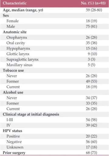
Table 1. Baseline characteristics of all patients
HPV, human papillomavirus.
Characteristic No. (%) (n=93)
Age, median (range, yr) 59 (28-80)
Sex
Female 18 (19)
Male 75 (81)
Anatomic site
Oropharynx 26 (28)
Oral cavity 35 (38)
Hypopharynx 15 (16)
Glottic larynx 9 (10)
Supraglottic larynx 3 (3)
Maxillary sinus 5 (5)
Tobacco use
Never 26 (28)
Former 49 (53)
Current 18 (19)
Alcohol use
Never 34 (37)
Former 33 (35)
Current 26 (28)
Clinical stage at initial diagnosis
I-III 54 (58)
IV 39 (42)
HPV status
Positive 20 (22)
Negative 56 (60)
Unknown 17 (18)
Prior surgery 68 (73)
Results
1. Clinical characteristics
A total of 93 patient tumors were included in 75 men and 18 women. Clinical data are summarized in Table 1; the median age of all patients was 59 years (range, 28 to 80 years)
and 39 patients (42%) had stage 4 disease at initial diagnosis.
Sixty-seven patients (72%) had smoking history and 59 patients (63%) had alcohol history. HPV status was known in 76 patients (82%), of whom 20 (22%) were positive. Sixty- eight patients (73%) had received prior surgery, and among patients who received surgery, 47 patients experienced recurrence: 14 (29%) with locoregional recurrence and 33 (71%) with distant metastasis. Surgery or radiotherapy was Fig. 1. (A) Mutational spectrum and copy number alterations in head and neck squamous cell carcinomas detected by tar- geted sequencing. Samples with a greater than 1% incidence of genetic alterations are shown, and are stratified by human papillomavirus (HPV) status and primary tumor anatomic site. Pos, positive; Neg, negative. (Continued to the next page)
TP53 CDKN2A CCND1 PIK3CA KMT2C FAT1 RELN FAT4 CDKN2B EGFR KMT2D NFE2L2 ADGRV1 NOTCH1 FAT2 CTTN MYC GNAS EPHB4 ASXL3 CHD4 SOX2 ASXL1 KEAP1 AR NOTCH2 KLHL6 TERT KRAS PTEN MAP3K9 CDH9 CDH1 HPV Anatomy 51 25
24 21 15 14 11 10 10 10 10 9 9 9 9 8 8 7 7 7 7 7 7 7 5 5 5 5 5 5 5 5 5 (%)
Amplification Deletion Pos
Alterations
HPV
Neg
AnatomyHypopharynx
Missense Nonsense Frame shift indel In-frame indel Splice site Larynx Oral cavity Oropharynx Other
12
0 4 6 2 10 8
A
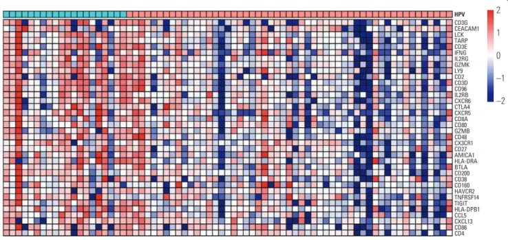
Fig. 1. (Continued from the previous page) (B) A heat map of 55 differentially expressed genes with an absolute fold change
2 and a false discovery rate (FDR) < 0.05. (C) Volcano plot showing the distribution of the fold changes in gene expression.
Genes with an absolute fold change 2 and FDR < 0.05 are indicated in red (high expression in HPV-positive tumors com- pared to HPV-negative tumors).
B
CD3GCEACAM1 LCKTARP CD3EIFNG IL2RG GZMKLY9 CD2CD3D CD96IL2RB CXCR6 CTLA4 CXCR5 CD8ACD80 GZMBCD48 CX3CR1 CD27AMICA1 HLA-DRA BTLACD200 CD38CD160 HAVCR2 TNFRSF14 TIGIT HLA-DPB1 CCL5CXCL13 CD86CD4 HPV
–2 2 1
–1 0
4
0 1 2
–2
Log fold change
–L og 10 q -v al ue
2
0 1
–1
C
3
Not significant
q-value < 0.05 LCK TARP
IFNG CEACAM1 CD3E
CD3D
CD80 GZMK LY9 CD96 CD2
CTLA4 IL2RB
CD8A CX3CR1
CXCR5 CXCR6
IL2RG
CD3G
CD86 CD4
CCL5 CXCL13 TIGIT CD160
CD200 CD38 CD27
HLA-DPB1 HLA-DRA AMICA1
GZMB
HAVCR2 TNFRSF14
CD48
BTLA
performed for locoregional recurrence, and systemic chemo- therapy was performed for metastatic disease. For the whole cohort, the median PFS and OS were 12.5 months (95% con- fidence interval [CI], 10.2 to 14.8) and 70 months (95% CI, 57.4 to 84.4), respectively, with a median follow-up of 20 months. Patients with HPV-positive oropharynx cancers
(n=16) showed a better 2-year OS rate than HPV-negative patients (n=10) (31% vs. 10%, respectively), although the dif- ference was not significant owing to the small number of cases.
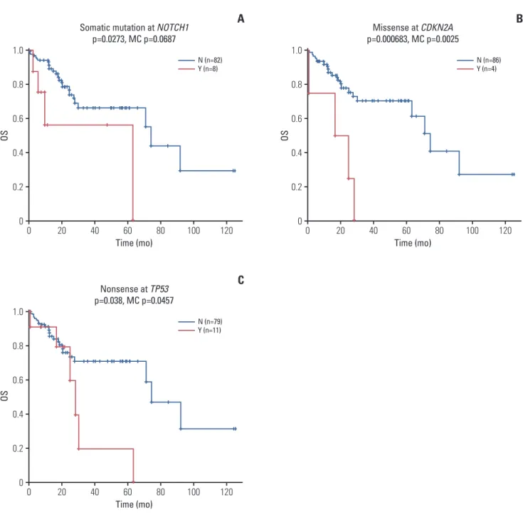
Fig. 2. Kaplan-Meier curves showing the association of single nucleotide variations and overall survival (OS) in patients.
(A) Patients with NOTCH1 somatic mutation had poorer overall survival (somatic mutation includes missense, nonsense, splice site mutations, frame shift indels, or in-frame indels). (B) Patients with CDKN2A missense mutations had poorer OS.
(C) Patients with TP53 nonsense mutation showed poorer OS.
1.0
0 0.2 0.4 0.8
0
Time (mo)
OS OS OS
Somatic mutation at NOTCH1 p=0.0273, MC p=0.0687
60 40
20 80 100 120
A
0.6
N (n=82) Y (n=8)
1.0
0 0.2 0.4 0.8
0
Time (mo) Nonsense at TP53 p=0.038, MC p=0.0457
60 40
20 80 100 120
C
0.6
N (n=79) Y (n=11)
1.0
0 0.2 0.4 0.8
0
Time (mo) Missense at CDKN2A p=0.000683, MC p=0.0025
60 40
20 80 100 120
B
0.6
N (n=86)
Y (n=4)
2. Overview of somatic mutations in HNSCC
A total of 2,315 somatic single nucleotide variations (SNVs) and 19 indels were identified from the targeted sequencing of the 92 tumors, which corresponds to a rate of 3.64 SNVs per 1 Mb. We found that TP53 was the most frequently mutated gene (n=47, 51%), followed by CDKN2A (n=23, 25%), CCND1 (n=22, 24%), and PIK3CA (n=19, 21%) (Fig. 1A).
As expected, smokers displayed a significantly higher TMB than non-smokers (4.16/Mb vs. 3.12/Mb, p=0.04) (S4 Fig.).
3. Comparison of HPV-positive vs. HPV-negative tumours
Of 92 patient tumors, 76 tumors (82%) had known HPV status and we compared molecular landscape of HPV-posi- tive and HPV-negative tumors. TMB counts were higher in
HPV-negative than HPV-positive tumors, although the dif- ference was not significant (4.16/Mb vs. 3.12/Mb, p=0.150) (S5 Fig.). TP53, CDKN2A, and CCND1 gene alterations were significantly more frequent in HPV-negative tumors (Fig. 1A).
As described previously, we observed TP53 mutations among HPV-negative tumors at higher rates than HPV-positive tumors (65.5% vs. 9.5%, p < 0.001). Inactivating mutations such as CDKN2A and CDKN2B deletions (n=6), and CCND1 amplification (n=17) were exclusively identified in HPV-neg- ative tumors. We also noted HPV-negative specific genetic alterations in receptor tyrosine kinases (RTKs) including EGFR, FGFR1/3, and platelet-derived growth factor receptor A (PDGFRA), which was consistent with a previous study [5]. PIK3CA mutations were more commonly found in HPV- positive tumors (23.8% vs. 16.4%, p > 0.05). Comparison of immune signatures between HPV-positive and HPV-nega- Fig. 3. Patients who received cisplatin-based chemotherapy were categorized into responders vs. non-responders and genetic alterations are shown in the order of frequency. Pos, positive; Neg, negative.
TP53 CCND1 CDKN2A PIK3CA FAT1 KMT2C RELN EGFR NFE2L2 NOTCH1 FAT2 CTTN FAT4 ADGRV1 GNAS EPHB4 ASXL1 CDH1 CDKN2B KMT2D KRAS MYC ASXL3 CHD4 SOX2 KEAP1 AR NOTCH2 PTEN CDH9 KLHL6 TERT MAP3K9 Cisplatin 59 35
27 20 20 18 18 14 12 12 12 12 10 10 10 10 10 10 8 8 8 6 6 6 6 6 6 6 6 6 4 4 4 (%)
Amplification Deletion Pos
Alterations
Cisplatin
Neg
Missense Nonsense Frame shift indel In-frame indel Splice site
tive tumors via nanostring assay revealed that HPV-positive tumors were significantly enriched with immune-related genes. HPV-tumors harbored higher levels of immune acti- vation: specifically, CD3 (p=6.010
–6), CECAM1 (p=4.910
–5) and IL2R (p=6.910
–5) expression (Fig. 1B and C).
4. Clinical correlation
We performed an exploratory analysis to correlate gene alterations (SNVs and CNAs) with survival (Fig. 2). In 90 patients with available survival data, genomic events asso- ciated with poorer OS were mutations in NOTCH1 (p=0.027),
CDKN2A (p < 0.001), and TP53 (p=0.038). The association between CDKN2A, TP53 mutations and poor OS was consis- tent with a previous analysis of The Cancer Genome Atlas (TCGA) database. CNAs were not associated with any gene alterations. In contrast to a previous report [19], PIK3CA amplification was not associated with worse OS (S6 Fig.).
Next, we analyzed gene alterations associated with cis- platin resistance by classifying patients who received cis- platin-based chemotherapy into responders and non-respon- ders. According to Response Evaluation Criteria in Solid Tumors (ver. 1.1), responders were patients who showed complete response, partial response or stable disease to cis- Fig. 4. Gene diagrams for a selection of key mutations in potentially targetable genes PIK3CA (A), CDKN2A (B), and TP53 (C). (D) Signaling pathway deregulation is shown. HPV, human papillomavirus. (Continued to the next page)
5
1 2 4
0
PIK3CA (Somatic mutation rate: 13.92%) NM_006218
M ut at io n
600 400
200 800 1,000 1,071
A
3
C2_PI3K_class_I_alpha PI3K_p85B
PI3K_rbd
PI3Ka_I PI3Kc PI3Kc_IA_alpha
Missense_mutation E545K
5
1 2 4
0
CDKN2A (Somatic mutation rate: 16.46%) NM_000077
M ut at io n
100
50 159
B
3
ANK ANK_5 Missense_mutation
Nonsense_mutation
R58X
platin-based chemotherapy, whereas non-responders were those with progressive disease [20]. Among 54 evaluable patients, 38 (70%) were responders, and 16 (30%) were non- responders. FAT1 gene mutations (5 missense and 5 non- sense) were highly enriched in cisplatin responders com- pared to non-responders (p < 0.05) (Fig. 3, S7 Fig.).
5. Targetable mutations and copy-number aberrations
We identified potentially targetable mutations in PIK3CA and CDKN2A. An established canonical mutation, PIK3CA E545K missense mutation were identified in five patients (5%) (Fig. 4A), while CDKN2A R58X nonsense mutation was identified in four patients (4%) (Fig. 4B). TP53 inactivating Fig. 4. (Continued from the previous page)
5
1 2 4
0
TP53 (Somatic mutation rate: 59.49%) NM_000546
M ut at io n
pathway MAPK activation
PI3K-AKT pathway activation
Apoptosis Transition G1-S
200
100 300 396
C
D
3 R209Q/W R243W/Q
P53 P53_TAD P53_tetramer
Missense_mutation
Nonsense_mutation Frame_Shift_Del Frame_Shift_Ins
HPV– incidence HPV+ incidence
EGFR 12.7% 0%
FGFR1
5.5% 0%
FGFR3 1.8% 4.3%
PIK3CA 16.4% 23.8%
AKT 3.6% 4.8%
MTOR
Oncogene
Tumor suppressor gene
1.8% 4.8%
MAPK1 3.6% 9.5%
CCND1 30.9% 0%
PTEN 5.5% 4.8%
RSA 12.7% 0%
ERBB2
1.8% 0%
cMET 1.8% 9.5%
CDKN2A 30.9% 4.8%
TP53
65.5% 9.5%
mutations (R209Q/W, R243W/Q) that cause cell cycle dereg- ulation occurred in eight patients (5%) (Fig. 4C).
Fig. 4D summarizes the deregulated signaling pathways.
Among RTKs, EGFR and cMET alterations were frequent, followed by FGFR3, FGFR1, and ERBB2. Among down- stream targets of the RTKs/RAS/PI3K pathway, PIK3CA dominated with occasional MAPK1 and MTOR mutations.
RAS and MAPK1 alterations occurred in 12.7% and 13.1% of patients, respectively. Alterations in tumor suppressors, TP53 and CDKN2A were notable in HPV-negative tumors, which were consistent with a recent report [21]. Overall, alterations in genes involved in cell death and PIK3CA/AKT/
MTOR pathway were predominant.
Discussion
Our umbrella trial suggests that using NGS for determin- ing treatment strategies for patients with HNSCC is feasible, and that translating genomic data into clinical care is attain- able. The most common genomic alterations (TP53, PIK3CA, CCND1, and CDKN2A) were identified at frequencies con- sistent with investigations of TCGA. Previous studies have characterized mostly surgically resected HNSCC samples, with a limited portion of HPV-positive samples. TCGA study, which is the largest cohort to date (n=279) is com- prised of surgically resected oral cavity or laryngeal squa- mous cell carcinoma patients, and treatment and survival data were limited [5]. Recently, Seiwert et al. [22] reported a large number of HPV-positive tumors, where they included 51 (42.5%) HPV-positive patients in a total of 120 patients.
Consistent with our finding, the mutational burdens in HPV- positive and -negative tumors were similar, while FGFR2 aberrations were exclusively identified in HPV-negative tumors.
Our study emphasizes how the application of NGS may be used as a prospective, master protocol tailored to each patient’s genotype. The turnaround time from patient sam- ple collection to NGS results was within 4 weeks, which is timely for patient enrolment. Similarly, another study recen- tly found it feasible to incorporate NGS into the clinical care of HNSCC patients [19]. Patients who received targeted ther- apy matched to their genotypes achieved a higher objective response rate than patients unmatched to therapy. However, they used two different NGS platforms with inconsistent mutation rates and actionable alterations. Additionally, the MOSCATO-01 trial showed that genomic analyses of 199 patients with advanced cancers produced improved out- comes with matched targeted therapy [23]. The ongoing NCI-MATCH trial is currently assessing whether molecular
markers can predict response to targeted therapies in patients with advanced cancer [24] and the results are awaited.
PI3K pathway aberrations are potential therapeutic targets in HNSCC patients. Prior studies identified that PIK3CA mutation or amplification was associated with various clini- cal outcomes. One study reported that PIK3CA amplification was associated with significantly decreased PFS, whereas PIK3CA mutation was not [19]. Another study demonstrated that PIK3CA mutations were correlated with poor prognosis in HPV-negative, locally advanced HNSCC [22]. A preclini- cal study also reported that patient-derived PIK3CA mutant HNSCC tumor grafts are potentially sensitive to PI3K/mTOR inhibitors [25]. In our cohort, patients with a PIK3CA hotspot mutation (E545K) will be treated with the PI3K pathway inhibitor (BYL719).
Deletion of CDKN2A or amplification of CCND1, which induces sustained CDK 4/6 activation, occurred at 27% and 22%, respectively, which were comparable to such cell-cycle related gene aberrations found in other studies [5,22]. Pre- clinical or clinical data regarding the activity of CDK inhibitor in HNSCC is limited, but our prospective trial may solve which genotypes will benefit from treatment with CDK inhibitors.
FAT atypical cadherin 1 (FAT1) was significantly enriched in cisplatin responders. FAT1 gene has been reported to be associated with various types of cancer, including HNSCC [5,26]. FAT1 gene acts as a tumor suppressor, in which loss- of-function activates Wnt pathway and tumorigenesis [27].
Recently, FAT1 mutation was significantly associated with better OS in HPV-negative patients from both the TCGA cohort and the International Cancer Genome Consortium (ICGC) data cohort [28]. The functional impact of the FAT1 mutation identified in our study requires further investiga- tion to determine its role as a prognostic or predictive bio- marker.
In our study, immune signatures were highly enriched in HPV-positive tumors, consistent with a previous finding that HPV-positive tumors have a distinct immune phenotype, characterized by more immune cell infiltration and higher levels of CD8
+T-cell activation [29]. As ongoing checkpoint inhibitor trials (NCT02105636, NCT01848834) showed prom- ising preliminary activities in HNSCC patients, improved outcome in HPV-positive patients may be related to their immunophenotype [30,31].
The accuracy and fidelity of genomic analysis are critical;
therefore, false-positive or false-negative genomic variants
should be carefully avoided. To that end, several technical
issues were noted in our study. First, the often inevitable low
tumour cellularity in samples, owing to normal cell contam-
ination, has a negative effect on the accuracy of calling of
SNVs and CNVs [32]. We found that, among 92 samples, 14
(15%) and five (5%) obtained via core needle biopsy and excision, respectively, showed low tumor cellularity (~30%).
As CNV analysis is directly affected by reduced cellularity, CNVs with ambiguous analysis scores may require confir- mation using alternative methods. Second, sequencing arte- facts can appear in every step of the NGS pipeline, which complicates the differentiation between true vs. false vari- ants. We observed an abnormally excessive number of low- level somatic mutations in a few samples (mutation-rate/Mb
> 100), which could only be removed using an oxoG filtering program [12]. Such false variants can distort the overall dis- tribution of somatic mutations and their relative burdens, and should be specially inspected via advanced bioinformat- ics analyses. Third, whole-exome or targeted sequencing for identifying CNVs remain secondary options, as more sensi- tive methods such as whole-genome sequencing or special- ized array-based methods are widely unavailable. As tar- geted sequencing based CNV analysis generally performs better in a larger cohort, size and sustainability of clinical tri- als should be considered when they are designed. Moreover, active participation of genome analysis experts is highly rec- ommended to manage such technical issues.
In conclusion, our large-scale targeted sequencing of HNS- CC patient samples identified potentially targetable alter- ations. Further prospective validation of NGS based mole- cularly targeted treatment is highly warranted.
Electronic Supplementary Material
Supplementary materials are available at Cancer Research and Treatment website (https://www.e-crt.org).
Conflicts of Interest
Conflict of interest relevant to this article was not reported.
Acknowledgments
This study was supported by a grant from the National R&D Pro- gram for Cancer Control, Ministry of Health and Welfare, Republic of Korea (1631050). The funder had no role in the study design; in the collection, analysis and interpretation of data; in the writing of the report; or in the decision to submit the article for publication.
Author Details
1
Division of Medical Oncology, Department of Internal Medicine, CHA Bundang Medical Center, Seongnam,
2Department of Hemato- Oncology, Chonnam National University Hwasun Hospital, Hwa- sun,
3Division of Hematology/Oncology, Department of Internal Medicine, Chung-Ang University Hospital, Chung-Ang University College of Medicine, Seoul,
4Department of Pharmacology, Sever-
ance Biomedical Science Institute, Yonsei University of College of Medicine, Yonsei Cancer Research Institute, JE-UK Laboratory of Molecular Cancer Therapeutics, Seoul,
5Division of Hematology and Medical Oncology, International St. Mary’s Hospital, Catholic Kwan- dong University College of Medicine, Incheon,
6Department of Med- icine, Samsung Medical Center, Sungkyunkwan University School of Medicine, Seoul,
7Department of Hemato-Oncology, Keimyung University Dongsan Medical Center, Daegu,
8Department of Inter- nal Medicine, Seoul National University Bundang Hospital, Seong- nam,
9Department of Internal Medicine, Uijeongbu St. Mary's Hospital, Uijeongbu,
10Department of Medical Oncology, Gachon University Gil Medical Center, Incheon,
11Divison of Medical Oncology, Department of Internal Medicine, Yonsei Cancer Center, Yonsei University College of Medicine, Seoul,
12HERINGS, The Institute of Advanced Clinical & Biomedical Research, Seoul,
13
Division of Medical Oncology, Department of Internal Medicine, Bucheon St. Mary's Hospital, College of Medicine, The Catholic Uni- versity of Korea, Bucheon,
14Department of Internal Medicine, Incheon St. Mary's Hospital, Incheon,
15Division of Hemato-Oncol- ogy, Department of Internal Medicine, Hallym University Kang- dong Sacred Heart Hospital, Hallym University College of Medicine, Seoul,
16Department of Internal Medicine, Konyang University Hos- pital, Daejeon,
17Division of Medical Oncology, Department of Internal Medicine, Korea University Guro Hospital, Korea Univer- sity College of Medicine, Seoul,
18Rare Cancers Clinic, Center for Specific Organs Cancer, National Cancer Center, Goyang,
19Depart- ment of Internal Medicine, SMG-SNU Boramae Hospital, Seoul,
20