Background: Various factors influencing the distribution of sensory blockade in epidural anesthesia have been identified; however, reports on the effects of gravity and different postures in thoracic epidural anesthesia have been rare. Medications may be injected with the lateral decubitus or the neutral position; however, it is unclear whether the distribution range of medication is similar or significantly different between these 2 postures.
Objective: We focused on identifying the effect of different postures on the distribution of local anesthetics using epidurography at the thoracic level.
Study Design: Prospective randomized trial.
Setting: An interventional pain management practice in South Korea.
Methods: A total of 68 patients were randomized to either the L group (n = 34, lateral decubitus with neck and hip flexion) or the P group (n = 34, prone position). After completing the insertion of the epidural catheter, the location of the catheter tip was adjusted between T7 and T8 and the patients were asked to change their posture according to their allocated group. Three mL of contrast medium was injected and the total number of segments, including the most cranial and caudal ends of the vertebra, was identified.
Results: The total number of vertebral segments confirmed by contrast medium spread was 7.4 ± 2.2 in group P and 9.2 ± 1.8 in group L. The total number and the number of vertebral segments covered in the caudad direction were higher in group L compared to
group P and this was statistically significant (P < 0.001).
Limitations: We made every effort to make the posture of group L like crouchback, we think that slight differences would present in the flexion angles of the neck and hip among the patients of group L.
Conclusion: Group L demonstrated a more extensive distribution of contrast medium for both the cranial and caudad directions compared to group P.
Key words: Epidural, thoracic, contrast medium, injections
Trial registry number: Clinical trial registry information service (NCT02865512).
Pain Physician 2017; 20:501-508
Randomized Trial
Posture Influences the Extent of Spread of Contrast Medium During Thoracic
Epidurography: A Prospective Randomized Trial
From: 1Department of Anesthesiology and Pain Medicine, Keimyung University DongSan Hospital, Dae Gu, Korea; 2Department of Psychiatry, Keimyung University DongSan Hospital, Dae Gu, Korea Address Correspondence:
Ji Hee Hong, MD, PhD Department of Anesthesiology and Pain Medicine, Keimyung University DongSan Hospital 56 Dal-Sung Ro, Jung gu, Dae
Gu, 700-712, Korea E-mail: [email protected]
Disclaimer: This work was supported by the National Research Foundation of Korea (NRF) Grant funded by the Korea Government (MSIP) (No.2014R1A5A2010008).
Conflict of interest: Each author certifies that he or she, or a member of his or her immediate family, has no commercial association (i.e., consultancies, stock ownership, equity interest, patent/licensing arrangements, etc.) that might pose a conflict of interest in connection with the submitted manuscript.
Manuscript received: 02-01-2017 Revised manuscript received:
03-09-2017 Accepted for publication:
03-30-2017 Free full manuscript:
www.painphysicianjournal.com
Ji Hee Hong, MD, PhD1, Sung Won Jung, MD, PhD2, and Ji Hoon Park, MD1
Thoracic epidural analgesia (TEA) is the preferred method to maintain intraoperative and postoperative analgesia, and proper pain control with TEA prevents postoperative complications
and improves pulmonary function (1). A better quality of postoperative pain relief has been reported with epidural injection of local anesthetics (LA) compared to intravenous or intraarticular injection of the medications
We hypothesized that the lateral decubitus posi- tion would result in a more extensive distribution of contrast medium compared to the neutral position.
This study was focused on analyzing the distribution of contrast medium spread under C-arm guidance and we attempted to compare the cranial and caudal end of the vertebral segment between lateral decubitus and the neutral position.
Methods
Patients
We performed a prospective, randomized, and comparative study. This study was approved by the Institutional Review Board (IRB) of our institution (2016-05-036) and was registered in the Clinical Re- search Information Service (NCT02865512). We gave a full explanation of the benefits, risks, and purposes of this study to the participants and acquired every writ- ten consent form from all participants. From May 2016 to November 2016, 73 participants who received C-arm guided epidural catheter insertion at the thoracic level were analyzed in this study. The inclusion criteria were patients who were scheduled to receive a Whipple operation, esophagectomy, laparoscopic gastrectomy, liver lobectomy, and lung lobectomy (Table 1). We performed a thoracic epidural catheterization by C-arm guidance one day before the elective surgery at the outpatient pain management clinic.
Exclusion criteria were patient refusal, pregnancy, acute infection, laboratory findings suggestive of co- agulopathy, infection, inflammation, allergy to contrast medium or LA, ankylosing spondylitis, and a previous history of spine surgery.
Before thoracic epidural catheterization, the pa- tients were randomized into 2 groups by computer randomization. Group L (lateral decubitus with neck and hip flexion, Fig. 1) received thoracic epidural cath- eterization under the prone position and the patient was asked to switch to lateral decubitus with neck and hip flexion. Before injecting the contrast medium to analyze the thoracic epidurography, we corrected the patient position once again in accordance with Fig.
1. Group P (prone position) received thoracic epidural catheterization under the prone position and 3 mL of contrast medium was injected under the prone position.
All patients in groups P and L maintained their respec- tive position for 3 minutes after injection of contrast medium and epidurography was assessed subsequently.
(2,3). Among various surgeries, thoracotomy is known as one of the most painful surgeries which leads to markedly decreased postoperative pulmonary functions and chronic postoperative pain in many patients (4,5).
Poor management of postoperative pain prohibits the patient from taking a deep breath which results in retention of secretions with atelectasis; therefore, it is one of the causative factors for decreased pulmonary function (6). In addition, during the postoperative period, the uncontrolled stress response and delay in mobilizing the patient due to poor pain control result in a hypercoagulable state which may cause deep vein thrombosis (7). Moreover, the incidence of chronic postoperative pain is very high after thoracotomy and it has been reported that the severity of acute postoperative pain is one of the causative factors of chronic postoperative pain (8,9).
Visser et al (10) suggested various factors influenc- ing the distribution of sensory blockade by LA in epi- dural anesthesia; they also concluded that reports on the effects of gravity and patient position in TEA were lacking. Nevertheless, another study demonstrated that significant cephalad spread of contrast medium was ob- served when high thoracic epidural injection with the neck flexion was performed, whereas limited cephalad spread was observed with the neck extension or neutral position (11).
The performance of TEA is possible either under fluoroscopic guidance or by the blind technique. If TEA is to be performed by the blind technique, the posi- tion of lateral decubitus with neck and hip flexion like crouchback is favored as this position helps the palpa- tion of the spinous process and confirmation of inter- spinous area. LA or other medication may be injected by lateral decubitus or the neutral position; however, it is unclear whether the distribution range of LA or medication is similar or significantly different between the 2 positions. LA or other medication is not injected solely in the neutral position; therefore, we think that analyzing the distribution range and pattern of epidu- rography among diverse patient positions and gravity is important.
In this study, we used the middle to lower level of thoracic epidural injection with 3 mL of contrast medi- um to predict the distribution range of LA. Yokoyama et al (12) concluded that epidural distribution of contrast medium corresponded well with the distribution of LA;
therefore, epidurography could be used as a reliable method for predicting the dermatomal distribution of the sensory anesthesia.
Procedures
All thoracic epidural catheterization (Arrow Inter- national CR, Czech Republic) was performed by an inter- ventional pain physician (J.H.) with more than 12 years experience in fluoroscopically guided interventions.
Groups P and L patients were asked to lie in the prone position on a table and were draped in a sterile fashion. Anteroposterior view (AP) was obtained to verify the interlaminar area of their ninth or tenth tho- racic vertebra. After anesthetizing the skin with 3 mL of 1% lidocaine, an 18 gauge Tuohy needle was advanced slowly toward the spinolaminar line of the ninth or tenth thoracic vertebra using the paramedian approach.
When the needle was firmly inserted, its depth was mea- sured using a lateral view. Loss of resistance technique with air was used to confirm the epidural space when the needle approached the targeted spinolaminar line.
Once the loss of resistance was felt, 1 mL of contrast me- dium was injected using a fluoroscopic image to verify the thoracic epidural space in the AP and lateral views.
After successful epidural injection was confirmed using 1 mL of contrast medium, an epidural catheter was care- fully inserted through the Tuohy needle, and advanced in the cranial direction. A small amount of contrast medium was injected to pass through the catheter before inserting through the Tuohy needle to improve confirmation of the catheter tip. When the catheter was curled within the epidural space, it was pulled out slightly to straighten the curling.
Before injecting 3 mL of contrast medium, we evalu- ated the final location of the catheter tip. We tried to modulate the final location of the catheter tip between the T7 and T8 vertebral body. The epidural catheter was sutured with nylon 5-0 around the skin and fixed firmly with an adhesive plaster. All procedures were performed in a pain management clinic and patients who completed the procedure were sent to their hospital room.
Analysis of Thoracic Epidurography
Epidurography was assessed by another pain physi- cian who was not involved in performing the previous procedure and was blinded to the patient groups.
After injection of the 3 mL contrast medium, the C-arm was moved in the cranial and caudal direction to confirm the most upper and lower level of vertebra.
Every image of the AP and lateral views was saved to the hard disc of the C-arm and they were transmitted to the picture archiving and communication system (PACS).
The pain physician who was blinded to the patient groups assessed the epidurography using PACS.
First, we tried to identify the number of spinal seg- ments and level of the vertebra covered by the contrast medium. The cranial and caudal ends of the spinal seg- ment on the AP and lateral views of groups P and L were investigated to evaluate the total spinal segment covered by the contrast medium (Fig. 2A - C and Fig.
3A - C). However, upper thoracic vertebra ranging from the first to third level were very difficult to investigate for the contrast spread pattern on the lateral view due to the obstruction by the shoulder joint or the upper humerus. As such, only an AP fluoroscopic image was used in such cases.
We tried to identify the unilateral or bilateral epidural spread by evaluating the AP fluoroscopic im- age (Fig. 2A and Fig. 3A). We made our own definite
Table 1. Demographic data and type of surgery in this study population.
Group P
(n = 34) Group L (n = 34)
Gender (male/female) 23/11 25/9
Age (yr) 63.6 ± 13.1 65.8 ± 11.5
Height (cm) 163.6 ± 10.8 161.5 ± 8.4
Weight (kg) 63.4 ± 11.1 60.4 ± 10.4
BMI 23.3 ± 3.1 22.7 ± 3.8
Type of surgery
Laparoscopic gastrectomy 8 8
Esophagectomy 0 2
Whipple operation 4 3
Lung lobectomy 15 11
Liver lobectomy 7 9
Values are mean ± SD or number of patients. There were no significant differences between groups P and L.
Fig. 1. Illustration showing the posture of lateral decubitus and neck and hip flexion (group L).
rules to more correctly determine which vertebral level should be included in counting to assess the total spinal segment covered by the contrast. We included in the counting spinal segments covered by contrast medium on the lateral view for more than half of the vertebral body height, and excluded those segments with the contrast medium spread covering less than half of the vertebral body height.
Statistical Analysis
According to our previous data, 65% of patients who were given 3 mL of contrast medium in the prone position demonstrated the contrast medium spread of more than 7 vertebral segments (13). Therefore, we thought that
at least 65% of patients of group P would demonstrate contrast medium spread of more than 7 segments in this study. Assuming the difference in incidence rate of con- trast medium spread showing more than 7 segments as 0.30 and an α error level of 0.05, a β error level of 0.02, 26 patients of TEA were required for each group for a power of 80%. The final sample size was 34 patients per group to allow for a 15% dropout rate.
Gender, age, height, weight, body mass index (BMI), and type of surgery between the P and L groups were compared using a t-test and a chi square test. The mean number and mean level of spinal segments cov- ered by contrast medium between the P and L groups were compared using a t-test.
Fig. 2. The cranial (A) and caudal end (B) of the spinal segment on the anteroposterior and lateral view were investigated to evaluate total spinal segment covered by contrast medium in group P.
Fig. 3. The cranial (A) and caudal end (C) of the spinal segment on the anteroposterior and lateral view were investigated to evaluate total spinal segment covered by contrast medium in group L.
Results
Seventy-three patients were enrolled in this study and 5 patients were excluded in this study due to refusal to participate. Ultimately, 68 patients were randomized to the P and L groups (34 patients per group) (Fig. 4).
Patient characteristics and type of surgery for which a TEA was performed are shown in Table 1. The number of men was more than double compared to women in both groups. Sixty-four patients in groups P and L were diagnosed with cancer of the stomach, esophagus, liver, gallbladder, pancreas, and lung and the remaining 4 patients were diagnosed with intra- hepatic duct stone, clonorchiasis, and donating for a liver transplantation. There was no significant dif- ference in patient characteristics and type of surgery (Table 1).
The mean value of the final catheter tip level was T7.6 for the group P and T7.9 for the group L. In case of group L, we confirmed the migration of the catheter tip level after the positional change and no patients had any migration of the catheter tip level.
The total number of vertebral segments confirmed
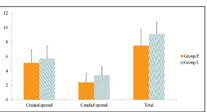
by contrast medium spread were 7.4 ± 2.2 for the group P and 9.2 ± 1.8 for the group L, and this spread was in both cranial and caudad directions. The total number and number of vertebral segments covered in the cau- dad direction was higher in patients in group L com- pared to group P and this was statistically significant (P < 0.001) (Table 2 and Fig. 5). For both groups P and L, the spread of contrast medium in the cranial direc- tion was more extensive than the caudad direction and this was statistically significant (P < 0.001). The cranial direction was compared between the P and L groups, and there was no significant difference (P = 0.059, Table 2 and Fig. 5). All patients in both groups demon- strated bilateral contrast medium distribution evenly.
The mean value of the uppermost and lowermost level of the thoracic vertebra were identified. Third thoracic vertebra was the uppermost level of both groups and the tenth and eleventh thoracic vertebra were the lowermost level of the P and L groups, respectively. The distribution of contrast medium spread into the cervical level was found in 2 patients in group P and 4 patients in group L.
Fig. 4. Consolidated Standards of Reporting Trials (CONSORT) diagram. L group: patients assessed with epidurography by the posture of lateral decubitus and neck and hip flexion; P group: patients assessed with epidurography by the prone postion.
Table 2. The mean vertebral segment of contrast medium spread.
Group P Group L Number of vertebral segments covered with
cranial spread 4.9 ± 1.7 5.7 ± 1.9
Number of vertebral segments covered with
caudad spread 2.3 ± 1.2 3.4 ± 1.0*
Total number of vertebral segments covered
with spread 7.4 ± 2.2 9.2 ± 1.8*
Values are mean ± SD. The spread of contrast medium to the cranial direction was more extensive than the caudad direction in both groups P and L. Total number and number of vertebral segments covered in the caudad direction was higher in patients with group L compared to group P and this was statistically significant. * P < 0.001
discussion
We designed this study under the estimation that the distribution of contrast medium during epidurog- raphy would coincide with the sensory blockade after injection of LA. Yokoyama et al (12) demonstrated that the distribution of contrast medium had an obvious correlation with the extent of sensory analgesia after injection of LA. In addition, Kim et al (14) reported that the viscosity of the contrast medium had minimal effect on its epidural spread.
Fig. 5. Histogram showing the number of vertebral segments covered with cranial, caudal, and total spread of contrast medium between groups P and L.
We hypothesized that a different patient position would result in a different level of distribution of con- trast medium during thoracic epidurography. Our study demonstrated that patients in group L received a more extensive distribution of contrast medium compared to group P. We think that the reduced epidural pressure due to the posture of neck and hip flexion in patients of group L may contribute to a more extensive distribution of contrast medium compared to group P. According to the study by Takahashi et al (15) epidural pressure dem- onstrated posture dependency in patients with lumbar spinal stenosis. They measured epidural pressure using a catheter transducer inserted into the lumbar epidural space and extension resulted in increased epidural pressure while flexion resulted in decreased epidural pressure. In addition, previous studies suggested that the dural sac cross-sectional area within the spinal canal demonstrates a dynamic effect, which varies according to the posture (16,17).
For both the P and L groups, the spread of contrast medium in the cranial direction was more extensive than in the caudad direction. The pressure gradient between the mid-thoracic and low-thoracic epidural space might explain a more extensive cranial spread (18). We found similar results in epidurography studies performed at the cervical and thoracic levels (13,19,20).
Our study also demonstrated that group L resulted in a more extensive distribution in the cranial direction compared to group P, although this did not show any statistical significance. Also, the number of patients who showed the spread of contrast medium up to the cervical level was 4 in group L and 2 in group P. Given the possibility of unwanted effects, extensive spread of contrast medium in the cranial direction or even in the cervical level is essential to consider. Cervical epidural anesthesia with 2% lidocaine could result in respiratory complications by weakening of the muscles of respira- tion (21). In addition, a greater cephalad spread of sen- sory analgesia above the T6 dermatome after injection of LA was inversely correlated with postoperative an- algesic consumption in the first 24 hours after uterine fibroid artery embolization (22).
Similar to the study by Lee et al (11) who reported extensive cranial spread to the cervical level with neck flexion, patients of group L were place in a posture of neck flexion in this study. We suppose that the neck flexion might be the main cause of extensive cranial spread. The distance between the spinal cord and the posterior arch of the cervical canal was widened by up to 89% during flexion, and narrowed by up to 17% during extension (23). Because the epidural space is included in this distance where the medication is injected during a TEA, this suggests that the spread of contrast medium in the cranial direction can be influenced by neck flexion.
Therefore, the dynamic variability of distances between the spinal cord and the posterior arch of the cervical spine can explain this cranial spread. Considering that previous studies demonstrated posture dependency and a dynamic nature for the lumbar or cervical spinal canal (15-17,23), we can expect that dynamic variability may also occur in the thoracic spinal canal. Although we did not measure the actual epidural pressure according to different postures, we suppose that subtle differences in the epidural pressure exist between the prone and the lateral decubitus positions. Gil et al (24) demonstrated that thoracic epidural pressures were lower in the sit- ting position than in the lateral decubitus position. In addition, enhanced thoracic curvature by the posture in group L might result in changes in the distance between the spinal cord and the posterior arch of the thoracic canal, similar to the results in the cervical spine demon- strated by Muhle et al (23).
We think that the posture of hip flexion of group L would be the main factor for increasing the caudal level. We suppose that subtle differences in epidural pressure in the posture of hip flexion compared to a neutral position and the dynamic variability which was observed in the cervical spine might also occur in the lower thoracic spine.
During the practice of lumbar epidural anesthesia, we might experience unilateral sensory anesthesia of one leg while having another leg with insufficient sensory anesthesia, although this is not frequent. We can suspect unilateral distribution of LA in such cases.
In this study, both groups showed bilateral and even distribution of contrast medium regardless of the dif- ferent postures.
This study includes several limitations. First, the study was performed under the supposition of dis- tribution of contrast medium correlating with the distribution of LA. It should be mentioned that out- comes grounded in the use of contrast medium may not always correspond to epidural distribution of LA.
However, to overcome this limitation, we proceeded with an additional study to verify the discrepancy be- tween contrast medium and LA. We could conclude that this difference between contrast medium and LA was within one dermatome. Second, we used a small volume of contrast medium to assure that the contrast was in the epidural space, not in the intravascular or intrathecal space, before injection of 3 mL of contrast medium. However, this study included only the main volume (3 mL) of contrast medium. Third, although we made every effort to make the posture of group L ac- cording to Fig. 1, we think that slight differences would be present in the flexion angles of the neck and hip among the patients of group L. Finally, more research is required to verify the actual differences in thoracic epi- dural pressure between different postures and gravity.
conclusion
In conclusion, group L demonstrated a more exten- sive distribution of contrast medium both in the cranial and caudad directions compared to group P. Therefore, we suggest that LA or other medication needs to be delivered in the neutral position to minimize an un- wanted effect.
RefeRences
1. Raveglia F, Rizzi A, Leporati A, Di Mau- ro P, Cioffi U, Baisi A. Analgesia in pa- tients undergoing thoracotomy: Epi- dural versus paravertebral technique. A randomized, double-blind, prospective study. J Thorac Cardiovasc Surg 2014;
147:469-473.
2. Park SK, Choi YS, Choi SW, Song SW. A comparison of three methods for post- operative pain control in patients un- dergoing arthroscopic shoulder surgery.
Korean J Pain 2015; 28:45-51.
3. Moslemi F, Rasooli S, Baybordi A, Gol- zari SE. A comparison of patient con- trolled epidural analgesia with intrave- nous patient controlled analgesia for postoperative pain management after major gynecologic oncologic surgeries:
A randomized controlled clinical trial.
Anesth Pain Med 2015; 5:e29540.
4. Hopkins KG, Hoffman LA, Dabbs Ade V, Ferson PF, King L, Dudjak LA, Zullo TG, Rosenzweig MQ. Postthoracotomy pain syndrome following surgery for lung cancer: Symptoms and impact on quality of life. J Adv Pract Oncol 2015;
6:121-132.
5. Biswas S, Verma R, Bhatia VK, Chaud- hary AK, Chandra G, Prakash R. Com- parison between thoracic epidural block and thoracic paravertebral block for post thoracotomy pain relief. J Clin Diagn Res 2016; 10:uc08-uc12.
6. Sengupta S. Post-operative pulmonary complications after thoracotomy. Indian J Anaesth 2015; 59:618-626.
7. Dentali F, Malato A, Ageno W, Impera- tori A, Cajozzo M, Rotolo N, Douketis J, Siragusa S, Crowther M. Incidence of venous thromboembolism in pa- tients undergoing thoracotomy for lung cancer. J Thorac Cardiovasc Surg 2008;
135:705-706.
8. Khelemsky Y, Noto CJ. Preventing post- thoracotomy pain syndrome. Mt Sinai J Med 2012; 79:133-139.
9. Reddi D, Curran N. Chronic pain after
surgery: Pathophysiology, risk factors and prevention. Postgrad Med J 2014;
90:222-227; quiz 226.
10. Visser WA, Lee RA, Gielen MJ. Factors af- fecting the distribution of neural block- ade by local anesthetics in epidural anes- thesia and a comparison of lumbar ver- sus thoracic epidural anesthesia. Anesth Analg 2008; 107:708-721.
11. Lee CJ, Jeon Y, Lim YJ, Bahk JH, Kim YC, Lee SC, Kim CS. The influence of neck flexion and extension on the distribu- tion of contrast medium in the high tho- racic epidural space. Anesth Analg 2007;
104:1583-1586; table of contents.
12. Yokoyama M, Hanazaki M, Fujii H, Mizobuchi S, Nakatsuka H, Takahashi T, Matsumi M, Takeuchi M, Morita K. Cor- relation between the distribution of con- trast medium and the extent of blockade during epidural anesthesia. Anesthesiol- ogy 2004; 100:1504-1510.
13. Hong JH, Oh JH, Park KB. Analysis of thoracic epidurography and correlating factors affecting the extent of contrast medium spread. Korean J Pain 2016;
29:255-261.
14. Kim YC, Lim YJ, Lee SC. Spreading pat- tern of epidurally administered contrast medium in rabbits. Acta Anaesthesiol Scand 1998; 42:1092-1095.
15. Takahashi K, Miyazaki T, Takino T, Matsui T, Tomita K. Epidural pressure measure- ments. Relationship between epidural pressure and posture in patients with lumbar spinal stenosis. Spine (Phila Pa 1976) 1995; 20:650-653.
16. Willen J, Danielson B, Gaulitz A, Nikla- son T, Schonstrom N, Hansson T. Dy- namic effects on the lumbar spinal canal:
Axially loaded CT-myelography and MRI in patients with sciatica and/or neuro- genic claudication. Spine (Phila Pa 1976) 1997; 22:2968-2976.
17. Hirasawa Y, Bashir WA, Smith FW, Mag- nusson ML, Pope MH, Takahashi K.
Postural changes of the dural sac in the
lumbar spines of asymptomatic individ- uals using positional stand-up magnetic resonance imaging. Spine (Phila Pa 1976) 2007; 32:E136-E140.
18. Visser WA, Gielen MJ, Giele JL, Scheffer GJ. A comparison of epidural pressures and incidence of true subatmospheric epidural pressure between the mid-tho- racic and low-thoracic epidural space.
Anesth Analg 2006; 103:1318-1321.
19. Hong J, Jung SW. Fluoroscopically guid- ed thoracic interlaminar epidural in- jection: A comparative epidurography study using 2.5 mL and 5 mL of contrast dye. Pain Physician 2016; 19:E1013-E1018.
20. Kim KS, Shin SS, Kim TS, Jeong CY, Yoon MH, Choi JI. Fluoroscopically guided cervical interlaminar epidural injections using the midline approach: An analy- sis of epidurography contrast patterns.
Anesth Analg 2009; 108:1658-1661.
21. Huang CH. Effect of cervical epidural blockade with 2% lidocaine plus epi- nephrine on respiratory function. Acta Anaesthesiol Taiwan 2007; 45:217-222.
22. Nader A, Kendall MC, Chrisman H, De Oliveira GS, Jr., Tureanu LM, McCarthy RJ. Greater cephalad extent of thoracic epidural sensory anesthesia after lido- caine and epinephrine test dose corre- lates with analgesic consumption and pain burden after uterine fibroid artery embolization. Reg Anesth Pain Med 2016;
41:56-64.
23. Muhle C, Wiskirchen J, Weinert D, Fal- liner A, Wesner F, Brinkmann G, Heller M. Biomechanical aspects of the sub- arachnoid space and cervical cord in healthy individuals examined with ki- nematic magnetic resonance imaging.
Spine (Phila Pa 1976) 1998; 23:556-567.
24. Gil NS, Lee JH, Yoon SZ, Jeon Y, Lim YJ, Bahk JH. Comparison of thoracic epidural pressure in the sitting and lat- eral decubitus positions. Anesthesiology 2008; 109:67-71.