© The Korean Society of Mycology Kor. J. Mycol. 36(2) : 123-129 (2008)
123
국내 해송에서 분리한 변색균 Ophiostoma quercus 의 균학적 특성
현민우·서동연·윤여홍·김성환*
단국대학교 미생물학과 및 기초과학연구소
Mycological Characteristics of Ophiostoma quercus , a Sap-staining Fungus Isolated from Japanese Black Pine in Korea
Min Woo Hyun, Dong Yeon Suh, Yeo Hong Yun and Seong Hwan Kim *
Department of Microbiology and Institue of Basic Sciences, Dankook University, Cheonan, Chungnam 330-714, Korea (Received November 26, 2008. Accepted December 24, 2008)
ABSTRACT: One of the sapstain fungal species isolated from the stained sapwood of a Japanese black pine in Korea was characterized. The fungal species is tolerant to cycloheximide and has Petosum -like synnema and Sporo- thrix type synanamorph which are found in the anamorphs of the Ophiostoma piceae complex group. But the spe- cies could not form perithecia on MEA. Based on cultural and morphological properties and analysis of the
β- tubulin gene sequence, the fungal species was identified as Ophiostoma quercus . Here, we report mycological char- acteristics of the anamorphic stage of Ophiostoma quercus isolated in Korea.
KEYWORDS : Japanese Black Pine, Ophiostoma piceae complex, O . quercus ,
β-tubulin gene
목재변색균과수목병원균을포함하는
Ophiostoma
균류 는 분류 지표로 볼 때 자낭균류문(Ascomycota),
각균강(Pyrenomycete),
장경자낭각균목(Ophiostomatales),
오피오스토마과
(Ophiostomataceae),
오피오스토마속( Ophios- toma )
에 속한다. Ophiostoma
속은 장경자낭각(perithecia;
long necked fruiting body)
과 분생포자경속(Synnema)
의형태적 특징이 있는 균류이다
.
이들 중O . piceae
complex
그룹에 속하는 종들은 그 형태가 매우 유사하여구분이 쉽지 않은 균류중의 하나이다
.
이들 간의 형태적구분은 분생포자경속
(synnema)
을 갖는Pesotum
과 균사 형의Sporothrix ,
분생자분지형(macronematous)
을 갖는Leptographium
등다양한무성세대의형태(anamorph)
와더불어 자낭포자
(ascospore)
의 모양,
크기,
그리고자낭각의색
,
길이와 넓이,
머릿구멍 균사(ostiolar hyphae)
의 유무등 유성세대의 형태
(telemorph)
에 의해 이루어진다(Hunt 1956; Upadhyay 1981; Wingfield et al ., 1993). O . piceae complex
그룹에속하는종은현재약13
종이알려져 있는데 이들은 형태적으로 구분이 매우 어려워
ITS rDNA
와 β-tubulin gene
등을 분석함으로서 구분하고있다
.
이들균류는전세계적으로널리분포하며나무의백목 질에서푸른색또는어두운변색을야기하기때문에목재 변색균 또는 청변균으로 알려져 있다
.
변색균은 목재의상업적가치를 떨어뜨리기때문에 임목산업에문제가되 는중요한균류이다
(De Beer et al ., 2003; Chung et al ., 2006).
목재변색균에의한푸른얼룩(blue stain)
은감염된백목질의도관 수지
(resin ducts)
와방사형유조직세포로집중된 곰팡이 균사에의해 야기된다
.
변색은변색균 균사의 세포벽에 존재하는 멜라닌과 변색균이분비하는 물 질과 목재에 존재하는 물질의 반응에 의해 일어난다
.
변색균은 목재에 침입 후 가도관 조직에집락을 형성한다
.
목재변색균은가도관을따라수직으로자라며유조직에서 변색을일으킨다
.
세포벽에직접적인침투와인접한구멍을통해점진적으로변색균이퍼져가면서이들유조직사 이에서 변색이 번지게된다
.
변색의 부위가 넓혀지면 종국에 목재의 질을 떨어뜨리게된다
(Braiser, 1979, 1991;
Braiser and Mehortra, 1995; Harrington, 1993; Lagerberg et al ., 1927).
이들
O. piceae complex
그룹균류중일부는활엽수에서도종종도관마름병을일으킨다
(Seifert, 1993).
이들균류를 전파하는 매개충으로는 나무좀과 딱정벌레
(bark beetles)
가 알려져있다(Wood and Bright, 1992).
도관마름병을 일으키는균류는 곤충을매개로도관에 침입하여 나무의 수분이동을방해 한다
.
그 중 가장 잘알려진 종으로는
O . ulmi
와O . novo - ulmi , O . himal - ulmi
가 있다(Braiser, 1979, 1990).
이들 종은느릅나무 마름병(Dutch elm disease)
의전염인자로북유럽과북미,
아시아일부에서발생하여 큰피해를 주었다
.
이는O. piceae complex
*Corresponding author <E-mail : [email protected]>
우리나라에서도소나무류목재에서변색을일으키는균 류에 대한 발생 보고가 있고
,
이를 방제하기 위하여O . quercus
백색변이균주(albino strain)
을 이용한 생물학적방제에대한 연구도이루어지고있다
(Lee and Oh, 2000;
Cho et al ., 2008).
그러나아직까지이들국내에서분리된Ophiostoma
균류의균학적인특성에대한보고가전무하다
.
본연구에서는2005
과2006
년사이에우리나라해송으로부터 분리한변색균의 하나인
O . quercus
균류에대하여 배양적
,
형태적 특성과 함께 유전자분석을 포함한불완전세대의균학적특성을보고하고자한다
. 재료 및 방법
균주의 분리 및 배양
균주의분리를위해서
2005
년과2006
년8
월사이에태안의 벌목된해송
( Pinus thunbergii )
의줄기로부터변색된부분을디스크형태로잘라비닐봉지에넣어채집한후실
험실로 가져와 살균된 정으로 잘게 칩
(chip)
으로 부수었다
.
마련된 칩은3
차 증류수에서1
분간 씻어준 후 다시70% ethanol
에서1
분, 90% ethanol
에서1
분간 처리 한후 상온에서 표면을 건조하였다
.
표면 살균된 칩은 단백질합성을저해하는항생제
cycloheximide(A.G. Scientific)
를
200 mg/ l
농도로 포함시킨2% malt extract agar(BD Sciences, USA)
가담긴페트리디쉬에놓인후25
oC
배양기에서
1
주일간배양되었다.
배양된칩에서뻗어나온균사의 끝을 취하여 새로운
MEA
배지에 옮김으로서분리균주를얻었다
.
분리균주를10
일이상배양후형성된포자를 살균수에현탁하여포자현탁액을 조제하였다
.
조제된 포자 현탁액을 희석하여
MEA
에다시 도말 접종하고배양
2
일후피어난균총의 균사를다시 새배지에접종함으로서단일포자균주를얻었다
(Braiser, 1981).
배양적 특성 조사 및
cycloheximide
저항성조사분리된 변색균의 배양적 특성을 알아보기 위해
cyclo-
heximide(200 mg/ l )
가 포함된2% MEA
배지에서 전 배양 된 균을
2% MEA(BD Sciences, USA), 2% OMEA (Oxoid, England), PDA(BD Sciences, USA)
배지에각각접종하여
20
oC
에서7
일간배양 후집락의형태와균사생장을 관찰 비교하였다
.
분리된 변색균의 성장을 위한 적2% Oxoid MEA
에접종하여25 C
에서7~8
일간배양하면서그형태를관찰하였다
.
형태관찰은위상차현미경(Karl Zeiss, Axioskop 40, Germany)
과 해부현미경(Olympus
SZ51, Japan)
을이용하여 관찰 대상 균류가형성한 분생포자경속
,
분생포자,
분생포자경속 상에 분생포자덩리의모양과색을관찰하고이들의크기를측정하였다
.
그리고주사전자현미경
(SEM, Hitachi 4300, Japan)
을 이용하여분생포자경속의발달과무성세대
,
분생포자의미세구조를관찰하였다
.
분자생물학적 방법을 이용한 변색균의 동정
Genomic DNA
의추출. DNA
추출은2% MEA
에균을정종하여
25
oC
암상태에서7
일동안 배양후형성된균주의 포자
100
µl
를 취하여멸균된3
차 증류수에 부유시킨다음멸균된셀로판
(BioRad, USA)
을깔은2% MEA (Difco, USA)
배지위에 도말하여25
oC
에서3
일 동안 배양하였다
.
배양 후 균사0.3 g
을 멸균된2 ml cryogenic vial
튜브에옮겼담은후TES buffer(50 mM Tris-HCl[pH 8.5], 50 mM EDTA, 3% sodium dodecyl sulfate) 200
µl
를 첨가 하여 균사를부유하였다
.
부유된균사는 드릴을이용하여 얼음위에서
2
분동안 분쇄하고2
분간대기 하였다가 다시
2
분간 분쇄 하였다(Kim et al ., 1999).
그리고 분쇄된균사에
3 mM sodium acetate(pH 5.2) 150
µl
를 첨가하고
,
−20
oC
에서10
분간 보관 후 냉장원심분리기(Sigma, Germany)
에서13,000
×g
로10
분간원심분리한다음상층액을취하여
1.5 ml
튜브에옮긴후동량의phenol : chlorform : isoamylalchol(25 : 24 : 1; Amresco, USA)
를첨가하여잘섞고다시냉장원심분리기로
5
분간원심분리하였다
.
원심분리된상층액을 취하여 다시 새튜브로 옮기고 동량의
isopropanol(Sigma, USA)
을 첨가하여 상온에서
5
분동안처리하였다.
처리된튜브를다시 원심분리하여
DNA
를 침전시켰다.
침전된DNA
는70% ethanol 750
µl
을 첨가하여 표면을 살짝 씻어준후4
oC
냉장원심분리기에서
13000
×g
로5
분간 원심분리한 다음ethanol
을 버리고나서 상온에서 건조시켰다
.
건조된 침전DNA
에
50
µl TE buffer(10 mM Tris[pH 8.0], 1 mM EDTA)
를첨가하여
DNA
를용해한후UV-VIS spectrophotometer (BIOmate)
를 이용하여DNA
양을 정량하였고PCR
분석에사용하였다
.
PCR
을 이용한 β-tubulin
유전자의 증폭과 염기서열분석
PCR
반응은Gene Amp-950 cycler(ABI, USA)
를이용하여수행하였다
. PCR
반응을위한primer
는Ophiostomatoid
곰팡이와 자낭균류에 특이적으로 부착하는
T10 forward primer(5'-ACGATAGGTTCACCTCCAGAC-3')
와BT12 reverse primer(5'-GTTGTCAATGCAGAAGGTCTC-3')
을사용하였다
(O’Donnel et al. , 1997; Schroeder et al ., 2001). PCR
반응물의조성은10
×reaction buffer 2.5
µl , 10 mM dNTPs 0.5
µl , 5
×Band doctor 2.5
µl , 20 pmol primer
각1
µl , template DNA 1
µl (fungal genomic DNA 100 ng/
µl ), 3
차 멸균 증류수17
µl , 2.5 U EF-Taq polymerase 0.5
µl (Solgent, Korea)
를첨가하여총25
µl
의 반응물을 만든 후 반응하였다.
반응 조건은 초기 변성(initial denaturation)
은94
oC
에서5
분실시하였고,
변성은(denaturation) 94
oC
에서1
분,
가열냉각(annealing)
은56
oC
에서
40
초,
증폭(extension)
은72
oC
에서1
분30
초로30
회반복하였다
.
그리고 마지막으로72
oC
에서5
분동안 증폭한 후
4
oC
에서 보관하였다.
증폭된PCR
반응산물은1%
agarose gel
상에서전기영동으로확인하였고Gel extraction kit
을 이용하여PCR
반응산물을 정제하였다(Solgent, Daejeon, Korea).
정제된 산물은pCR2.1-TOPO vector (Invitrogen, USA)
에cloning
한 후Macrogen
사(Seoul,
Korea)
에 염기서열 분석을 의뢰하였다.
염기서열 분석은ABI PRISM 3700 DNA Sequencer(ABI, USA)
을이용하고
BigDyeTM terminator
방법을 사용하여 분석되었다.
결정된 염기서열은
NCBI GenBank database(http://
blast.ncbi.nlm.nih.gov/Blast.cgi)
에 등록된 알려진 균류의염기서열과상동성을비교하였다
. 결과 및 고찰
본연구에서분리된
3
개변색균주(DKM0511, DKM0512, DKM0513)
에대한배양적특성,
형태적특성,
유전자염기서열 구조 특성을조사한결과 서로 동일한양상을나
타내어 이중의 한 균주인
DKM0511
을 대표균주로 하여그조사된특성을아래와같이제시하였다
.
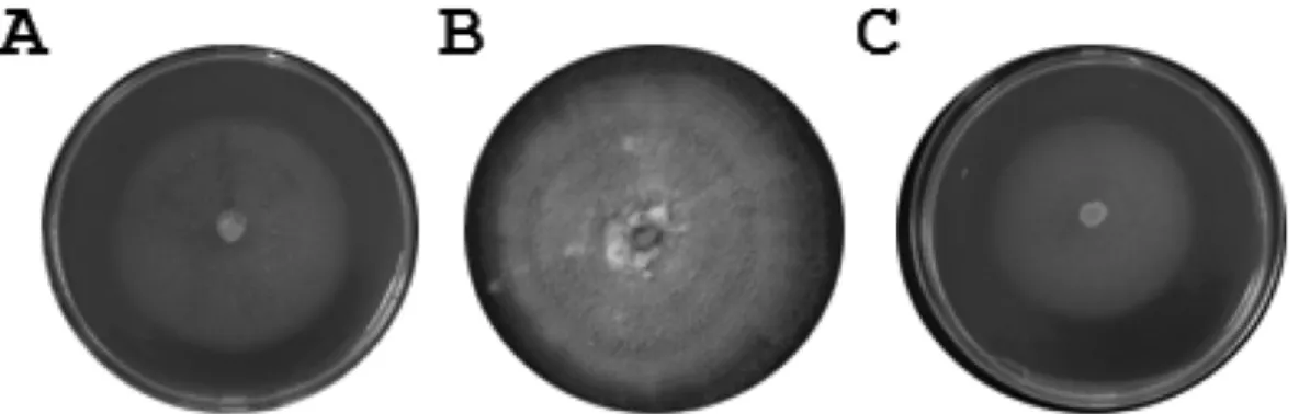
배양특성 및
cycloheximide
저항성20
oC
에서7
일간 배양 후2% MEA, 2% OMEA, PDA
배지상에서나타나는균총의형태학적특징을관찰한분
리균주는
2% MEA
상에서는 균사 색이 투명하였고 분생포자경속을 많이 만들었다
(Fig. 1A).
이에 반해2%
OMEA
배지상에서는고리모양의균총을만들며성장하는것을관찰 할수있었다
.
그러나분생포자경속의형성은거의보이지않았다
(Fig. 1B). PDA
배지 상에서의균총 역시 분생포자경속 형성이 관찰되지 않았다
.
하지만특이하게 하얀색의 솜털 같은 균사를 많이 만드는 것을
관찰 할수있었다
(Fig. 1C).
전반적으로배지 별생장에있어서는
2% OMEA
와PDA
보다2% MEA
에서균사가활발하게생장하는 것을확인할수있었다
.
짙은갈색을띠는 분생포자경속
(synnema)
을 형성함에따라Ophiosto-
matoid
에 속하는 균류로 추정되었다.
생장을 위한 적정온도는
7
일간 각각 다른 온도에서 배양시키면서 균사의생장길이를측정하였다
.
각온도에서의생장을보면10
oC
에서는
5 mm 15
oC
에서는25 mm, 20
oC
에서는30 mm, Fig. 1. Colony features of the isolate DKM 0511 grown for 7 days at 20
oC on 2% MEA (A), 2% OMEA (B), and PDA (C).
Fig. 2. Mycelial growth of the isolate DKM 0511 grown for 7
days on 2% MEA at different temperatures.
Fig. 3. Anamorphic stages of Ophiostoma quercus shown by the isolate DKM 0511. A, mononematous synnema after lactophenol
blue staining (scale bar = 50
µm); B, white conidial mass on individual synnema (scale bar = 50
µm); C, Pesotum- like
synnema (scale bar = 50
µm); D-E, arrows indicate apex of conidiogeneous cells with denticles (scale bar = 20
µm and
5
µm); F-H, conidiophores and conidia of Sporothrix synanamorph (scale bar = 20
µm, 10
µm, and 10
µm); I, arrows
indicate morphogenesis of Sporothrix anamorph in the middle of conidiophores (scale bar = 5
µm); J, arrows indicate
sympodial and annellidic conidiogeneous cells with synnematous conidiophores (scale bar = 10
µm); K~L, Petosum- like
synnema and conidia (scale bar = 100
µm and 10
µm).
Kirk(2001)
등이 기술한현미경관찰법과Seifert(1993)
와Okada(1998)
그리고Jacobs
와Wingfield(2001)
가 기술한Ophiostoma
의 특징과 무성세대의 분류 문헌을 참고하여관찰하였다
.
그 결과 본 연구에서 분리된 균류로 부터Ophiostoma
균류의 무성세대로 알려진Pesotum
의 형태학적 특징인 단생분생포자경
(mononematous synnema)
과어두운갈색의분화분생포자경
(macronematous synnema)
를 관찰하였으며
(Fig. 3A, C),
해부현미경을 통해 하얀색을 띄는 둥근 점액질덩어리모양을가진 분생포자 덩어 리를확인하였다
(Fig. 3B).
그리고Ophiostoma
균류의또다른 무성세대 형태인
Sporothrix
의 모습도 관찰하였다.
무성세대이명
(synanamorph) Sporothrix
의특징인분생포 자경(conidiophore)
의상단끝에분생포자와작은denticle-
like
구조를형성하는것을주사전자현미경관찰을통하여확인하였다
(Fig. 3E~H).
특이하게 균사의 중간 부분에서무성세대
Sporothrix
의형태를형성하는것도관찰되었다(Fig. 3I).
형성된 포자의 모양은 원통형(Cylidrical)
과 도란형
(Obovoid)
모양이존재하였다(Fig. 3G, L).
무성세대Pesotum
이갖는분생자병속의 미세적구조와분생포자경속을 이루는각 각의 분생포자경에서분생포자의형태형 성을관찰한결과분생포자경에서분생포자의형태형성이 가축
(sympodial)
의 환형(annellidic)
모습으로 이루어지는 것을 확인하였다
(Fig. 3J, K).
하지만 완전세대 형태(Telemorph)
의특징인자낭각(perithecium)
형성은관찰하지못하였다
.
이상의관찰결과를토대로할때분리된균은전반적으로
O. piceae complex
균류의무성세대 특징을지니고있음을알수있었다
.
O. piceae complex
균류에속하는종으로는13
종정도가 알려져있으며이들에대한 몇 가지분류학적 지표를
줄 수 있는 유전자 정보가
NCBI GenBank
에 등록되어Fig. 4. Phylogenetic relationships of the fungal isolate DKM 0511 to other Ophiostoma species. Phylogram based on the analysis
of partial nucleotide sequence of the
β-tubulin gene was generated from the neighbor-joining analysis. Numbers at nodes
represent percentage of bootstrap resampling based on 1000 replicates. Ceratocystis coerulescens was used as an outgroup.
있는바 본연구에서 분리된균의 분자적동정을 또한시 도하였다
.
이에 따라O. piceae complex
균류의 분류에가장 적절하게 활용되는 유전자인 β
-tubulin
유전자 염기서열분석을시도하였다
. DKM0511, DKM0512, DKM0513
등
3
개균주들의genomic DNA
에대하여PCR
을수행하여 증폭된
DNA
밴드를 클로닝하여 염기서열을 분석한결과
3
개균주모두842 bp
의크기의서로동일한염기서열 정보를 가지고 있었다
.
이에 따라DKM0511
균주로부터 얻어진염기서열정보를
GenBank DNA database
상에서
BlastN
프로그램을이용하여 상동성을 지닌 균류를탐색한 결과
O. piceae complex
에 속하는13
종의 균류중에서
Ophiostoma quercus
의β-tubulin
유전자염기서열들
(Accession number: AY789156, AY789157)
과99~
100%
상동성을보였다.
이에따라본실험에서DKM0511, DKM0512, DKM0513
균주로부터얻어진β-tubulin
유전자의 염기서열을
NCBI GenBank
에 등록하였다(DKM0511:
Accession number FJ573040, DKM0512: Accession number FJ573041, DKM0513: Accession number FJ573042).
이결과를 토대로
PAUP* 4.0
프로그램(Swofford, 2003)
을이용하여 다른
Ophiostoma
종들의 β-tubulin
유전자염기서열과 비교를 통해 계통분석을 실시하였다
.
그 결과 분리주
DKM0511
는O. quercus
와 함께 계통유전적 관계 를이루는것으로나타났다(Fig. 4).
O. quercus
는O. piceae complex
에 속하는 종으로O.
piceae
와는 형태적으로 구분이 매우 어려운바Braiser
와Kirk(1993)
에 의해sibling species
로 분류되었고Braiser
와
Stephens(1993)
에 의해 온도에따른 생장비교 연구가이루어졌으며
Harrington(2001)
에의하여그형태적특성이자세히비교정리된바있다
.
이에따라본연구에서분리된 균의 형태적특성을
Harrington(2001)
이 정리한O.
piceae
와O. quercus
의 형태특성과 비교하여Table 1
에제시하였다
.
본연구에서분리된변색균의조사된형태적특성은보고된
O. quercus
의무성세대에나타나는형태적특성과거의일치하였다
.
이상의 본 연구에서 분리된 균류에 대한 형태적 특성
,
배양특성
,
유전자분석 결과를 바탕으로 분리균류는O.
quercus
의 무성세대인 것으로 동정되었다. O. quercus
는 주로 활엽수에서 분리되는 변색균류로 알려졌지만 일부 침엽수에서도 분리되고 있다(Schirp et al ., 1999).
본 연구에서 사용된
O. quercus
도 우리나라 해송(Japanease
black pine)
에서 분리되었고이전에 국내의잣나무에서도분리된 보고
(Lee and Oh, 2000)
가 있는바,
국내의 서로다른 침염수종에 대한 이 변색균의 분포가 다시금 확인 되었다
.
그러나아직 국내 활엽수에 존재하는 보고는 없어이에대한 조사가확대되어야할것이다
.
더불어국내분리
O. quercus
의완전세대가지니는 균학적특성이아직밝혀지지않았으므로유전적교배를통하여자낭을형 성할수있는 다른극성을지닌 교배형
(mating type)
균주의발굴작업도병행되어야할것이다
.
적 요
한국에서 해송의 줄기변색 부위에서 분리된 변색균류 중한종에대하여그특성을조사하였다
. Cycloheximide
에저항성인
Ophiostoma piceae complex
그룹에서 보여지는 무성세대
Petosum
의분생포자경과또다른무성세대Sporothrix
의형태를지니고있었다.
그러나완전세대의모습인자낭각을형성하지못하였다
.
이균류는배양적특성,
형태적특성그리고 β
-tubulin
유전자염기서열 분석을통하여
Ophiostoma quercus
로 동정되었다.
본 연구에서는한국에서 분리된
O . quercus
의 균학적 특성을 처음으로보고하고자한다
.
감사의 글
이논문은
2005
년정부(
교육인적자원부)
의재원으로한국학술진흥재단의지원을받아 수행된연구임
(R05-2004- 000-12579-0).
Sexual behavior heterothallic heterothallic heterothallic
Synnema conidial mass White White White
Host Various Oak and hardwood Japanese black pine
A,B