Hesperetin Ameliorates Inflammatory Responses in Lipopolysaccharide-stimulated RAW 264.7 Cells via p38 MAPK and ERK1/2
Seung-Hoon Lee
1, Eun-Joo Lee
1, Chungwook Chung
1, Ho-Yong Sohn
2and Jong-Sik Kim
1*
1Department of Biological Sciences, Andong National University, Andong 36729, Korea
2Department of Food and Nutrition, Andong National University, Andong 36729, Korea Received December 7, 2018 /Revised January 4, 2019 /Accepted January 4, 2019
In a previous study, we isolated 11 different kinds of compounds from ethyl acetate fractions of lees (jubak) which is a by-product of Korean traditional wine production. These compounds were identi- fied as caffeic acid, coumaric acid, D-mannitol, ferulic acid, hesperetin, hesperidin, naringenin, nar- ingin, sinapic acid, syringic acid, and vanilic acid. To evaluate their anti-inflammatory activities in an
in vitro model, nitric oxide (NO) production was measured in lipopolysaccharide (LPS)-stimulatedRAW 264.7 cells after the treatment of these cells with each compound. Among the various chemicals, hesperetin and naringenin showed the highest inhibition of NO production in the LPS-activated RAW 264.7 cells. Hesperetin was chosen for further study because of its strong anti-inflammatory activity and because the mechanisms underlying its anti-inflammatory properties still remain unclear. Our re- sults showed that hesperetin dramatically inhibited NO production in a dose-dependent manner as compared with in an LPS-only treated group, without affecting cell viability. In addition, hesperetin reduced the protein expression of the pro-inflammatory gene inducible nitric oxide synthase (iNOS) in a dose-dependent manner, whereas it did not affect cyclooxygenase-2 (COX-2) expression. Further- more, hesperetin inhibited phosphorylation of p38 mitogen- activated protein kinase (MAPK) and ex- tracellular signal regulated kinase (ERK) 1/2, whereas it did not affect phosphorylation of c-jun N- termi- nal kinase (JNK). The results indicated that hesperetin regulated the LPS-induced inflammatory re- sponse by suppressing p38 MAPK and ERK1/2 signaling. Overall, our results may help to understand the mechanisms underlying the anti-inflammatory activity mediated by hesperetin.
Key words : ERK1/2, hesperetin, inflammation, iNOS, p38 MAPK
*Corresponding author
*Tel : +82-54-820-5798, Fax : +82-54-820-7705
*E-mail : [email protected]
This is an Open-Access article distributed under the terms of the Creative Commons Attribution Non-Commercial License (http://creativecommons.org/licenses/by-nc/3.0) which permits unrestricted non-commercial use, distribution, and reproduction in any medium, provided the original work is properly cited.
Journal of Life Science 2019 Vol. 29. No. 1. 129~134 DOI : https://doi.org/10.5352/JLS.2019.29.1.129
서 론
Hesperetin은 감귤 등의 Citrus 속 식물의 과피에 hesper- etin 7-rutinoside 인 hesperidin 배당체 형태로 1.5~3.0% 함유 되어 있으며 현재까지 약 4,000 여종 이상 밝혀진 flavonoid의 한 계열인 flavanone으로 분류되고 있다. 천연물을 이용한 생 리활성 연구가 진행됨에 따라 이들 flavonoid에 의한 항암[24], 항산화[17], 그리고 항균 활성[9, 18]등 다양한 생리활성이 보 고된 바 있다. 특히, hesperidin은 항암[1], 항산화[11, 13] 그리 고 항염증[20]등에서 강한 활성을 나타내는 대표적인 fla- vonoid 물질로 연구되고 있으며 또한 전구체인 chalcone 계 물질 역시 mitogen-activated protein kinases (MAPKs) 및 NF-κB (nuclear factor-kappaB) 경로를 억제하고[6], 염증 매개
인자인 inducible nitric oxide synthase (iNOS)의 발현과 nitric oxide (NO)의 합성을 억제한다는 것이 보고된 바 있다[4]. 반 면, 핵심 구조체인 hesperetin에 의한 lipopolysaccharide (LPS) 염증 모델에서의 항염증 활성 연구는 미미한 수준이다.
염증이란 다양한 외부자극으로 인해 발생되는 신체 방어기 작의 하나로서, lipopolysaccharide (LPS)와 같은 세균 유래 내독소(endotoxin)는 대표적인 염증 유도 물질로 알려져 있다 [2]. LPS는 그람 음성세균의 세포벽 구성성분으로서, 대식세포 에 의한 염증반응을 유도하는 성분으로 알려져 있으며, 대표 적인 pattern recognition receptor인 toll-like receptor 4 (TLR4)와 MD-2의 복합체에 의해 인식된다. 그 결과, 세포 내 주요 신호전달 경로 중 하나인 MAPK 경로를 활성화 시키게 된다[3]. 이를 구성하는 p38 MAPK, ERK1/2 (extracellular signal-regulated kinase 1/2) 그리고 JNK (c-jun N-terminal kinase)는 전사 조절 인자인 ATF2 [7], CREB [10], STAT3 [8]
그리고 NF-κB [19] 등 다양한 경로를 통해 염증 유전자인 in-
ducible nitric oxide synthase (iNOS) 및 tumor necrosis fac-
tor-alpha (TNF-α), cyclooxygenase-2 (COX-2) 등의 발현을 조
절하는 것으로 알려져 있다. 최종적으로 iNOS와 COX-2로부
터 생성 유도된 nitric oxide (NO)와 prostaglandin E
2(PGE
2)
- Note -
는 다양한 염증 반응을 매개하는 것으로 알려져 있어 이들의 발현 여부는 염증 반응 정도를 판단하는 중요한 척도로 이용 되고 있다[12]. 또한, 2차 면역 반응을 유도하여 방어기작에 관여하는 것으로 알려져 있으나[21], 감염원과 반응기전에 따 라 다양한 염증성 질환의 원인이 되기도 한다[15]. 따라서, 이 러한 염증 반응을 억제하는 것이 염증관련 질환을 예방하는데 매우 중요하며, 최근 다양한 천연물을 이용한 염증 반응의 조 절에 대한 연구가 활발하게 진행되고 있다.
본 연구에서는 전통주 제조시 부산물로 발생되는 주박으로 부터 분리 동정된 순수물질 11종에 의한 NO 생산 저해 활성을 연구하였으며, 이 중 저해 활성이 높은 hesperetin에 의한 항염 증 기전을 연구하기 위해 NO 생산 저해 활성, pro-inflamma- tory 단백질의 발현 분석 및 MAPKs 경로와의 상관관계에 대 해 연구하였다.
재료 및 방법
11종의 순수물질 및 hesperetin
11종의 순수물질은 ㈜국순당에서 제공받은 주박에서 분리 하여 동정하였다. 즉, 실험에 사용한 각 순수물질은 ㈜국순당 에서 제공받은 주박 1.5 kg에 95% 에탄올 6 l를 가하여 상온에 서 3일간 2회 추출하였고, 여과 후 감압 농축하여 분말로 제조 하였다. 추출된 추출물 4 g을 물에 현탁 한 후, n-hexane, eth- yl-acetate 및 butanol을 이용하여 순차적으로 분획하고 최종 적으로 물 잔류물을 회수하여 HPLC 를 통해 각각 분리한 후 standard curve 비교를 통해 각 순수물질로 동정하였다. 11종 의 순수물질은 1차 선별과정에서만 사용하였고, 이후 실험에 사용한 hesperetin은 Sigma 사(St. Louis, MO, USA)에서 구입 하여 사용하였다.
마우스 대식세포 RAW 264.7 배양 및 염증반응 유도 마우스 대식세포 RAW 264.7 세포주는 American Type Culture Collection (ATCC, Fredrick, MD, USA) 에서 구입하 였다. 세포주 배양은 10% Fetal Bovine Serum (FBS, Gibco, Grand Island, NY, USA), 1% penicillin 및 streptomycin (WelGene, Korea)이 첨가된 Dulbecco’s Modified Eagle Medium (DMEM, Gibco)를 사용하였다. 염증 반응의 유도는 Gram 음성 세균의 외막 구조물인 lipopolysaccharide (LPS, Sigma, USA)를 이용하였다.
Nitric oxide (NO) 생성 분석
LPS를 처리하여 염증 유도된 마우스 대식세포 RAW 264.7 세포주에서 주박에서 분리한 11가지 순수물질과 hesperetin에 의한 NO 생산 수준을 측정 하였다. 즉, 96 well plate의 각 well에 RAW 264.7 세포주를 2×10
5개 접종한 후 19시간 동안 배양하였다. 그 후 0.2 μg/ml 농도의 LPS (Sigma)를 1시간
동안 처리하였다. 그 다음 11종의 순수물질들과 hesperetin을 농도별로 처리한 뒤 16시간 동안 배양하였다. 그 후 상등액 100 μl와 Griess reagent (Sigma) 100 μl를 혼합하여 15분간 상온에서 반응시키고, Tecan Infinite M200 plate reader (Tecan Austria GmbH, Austria)를 사용하여 540 nm에서 흡광 도를 측정하였다. NO 생성 분석은 독립적인 4개의 well에서 수행되었으며, 통계 분석은 Sigma plot을 이용하여 mean ± SD 값으로 나타내었다.
세포 생존율 분석
세포 생존율 분석은 CellTiter 96® AQueous One Solution Cell Proliferation Assay kit (Promega, Madison, WI, USA)를 이용하여 수행하였다. 즉, 96 well plate의 각 well에 RAW 264.7 세포주를 2×10
5개 접종한 후 19시간 동안 배양하였다.
그 후 0.2 μg/ml 농도의 LPS (Sigma)를 1시간 동안 처리한 후 순수물질과 hesperetin을 농도별로 처리한 뒤 16시간 동안 배양하였다. 그 후 RAW264.7 세포주에 20 μl의 MTS 용액을 처리하여 동물세포 배양기서 4시간 동안 반응시키고, Tecan Infinite M200 plate reader (Tecan Austria GmbH, Austria)를 사용하여 480 nm에서 흡광도를 측정하였다. 세포생존율 분석 은 독립적인 4개의 well에서 수행되었으며, 통계 분석은 Sigma plot을 이용하여 means ± SD 값으로 나타내었다.
Western Blot Analysis
마우스 대식세포 RAW 264.7 세포주를 60 mm dish에 접종 한 후 19시간 동안 배양하였다. 그 후 0.2 μg/ml 농도의 LPS (Sigma)를 처리하고 1시간 동안 대기한 다음 hesperetin을 주 어진 농도 별로 처리한 뒤 8시간 동안 배양하였다. MAPKs와 phospho-MAPKs 단백질의 경우 각 추출물을 serum free me- dia에 혼합하여 처리하고 4시간 후, 1.0 μg/ml 농도의 LPS를 처리하고 30분 후 세포를 수확하였다. 수확한 세포는 4℃에서 세포활동을 정지시킨 뒤 10x RIPA buffer (Cell signaling, Beverly, MA, USA)를 4x로 희석하여 처리한 후 sonication 하였다. Sonication 된 cell lysate는 15분간 3,000 rpm에서 원 심분리하고 상층액을 취하여 사용하였다. 단백질 정량은 Bradford assay (Bio-Rad, Hercules, CA, USA) 방법을 이용하 였다. 본 실험에서 사용된 항체 중 iNOS, COX-2, p38, p-p38, ERK1/2. p-ERK1/2, JNK, p-JNK에 대한 항체는 모두 Cell sig- naling사(USA)에서 구입하였으며, Actin 항체와 2차 항체는 모두 Santa Cruz 사(Santa Cruz, CA, USA)로부터 구입하였다.
통계 분석
모든 실험은 최소 3회 이상 실시하였으며, 실험결과는 mean
± SD로 나타내었고, 각 실험결과의 유의성 검토는 시료가 포
함되지 않은 대조구와 비교하여 Student’s t-test에 의해 판정
하였으며 p 값이 0.05 미만일 때 유의성이 있다고 판단하였다.
A B
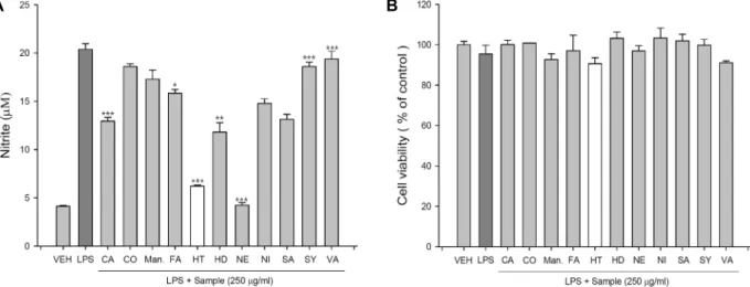
Fig. 1. Effects of 11 different kinds of pure compounds on NO production and cell viability in LPS-stimulated RAW 264.7 cells.
RAW 264.7 cells were plated at 2x10⁵ cells/well in a 96-well plate and incubated with 250 μg/ml of each pure compound for 16 hr. And then, (A) NO production was measured by NO production assay. (B) Cell viability was measured using MTS proliferation assay kit. Values indicate means
±
SD (n=4). *p<0.05, **p<0.01, ***p<0.001. CA; caffeic acid, CO; coumaric acid, Man.; D-mannitol, FA; ferulic acid, HT; hesperetin, HD; hesperidin, NE; naringenin, NI; naringin, SA; sinapic acid, SY; syringic acid, VA; vanilic acid.A B
Fig. 2. Effects of hesperetin on NO production and cell viability in LPS-activated RAW 264.7 cells. RAW 264.7 cells were plated at 2×10⁵ cells/well in a 96-well plate and incubated with 0.25 or 0.5 mM of hesperetin for 16 hr. (A) And then, NO production was measured by NO production assay. (B) Cell viability was measured using MTS proliferation assay kit. Values indicate means
±
SD (n=4). ***p<0.001.결과 및 고찰
11가지 순수물질과 hesperetin에 의한 NO 생성 억제 및 세포 생존율 변화 분석
전통주 주박 ethyl acetate 추출물로부터 분리 동정한 11개 의 순수 물질이 LPS로 활성화된 RAW 264.7 세포에서 nitric oxide (NO) 생산과 세포성장에 미치는 영향을 연구하였다.
LPS로 활성화된 RAW 264.7 세포주에 250 mg/ml의 11개의 순수물질을 각각 처리한 결과, hesperetin과 naringenin 처리 에 의해 현저하게 NO의 생산이 감소하는 것을 확인하였다 (Fig. 1A). 한편, 같은 조건에서 세포생존율 변화를 측정한 결 과 처리한 모든 순수물질들이 세포 생존율에는 큰 영향을 주
지 않은 것으로 측정되었다(Fig. 1B).
세포 생존율에는 큰 영향을 주지 않으면서 가장 높은 NO
생산 저해율을 보여준 순수물질 중 hesperetin을 구입하여 추
가 실험을 진행하였다. Hesperetin에 의한 NO 생성 저해 여부
를 확인하기 위하여 LPS로 활성화된 RAW 264.7 세포에 0.25
mM과 0.5 mM의 농도로 hesperetin을 처리한 후 NO 생성과
cell viability를 측정하였다. 그 결과 Fig. 2에서 보는 바와 같이
hesperetin 처리는 세포 생존율에는 영향을 미치지 않으면서
(Fig. 2B), 처리한 hesperetin의 농도의존적으로 NO 생성을 억
제함을 확인하였다(Fig. 2A). 따라서, hesperetin은 LPS로 자극
된 RAW 264.7 세포에서 세포 생존율에는 영향을 미치지 않으
면서 NO 생산을 농도 의존적으로 저해함을 확인하였다. 최근
Fig. 3. Down-regulation of iNOS protein by hesperetin. LPS- stimulated RAW 264.7 cells were treated with 0.25 or 0.5 mM of hesperetin for 16 hr. And then, cell lysates were prepared and subjected to Western blotting by us- ing antibodies against COX-2, iNOS and Actin. Actin was used as a loading control.
Fig. 4. Suppression of the phosphorylation of p38 MAPK and ERK1/2 by hesperetin treatment. RAW 264.7 cells were pre-treated with 0.25 or 0.5 mM of hesperetin for 4 hr, and stimulated with LPS (1 μg/ml) for 30 min. And then, cell lysates were prepared and subjected to Western blot- ting by using antibodies against p38, phopho-p38, ERK- 1/2, phospho-ERK-1/2, JNK, phospho-JNK, and Actin.
Actin was used as a loading control.
hesperetin 유도체인 HD-14가 급성 간조직 상처부위나 LPS 처리된 RAW 264.7 세포에서 항염증 활성을 보여준다는 보고 가 있었다[5]. 또한 Wang 등에 의해 16개의 신규 hespertin의 유도체 제조방법과 유도체에 의한 함염증 활성에 대한 보고가 있었다[22]. 따라서, 천연물질인 hesperetin은 그 자체가 항염 증 활성을 가질뿐 만 아니라 다양한 유도체의 개발에 따라 좀 더 활성이 높은 천연물 신약의 개발도 가능하다고 생각된다.
Hesperetin에 의한 iNOS 단백질 발현 감소
Hesperetin에 의한 pro-inflammatory 유전자 iNOS와 COX- 2의 단백질의 발현 분석을 수행하였다. 즉, LPS로 활성화된 RAW 264.7 세포주에 각각 0.25 mM과 0.5 mM의 농도로 hes- peretin을 염증 유전자인 iNOS와 COX-2 단백질의 발현을 분 석하였다. 그 결과, iNOS 단백질의 발현은 hesperetin에 의해 농도 의존적으로 발현이 감소하였으나, COX-2 단백질의 경우 hesperetin에 의해 발현 변화가 관찰되지 않았다(Fig. 3). Wu 등은 한약재인 단삼(Salvia miltiorrhiza Bunge) 유래의 천연물 인 neocryptotanshinone도 COX-2의 발현에는 영향없이 iNOS 의 발현을 감소시킨다고 보고 하였다[23]. 반면 천연물인 oxy- resveratrol에 의해서는 iNOS와 COX-2의 유전자와 단백질의 발현이 모두 감소되었다[14]. 이는 천연물에 따라 항염증 활성 의 작용의 기전이 다를 수 있음을 시사한다.
Hesperetin에 의한 p38 MAPK와 ERK1/2 신호 경로 저해
Hesperetin에 의한 염증반응 조절에 MAPKs 신호경로의 관련여부를 확인하기 위하여 hesperetin을 처리한 후, 3가지 MAPKs의 총 단백질 발현과 각 단백질의 인산화를 Western blot으로 분석하였다. 즉, Hesperetin을 0.25 mM과 0.5 mM의 농도로 RAW 264.7 세포에 4시간 동안 처리한 후 LPS로 15분 동안 자극하여 p38 MAPK, ERK1/2와 JNK 총 단백질과 각 MAPK의 인산화의 변화를 분석하였다. 그 결과 0.25 mM 과 0.5 mM 농도의 모든 처리군에 비해 p38 MAPK, ERK1/2 및 JNK의 총 단백질의 발현 변화는 관찰되지 않은 반면, p38
MAPK와 ERK1/2 단백질의 인산화는 저해되었으며, JNK의 인산화는 저해되지 않았다(Fig. 4). 이러한 연구 결과는 hes- peretin이 p38 MAPK 와 ERK1/2 신호전달경로를 억제함으 로써 LPS로 유도된 RAW 264.7 세포에서 염증반응을 억제한 다는 사실을 시사하는 것이다. 본 연구 결과와 유사하게 다양 한 천연물질의 항염증 활성이 MAPK 신호경로와 NF-κB 경로 를 억제함으로써 이루어진다는 것이 보고되었다[16, 25].
총괄적으로 본 연구에서는 주박에서 분리 동정한 11개의 순수물질 중 hesperetin과 naringenin에 의한 염증 지표 물질 중 하나인 NO 생산이 가장 높게 저해되었으며, 이 중 hesper- etin의 항염증 활성은 p38 MAPK와 ERK1/2 경로를 억제함으 로써 조절될 수 있음을 시사한다. 이러한 연구 결과는 주박에 서 분리 동정된 천연물 중 하나인 hesperetin에 의한 항염증 활성 및 작용기전을 이해하는데 도움을 줄 것으로 기대된다.
감사의 글
이 논문은 안동대학교 기본연구지원사업에 의하여 연구되 었음.
References
1. Ahmadi, A. and Shadboorestan, A. 2015. Oxidative stress and cancer: the role of hesperidin, a Citrus natural bio- flavonoid, as a cancer chemoprotective agent. Nutr. Cancer 18, 1-12.
2. Akira, S. and Takeda, K. 2004. Toll-like receptor signaling, Nat. Rev. Immunol. 4, 499-511.
3. Anderson, N. and Borlak, J. 2008. Molecular mechanisms and therapeutic targets in steatosis and steatoheptitis.
Pharmacol. Rev. 60, 311-357.
4. Chen, W., Ge, X., Xu, F., Zhang, y., Liu, Z., Pan, J., Song, J., Dai, Y., Zhou, J., Feng, J. and Liang, G. 2015. Design, synthesis and biological evaluation of paralleled aza resver- atrol-chalcone componds as potential anti-inflammatory agents for the treatment of acute lung injury. Bioorg. Med.
Chem. Lett. 25. 2998-3004.
5. Chen, X., Ding, H. W., Li, H. D., Huang, H. M., Li, X. F., Yang, Y., Zhang, Y. L., Pan, X. Y., Huang, C., Meng, X. M.
and Li, J. 2017. Hesperetin derivative-14 alleviates in- flammation by activating PPAR-γ in mice with CCl4-induced acute liver injury and LPS-treated RAW264.7 cells. Toxicol.
Lett. 274, 51-63.
6. Fang, Q., Deng, L., Wang, L., Zhang, Y., Weng, Q., Yin, H., Pan, Y., Tong, C., Wang, J. and Liang, G. 2015. Inhibition of mitogen-activated protein kinases/nuclear factor κB-de- pendent inflammation by a novel chalcone protects kidney from high fat diet-induced injuries in mice. J. Pharmacol. Exp.
Ther. 355, 235-246.
7. Jacobsen, M. C., Dusart, P. J., Korowicz, K., Bajaj-Eliott, M., Hart, S. L., Klein, N. J. and Dixon, G. L. 2016. A critical role for ATF2 transcription factor in the regulation of E-se- lectin expression in response to non-endotoxin components of Neisseria meningitides. Cell Microbiol. 18, 66-79.
8. Ju, A., Cho, Y. C. and Cho, S. 2015. Methanol extracts of Xanthium sibiricum roots inhibit inflammatory response via the inhibition of nuclear factor-κB (NF-κB) and signal trans- ducer and activator of transctiption 3 (STAT3) in murine macrophages. J. Ethnopharmacol. 174, 74-81.
9. Khalifa, H. O., Kamimoto, M., Shimamoto, T. and Shimamo- to, T. 2015. Antimicrobial effects of blueberry, raspberry, and strawberry aqueous extracts and their effects on viru- lence gene expression in Vibrio cholerae. Phytother. Res. 29, 1791-1797.
10. Kim, H. G., Shi, C., Bode, A. M. and Dong, Z. 2015. p38α MAPK is required for arsenic-induced cell transformation.
Mol. Carcinog. 55, 910-917.
11. Kuwano, T., Watanabe, M., Kagawa, D. and Murase, T.
2015. Hydrolyzed methyl-hesperidin induces anti-oxidant enzyme expression via the Nrf2-ARE pathway in normal human epidermal keratinocytes. J. Agric. Food Chem. 63, 7937- 7944.
12. Lee, E. S., Ju, H. K., Moon, T. C., Lee, E., Jahng, Y., Lee, S. H. and Chang, H. W. 2004. Inhibition of nitric oxide and tumor necrosis factor-alpha (TNF-alpha) production by pre- penone compound through blockade of nuclear factor (NF)- κB activation in cultured murine macrophages. Biol. Pharm.
Bull. 27, 617-620.
13. Lee, H. J., Lee, W. J., Chang, S. E. and Lee, G. Y. 2015.
Hesperidin, A popular antioxidant inhibits melanogenesis via ERK1/2 mediated MITF degradation. Int. J. Mol. Sci. 16, 18384-18395.
14. Lee, H. S., Kim, D. H., Hong, J. E., Lee, J. Y. and Kim, E.
J. 2015. Oxyresveratrol suppresses lipopolysaccharide-in- duced inflammatory responses in murine macrophages.
Hum. Exp. Toxicol. 34, 808-818.
15. Li, G., Wulan, H., Song, Z., Paik, P. A., Tsao, M. L., Good- man, G. M., MacEachern, P. T., Downey, R. S., Jankowska, A. J., Rabinowitz, Y. M., Learch, T. B., Song, D. Z., Yuan, J. J., Zheng, S. and Zheng, Z. 2015. Regulatory B cell function is suppressed by smoking and obesity in H. pylori-infected subjects and is correlated with elevated risk of gastric cancer. PLoS One 10, e0134591.
16. Li, H., Zhang, Q., Jin, X., Zou, X., Wang, Y., Hao, D., Fu, F., Jiao, W., Zhang, C., Lin, H., Matsuzaki, K. and Zhao, F. 2018. Dysifragilone A inhibits LPS-induced RAW264.7 macrophage activation by blocking the p38 MAPK signaling pathway. Mol. Med. Rep. 17, 674-682.
17. Mrvova, N., Skandik, M., Kuniakova, M. and Rackova, L.
2015. Modulation of BV-2 microglia functions by novel quercetin pivaloyl ester. Neurochem. Int. 90, 246-254.
18. Oliveira, A. M., Mesquita, M. S., Silva, G. C., Oliveria, L.
E., Medeiros, P. L., Paiva, P. M., Souza, I. A. and Napoleao, T. H. 2015. Evaluation of toxicity and antimicrobial activity of an ethanolic extract from leaves of Morus alba L. (Moa- ceae). Evid. Based Complement Alternat. Med. 2015, 513978.
19. Sahu, B. D., Kumar, J. M. and Sistla, R. 2015. Baicalein, a bioflavonoid, prevents cisplatin-induced acute kidney injury by up-regulating antioxidant defense and down-regulating the MAPKs and NF-κB pathways. Plos One 10, e0134139.
20. Sung, M. J., Davaatseren, M., Kim, S. H., Kim, M. J. and Hwang, J. T. 2013. Boehmeria nivea attenuates LPS-induced inflammatory markers by inhibiting p38 and JNK phosphor- ylation in RAW264.7 macrophages. Pharm. Biol. 51, 1131- 1136.
21. Vlkova, M., Ticha, O., Nechvatalova, J., Kalina, T., Litzman, J., Mauri, C. and Blair, P. A. 2015. Regulatory B cells in CVID patients fail to suppress multi-functional IFN-gam- ma+TNF-alpha+CD4+ T cells differentiation. Clin. Immunol.
29, 292-300.
22. Wang, Q. Q., Shi, J. B., Chen, C., Huang, C., Tang, W. J.
and Li, J. 2016. Hesperetin derivatives: Synthesis and anti- inflammatory activity. Bioorg. Med. Chem. Lett. 26, 1460-1465.
23. Wu, C., Zhao, W., Zhang, X. and Chen, X. 2015. Neocrypto- tanshinone inhibits lipopolysaccharide-induced inflammation in RAW264.7 macrophages by suppression of NF-κB and iNOS signaling pathways. Acta Pharm. Sin. B. 5, 323-329 24. Zamora-Ros, R., Guino, E., Henar, A. M., Vidal, C., Barenys,
M., Soriano, A. and Moreno, V. 2015. Dietary flavonoids, lignans and colorectal cancer prognosis. Sci. Rep. 5, 14148.
25. Zhang, Y., Liu, D., Fang, L., Zhao, X., Zhou, A. and Xie, J. 2018. A galactomannoglucan derived from Agaricus brasi- liensis: Purification, characterization and macrophage acti- vation via MAPK and IκB/NFκB pathways. Food Chem. 239, 603-611.
초록:마우스 대식세포 RAW 264.7 세포주에서 hesperetin에 의한 p38 MAPK와 ERK1/2를 통한 염증반응 조절
이승훈
1․이은주
1․정정욱
1․손호용
2․김종식
1*
(1국립안동대학교 생명과학과, 2국립안동대학교 식품영양학과)