Fatty Acid Composition and Antiproliferative Activity of Extracts from Euphorbia Supina
Hyang Mi Choi and Sun Young Lim*
Division of Marine Environment & Bioscience, Korea Maritime and Ocean University, Korea
Received November 18, 2013 /Revised December 27, 2013 /Accepted December 30, 2013The objective of this study was to determine the fatty acid composition and the antiproliferative effect of extracts and fractions from Euphorbia supina. With regards the fatty acid composition, the percen- tages of 18:3n-3 in acetone/methylene chloride (A+M) and methanol (MeOH) extracts were 53.4 and 42.1%, respectively. Among the fractions, an 85% aqueous methanol (85% aq. MeOH) fraction con- tained the highest percentage of 18:3n-3. Treatments with crude extracts and fractions significantly in- hibited the growth of HT-29 and AGS human cancer cell lines (p<0.05). The A+M extract showed a higher inhibitory effect on the growth of both cancer cells compared to MeOH extract. Among the fractions, the 85% aq. MeOH and n-hexane fractions exerted a greater inhibitory effect on the pro- liferation of both types of cancer cells. Our results suggest that 85% aq. MeOH and n-hexane fractions exert potent inhibitory effects on the proliferation of human cancer cells.
Key words : Antiproliferative, Euphorbia supina, fatty acid composition, human cancer cell
*Corresponding author
*Tel : +82-51-410-4757, Fax : +82-51-404-4750
*E-mail : [email protected]
This is an Open-Access article distributed under the terms of the Creative Commons Attribution Non-Commercial License (http://creativecommons.org/licenses/by-nc/3.0) which permits unrestricted non-commercial use, distribution, and reproduction in any medium, provided the original work is properly cited.
Journal of Life Science 2014 Vol. 24. No. 1. 74~80 DOI : http://dx.doi.org/10.5352/JLS.2014.24.1.74
서 론
비단풀은 대극과(Euphorbiaceae) 대극속에 속하는 한해살이 풀땅빈대로서 전 세계적으로 약 1,600종이 자라고 있으며, 우 리나라에는 약 11종이 분포되어 있다[3]. 비단풀은 땅바닥을 비단처럼 곱게 덮었다는 어원이 있으며 땅바닥을 기면서 자라 고 도시의 화단, 운동장, 주차장 가장자리 등에서 쉽게 볼 수 있다. 비단풀에는 3가지가 있는데 애기땅빈대(Euphorbia supi- na Raf.)와 땅빈대(Euphorbia humifusa Willd.)는 땅에 바짝 붙 어서 자라고 큰땅빈대(Euphorbia maculate L.)는 키가 20-60 cm 까지 자란다. 개화기는 땅빈대(E. humifusa)와 큰땅빈대(E.
maculata)가 8-9월이며 잎에 점이 없고 애기땅빈대(E. supina) 의 개화기는 6-8월이며 잎에 점이 있는 것이 특징이다. 비단풀 의 원줄기는 지면을 따라 퍼지며 길이 10-25 cm이고, 잎과 더 불어 털이 다소 있고, 중앙부에는 붉은빛이 도는 갈색반점이 있다. 비단풀의 줄기나 잎에 상처가 생기면 흰색의 끈적한 유 즙이 나오는데 칼로 베인 상처나 가시에 찔리거나 풀 등에 베인 상처에 바르면 효과가 있다고 하여 민간요법에서는 외상 출혈, 토혈, 빈혈에 효능하고 신장결석, 방광결석, 신장염, 항 암, 항균, 진정작용, 해독작용, 혈액순환 등에 활용되고 있다
[19]. 또한 마음을 편안하게 하고 통증을 멎게 하는 작용이 있 으며 독성은 전혀 없다[6]. 애기땅빈대의 성분에 관한 연구로 는 tannins [1, 20], phenol성 물질 및 flavonoids [3, 14], terpe- noids [8, 27] 등에 관한 연구가 보고되어 있다[18, 26]. Cha [4] 등은 땅빈대 MeOH 엑기스는 사람 뇌종양세포에 대해 농 도의존적으로 세포독성을 나타낸다고 보고하였으며 Chen [5]
등은 등대풀(Euphorbia helioscopia)이 마우스 폐선암세포에서 농도 의존적으로 세포독성을 나타낸다고 보고하였다.
경제성장에 따른 소득수준의 증가와 가공식품 급식 및 외식
의 증가에 따른 식생활 환경변화, 신체활동량의 감소 등이 비
만인구의 빠른 증가를 가져왔다. 지방의 섭취와 암발생간의
역상관관계가 알려지면서 현대인의 건강한 삶을 위해 포화지
방산의 섭취량을 줄이고 다가불포화지방산의 섭취량을 늘려
야 한다고 권장되고 있다[28]. 포화지방산과 n-6계 linoleic
acid의 과도한 섭취는 특히 유방암과 대장암의 발생이 촉진되
는 반면 α-linolenic acid, eicosapentaenoic acid (EPA)와 doco-
sahexaenoic acid (DHA) 등 n-3계 다가불포화지방산에 의해
서는 대장암 발생 및 암세포 증식이 억제된다고 보고하였다[9,
24]. 지방질 섭취량이 총 열량의 20% 내외에서 40-45% 내외로
변화하게 되면서 이러한 암에 걸릴 수 있는 확률은 최소 2-4배
정도 높아짐을 동물실험과 역학조사에서 살펴 볼 수가 있다
[7]. 또한 암치료제를 개발하기 위해 전세계적으로 다양한 연
구가 시도되고 있는데, 현재까지 승인된 암치료제의 60% 정도
는 식물유래화합물(vinceristine, taxanes) 또는 미생물(dacti-
nomicine, anthracyalines) 등의 자연산물을 이용해 개발되었
다[16]. 비단풀은 우수한 항암약초자원으로 뇌종양, 췌장암, 위
암, 폐암, 직장암, 대장암, 신장암, 간암 등에 좋으며 특히 뇌종
E. supina
(520 g)← Acetone + Methylene chloride Acetone + Methylene chloride
extract (15.9 g)
Residue
← Methanol Methanol extract
(35.5 g)
Residue
Crude Extract
partition
Methylene chloride Water
n
-Hexane+85% aq. MeOH → ←n
-BuOHpartition
n
-Hexane fraction(5.6 g) 85% aq. MeoH
fraction (2.7 g)
n
-BuOH fraction(6.7 g) Water fraction (1.9 g) Fig. 1. The procedure for solvent extraction and fraction from
E. supine.
양과 췌장암에 뛰어난 효과가 알려져 있으나 문헌이 전하는 기록이 많지 않은 이유로 대부분 민간에서 우수한 약초자원으 로 활용되고 있다. 따라서 본 연구에서는 다양한 약효가 있다 고 알려진 애기땅빈대의 항암능력을 살펴보았고 용매 추출물 및 분획물의 지방산 조성을 분석하였다.
재료 및 방법
재료 및 시약
본 실험에 사용된 애기땅빈대(E. supina)는 부산 한국해양대 학교의 바위옆, 들판 및 보도블럭 틈새에서 7-8월경 집중적으 로 채취하여 햇볕에 건조하였다.
추출 및 분획
건조된 애기땅빈대은 실험 사용 전까지 -75℃의 deep freez- er (NF-400SF, NIHON FREEZER, Tokyo, Japan)에 냉동 보관 하였다가 유기용매 추출을 위하여 acetone:methylene chlor- ide를 1:1 비율로 혼합하여 비단풀이 충분히 잠기도록 하여 24시간 방치한 후 추출하였다. 이 과정을 2회 반복하여 얻은 여액은 40℃ 수욕상에서 rotary vacuum evaporator (N-1000, EYELA, Tokyo, Japan)로 농축하여 acetone/methylene chlor- ide 추출물(A+M)을 얻었다. A+M 용매로 추출되지 않은 성분 을 methanol (MeOH)로 추출하고 남은 잔사물에 A+M와 동량 의 MeOH로 위와 동일한 방법으로 2회 반복한 후 농축하여 MeOH 분획물을 얻었다. 두 용매로부터 최대로 수득한 추출
물을 혼합하여 다시 용매극성에 따라 순차적으로 분획하여 n-Hexane, 85% aqueous MeOH (85% aq. MeOH), n-butanol (n-BuOH) 및 water 분획물을 얻었다(Fig. 1). 실험에는 각각의 추출물들을 dimethyl sulfoxide (DMSO)에 녹여 세포배지로 필요한 농도로 희석하여 실험에 사용하였다.
지질 및 지방산 추출
지질추출은 Folch 등[15]의 방법을 변형하여 실시하였으며 간단히 요약하면 다음과 같다. 건조된 애기땅빈대와 애기땅빈 대 추출물과 분획물들은 butyl hydroxy toluene (BHT)을 함께 함유한 methanol로 교반하여 균질화하였다. 균질물을 1 ml 취한 후 chloroform 2 ml와 0.2 M NaH
2PO
41.4 ml를 넣고 교반하여 4℃, 3,000 rpm에서 3분간 원심분리 후 지질층을 얻 었다. 이와 같은 방법을 한번 더 진행한 뒤 질소가스를 이용하 여 지질층의 유기용매를 완전히 날린 다음 지질을 얻었다.
Morrison과 Smith의 방법[22]에 따라 추출된 지질에 methyl- ation용 시약인 boron trifluoride (BF
3) methanol 1 ml와 n-hexane 0.4 ml를 가한 후 1시간 동안 100℃에서 가열하였다.
1시간 후 실온까지 충분히 냉각시킨 다음 n-hexane 2 ml와 증류수 2 ml를 가한 후 다시 4℃, 3,000 rpm에서 3분간 원심분 리 후 상등액을 얻었다. 이 상등액을 질소가스를 이용하여 유 기용매를 날린 후 얻은 지방산은 지방산 분석 전까지 -75℃
deep freezer (NF-400SF, NIHON FREEZER, Tokyo, Japan)에
보관하였다.
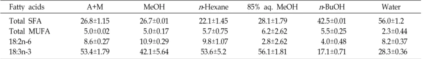
Table 1. Several fatty acid compositions (% area) of extracts and fractions from
E. supina
Fatty acids A+M MeOH
n
-Hexane 85% aq. MeOHn
-BuOH WaterTotal SFA Total MUFA 18:2n-6 18:3n-3
26.8±1.15 5.0±0.02 8.6±0.27 53.4±1.79
26.7±0.01 5.0±0.17 10.9±0.29 42.1±5.64
22.1±1.45 5.7±0.75 9.8±1.07 53.6±5.2
28.1±1.79 6.2±2.62 2.8±2.62 56.1±1.81
42.5±0.01 5.5±0.25 4.0±0.48 17.1±0.71
56.0±1.2 2.3±0.44 8.2±0.37 28.3±0.36 SFA, saturated fatty acids; MUFA, Monounsaturated fatty acids; A+M, acetone with methylene chloride extract; MeOH, methanol extract;
n
-Hexane,n
-hexane fraction; 85% aq. MeOH, 85% aqueous methanol fraction;n
-BuOH,n
-butanol fraction; Water, water fraction.Gas chromatography를 이용한 지방산 분석
시료에서 분리된 지방산을 1 μl 취하여 지방산 분석용 gas chromatography에 주입하여 지방산을 분석하였다[25]. 지방 산 분석에 사용한 표준용액은 미국 NU-CHEK-PREP사의 462 standard였으며, 이용된 column은 silica capillary column (CP-7856, 60 m × 0.32 mm inner diameter × 0.10 μm film thickness)이다. 기기의 분석조건은 injector 250℃, detector (FID) 250℃, oven (initial 130℃, 분당증가율은 175℃까지 4℃
/min, 210℃까지 1℃/min, 245℃까지 30℃/min), carrier gas 는 헬륨을 사용하였다. 지방산 분석은 표준용액의 retention time과 비교하여 정성하였고, 내부표준물질(22:3n-3, methyl ester)을 이용하여 총지방산을 정량하였으며 개개의 지방산들 은 전체 peak area의 퍼센트로 산출하였다.
세포배양
한국세포주은행(Seoul, Korea)으로부터 인체 결장암세포 (HT-29), 인체 위암세포(AGS)를 분양받아 본 실험실에서 배양 하면서 실험에 사용하였다. HT-29 세포와 AGS 세포는 100 unit/ml의 penicillin-streptomycin (GIBCO, Gaithersburg, MD, USA)과 10% FBS (Hyclone, Logan, UT, USA)가 함유된 RPMI 1640 (GIBCO)을 사용하여 37℃, 5% CO
2incubator (MCO-15AC, SANYO Electric Biomedical Co., Tokyo, Japan) 에서 배양하면서 배양 중인 세포를 일주일에 2번 새로운 배지 로 바꿔주었다. 일주일 후 phosphate buffered saline (PBS)으 로 세척한 뒤 0.05% trypsin-0.02% EDTA (GIBCO)로 부착된 세포를 분리하여 원심분리한 후 집적된 암세포에 배지를 넣고 피펫으로 암세포가 골고루 분산되도록 잘 혼합하여 cell cul- ture flask에 10 ml씩 일정한 수로 분할하여 주입하고, 6-7일마 다 계대배양하면서 실험에 사용하였다.
MTT assay
배양된 암세포는 96-well cell culture plate에 5×10
4cells/
ml이 되도록 100 μl씩 분주하여 37℃, 5% CO
2incubator에서 24시간 배양한 후 배지는 제거한 뒤 각 시료를 배지로 희석하 여 각 well 당 100 μl씩 첨가하고, 대조군에는 시료대신 PBS를 100 μl씩 첨가하였다. 이 plate를 다시 37℃, 5% CO
2incubator 에서 24시간 배양하였다. 배양 후 3-(4,5-dimethylthiazol)-2,
5-diphenyltetrazolium bromide (MTT) assay [10]를 위하여 MTT 시약 5 mg을 PBS 1 ml로 녹인 후, 10% FBS가 함유된 배지 9 ml와 희석하여 100 μl를 첨가하고 3-4시간 동안 더 배양 하여 MTT가 환원되도록 하였다. 배양종료 후 생성된 for- mazan 결정을 가라앉힌 후 각 well에 형성된 결정이 흐트러지 지 않도록 주의하면서 반응 후 남은 MTT가 처리된 배지를 제거하였다. 배지가 제거된 각 well에 formazan 결정을 용해 시키기 위하여 DMSO를 100 μl씩 분주하여 5-10분간 반응시켜 microplate reader (VICTOR3, Perkin Elmer, Waltham, MA, USA)로 540 nm에서 흡광도를 측정하였다. 이 흡광도는 MTT 가 세포에 의해서 환원된 양을 나타내며, 따라서 각 well에 존재하는 세포의 생존수와 비례한다.
통계분석
실험결과는 Mean ± SEM (Standard Error of Mean)으로 나타내었고 분석된 실험데이터는 대조군과 각 시료로부터 얻 은 실험자료로부터 ANOVA를 실시하여 유의성이 있을 경우 에 post-hoc test로 Duncan’s multiple range test를 실시하여 95% 수준에서 유의성을 검증하였다.
결과 및 고찰
애기땅빈대(E. supina) 추출물 및 분획물의 지방산 조성
건조된 애기땅빈대는 2.3%의 총 포화지방산(saturated fatty
acids, SFA), 24.5%의 총 단일불포화지방산(monounsaturated
fatty acids, MUFA), 2.9%의 18:2n-6 및 53.2%의 18:3n-3를 함
유하였으므로 식물체로서는 높은 n-3 지방산을 함유하고 있음
을 알 수가 있다. 애기땅빈대 추출물 및 분획물들의 지방산
조성을 Table 1에 나타내었다. A+M 추출물과 MeOH 추출물
의 지방산 조성 패턴은 유사했으나 MeOH 추출물과 비교했을
때 A+M 추출물는 낮은 함량의 18:2n-6와 높은 함량의 18:n-3
를 나타내었다. 분획물들 중 85% aq. MeOH와 n-Hexane 분획
물들은 n-BuOH와 Water 분획물들과 비교했을 때 낮은 함량
의 총 SFA를 나타내었고 상당히 높은 함량의 18:3n-3를 나타
내었다. n-Hexane 분획물과 비교했을 때 85% aq. MeOH 분획
물은 낮은 함량의 18:2n-6와 약간 높은 함량의 18:n-3을 나타내
었다. Pascual-Villalobos 등[23]은 대극과에 속하는 Euphorbia
Fig. 2. Effect of acetone/methylene chloride (A+M) and meth- anol (MeOH) extracts from
Euphorbia supina
on the growth inhibition of HT-29 human colon cancer cells.*
p
<0.05, significant between the control and each extract.Fig. 3. Effect of acetone/methylene chloride (A+M) and meth- anol (MeOH) extracts from
Euphorbia supina
on the growth inhibition of AGS human gastric adenocarcinoma cells. *p
<0.05, significant between the control and each extract.lagascae.와 Euphorbia lathyris의 잎으로부터 지방산을 추출한 결과 다른 지방산들보다 18:3n-3의 함량이 높은 것을 보고하 였다.
애기땅빈대 추출물 및 분획물의 인체 암세포 증식 억제효과 MTT assay를 행하여 애기땅빈대 추출물 및 분획물의 인체 암세포 증식 억제효과를 살펴보았다. DMSO에 의한 독성은 값이 거의 변화하지 않았으므로 DMSO에 의한 독성은 세포의 생존율에 아무런 영향을 미치지 않았다. Fig. 2는 애기땅빈대 의 A+M 및 MeOH 추출물을 인체 결장암세포(HT-29)에 농도 별로 처리했을 때 증식 억제효과를 나타낸 것이다. 애기땅빈 대 A+M 및 MeOH 추출물은 대조군과 비교했을 때 HT-29 세포의 증식을 유의적으로 억제시켰다(p<0.05). A+M 추출물 을 0.5 및 0.25 mg/ml 첨가농도로 처리했을 때 88%의 높은 억제효과를 나타내었으며, IC
50은 0.05 mg/ml이었다. MeOH 추출물의 경우(0.5 mg/ml 첨가농도), 91%의 암세포 증식 억제 효과를 나타내었고, IC
50은 0.22 mg/ml이었다. Fig. 3는 인체 위암세포(AGS)에 대한 결과를 나타낸 것으로, A+M 추출물은 0.5 mg/ml의 첨가농도에서 91%의 높은 억제효과를 나타내었
으며, IC
50은 0.09 mg/ml이었다. MeOH 추출물의 경우, 0.5 mg/ml의 첨가농도에서 89%의 암세포 증식 억제효과를 나타 내었고, IC
50은 0.15 mg/ml이었다. 따라서 HT-29 및 AGS 세포 모두에서 MeOH 추출물과 비교했을 때 A+M 추출물에 의한 암세포 증식 억제효과가 높았고 두 종류의 암세포 중 AGS 세포의 증식 억제효과가 더 높았음을 살펴 볼 수가 있었다.
한편, 애기땅빈대의 각 분획물을 농도별로 HT-29 암세포에 처리하였을 때, 농도의존적으로 암세포의 증식을 억제하였고, 특히 85% aq. MeOH 분획물에 의한 저해활성이 가장 높았다 (Fig. 4). 85% aq. MeOH 분획물의 경우 0.1 mg/ml 이상의 농도에서 90% 이상의 높은 암세포 증식 억제효과를 나타내었 으며, IC
50은 0.05 mg/ml이었다. n-Hexane 분획물의 경우, 0.11 mg/ml의 IC
50값을 나타내었다. Fig. 5는 애기땅빈대 분획 물들의 AGS 암세포 증식에 대한 억제효과를 나타낸 것으로 HT-29와 유사하게 85% aq. MeOH 분획물에 의한 저해활성이 높았다. 85% aq. MeOH 분획물의 경우 0.1 mg/ml 이상의 농 도에서 95% 이상의 높은 암세포 증식 억제효과를 나타내었으 며, IC
50은 0.03 mg/ml이었다. n-Hexane 분획물의 IC
50은 0.03 mg/ml이었으며 HT-29 세포와 비교했을 때 높은 암세포 억제 효과를 나타내었다. An 등[2]은 비단풀의 물 추출물에서 항암 효과를 살펴 본 결과 HT-29에서는 10 μg/ml 농도에서 49%의 생존율을 나타내었고, 유방암유래의 암세포주 MCF-7에서는 60%의 생존율을 나타낸다고 보고하였다. Montopoli 등[21]은 대극과인 Crotone lechleri의 항암효과를 살펴 본 결과 HT-29에 서 10 μg/ml 농도에서 가장 높은 암세포 증식 억제효과가 나 타났으며 IC
50은 0.8 μg/ml 이었다고 보고하였다. da Mota 등 [11]은 대극과인 Synadanium umbellatum을 Dalton’s 림프종에 처리했을 때 caspase 3를 활성화시켜 apoptosis를 유도하였다 고 보고했다. Yu 등[29]은 Euphorbia kasui 추출물은 농도의존 적으로 종양세포의 성장억제율이 증가한다고 보고하였다. 이 상의 결과로부터 애기땅빈대에 의한 암세포 증식 억제효과는 MeOH 추출물보다는 A+M 추출물에서 그리고 분획물들 중에 서는 n-Hexane과 85% aq. MeOH 분획물들에서 활성이 높은 것을 살펴 보았으며 18:3n-3 지방산의 함량을 고려했을 때도 이들 A+M 추출물과 n-Hexane과 85% aq. MeOH 분획물들에 서 높은 함량의 18:3n-3 지방산의 확인되었다. Dai 등[12]은 in vitro 실험에서 18:3n-3 처리는 위암세포(MGC 및 SGC)의 증식을 억제하였고 apoptosis을 유도하였다고 보고하였다.
Dommels 등[13]은 18:3n-3 처리는 인체 결장암 세포(Caco-2)
의 증식을 억제하였다고 보고하였고 Habermann 등[17]도
18:3n-3를 인체 결장암세포(LT97 및 HT-29)에 처리했을 때 두
결장암세포의 증식을 억제하였다고 보고하였다. 따라서 애기
땅빈대의 항암효과는 n-Hexane과 85% aq. MeOH 분획물들
속에 있는 활성 성분과 관련이 있는 것으로 사료되며 향후
정제하여 규명할 필요가 있다.
0.5
Fig. 4. Effect of solvent fractions from
Euphorbia supina
on the growth inhibition of HT-29 human colon cancer cells. *p
<0.05, significant between the control and each extract; (A)n
-Hexane,n
-hexane fraction; (B) 85% aq. MeOH, 85% aqueous methanol fraction;(C)
n
-BuOH,n
-butanol fraction; (D) Water, water fraction.0.5
Fig. 5. Effect of solvent fractions from
Euphorbia supina
on the growth inhibition of AGS human gastric adenocarcinoma cells. *p
<0.05, significant between the control and each extract; (A)n
-Hexane,n
-hexane fraction; (B) 85% aq. MeOH, 85% aqueous methanol fraction; (C)n
-BuOH,n
-butanol fraction; (D) Water, water fraction.감사의 글
본 과제(결과물)은 해양수산부의 지원으로 수행한 해양에 너지전문인력양성사업과 2013년도 정부(교육부)의 재원으로 한국연구재단의 지원을 받아 수행된 기초연구사업(NRF- 2013R1A1A2004694)의 연구결과입니다.
References
1. Agata, I., Hatano, T., Nakaya, Y., Sugaya, T., Nushibe, S., Yochida, T. and Okuda, T. 1991. Tannins and related poly- phenols of Euphorbiaceous plants. Eumaculin A and eusu- pinin A, and accompanying polyphenols from
Euphorbia maculate
L. andE. supine
Rafin.Chem Pharm Bull
39, 881-883.2. An, D. H., Cho, S. J., Jung, E. S., Lee, H. J. and Hwang, J. H. 2006. Antioxidant and anticancer activities of water extracts from
Ceramium kondoi
.J Korean Soc Food Sci Nutr
35, 1304-1308.3. An, R. B., Kwon, J. W., Kwon, T. O., Chung, W. T., Lee, H. S. and Kim, Y. C. 2007. Chemical constituents from the whole plants of
Euphorbia supine
Rafin.Korean J Pharmacogn
38, 291-295.4. Cha, B. C., Kim, J. A. and Lee, Y. S. 1996. Cytotoxic activities of Panax ginseng and
Euphorbia humifusa
in human brain tumor cells.Korean J Pharmacogn
27, 350-353.5. Chen, H., Wang, Z. and Yang, L. 2011. Analysis of euphor- nin in
Euphorbia helioscopia
L. and its cytotoxicity to mice lung adenocarcinoma cells.Nat Prod Res
1, 1-5.6. Choi, J. G. 2006. The medicine of grasses, flowers and trees.
pp. 193-201, Hanmunsa, Seoul.
7. Choi, M. 1991. Dietary fats and cancer.
J Korean Soc Food Nutr
20, 513-518.8. Chung, B. S. and Kim, H. G. 1985. Studies on the terpenoid constituents of
Euphorbia supine
Rafin. Korean J Pharmacogn
16, 155-159.9. Cognault, S., Jourdan, M. L., Germain, E., Pitavy, R., Morel, E., Durand, G., Bougnoux, P. and Lhuillery, C. 2000. Effect of an α-linolenic acid-rich diet on rat mammary tumor growth depends in the dietary oxidative status.
Nutr Cancer
36, 33-41.10. Denizot, F. and Lang, R. 1986. Rapid colorimetric assay for cell growth and survival: Modifications to the tetrazolium dye procedure giving improved sensitivity and reliability.
J Immunol Methods
89, 271-277.11. da Mota, M. F., Benfica, P. L., Batista, A. C., Martins, F.
S., Paula, J. R. and Valadares, M. C. 2012. Investigation of Ehrlich ascites tumor cell dealth mechanisms induced by
Synadanium umbellatum
Pax.J Enthnopharmacol
139, 319-329.12. Dai, J., Shen, J., Pan, W., Sehn, S. and Das, U. N. 2013. Effect of polyunsaturated fatty acids on the growth of gastric can- cer cells
in vitro
.Lipids Health Disease
12, 71-86.13. Dommels, Y. E. M., Alink, G. M., Linssen, J. P. H. and Ommen, B. V. 2002. Effects of n-6 and n-3 polyunsaturated fatty acids on gap junctional intercellular communication
during spontaneous differentiation of the human colon ad- enocarcinomacell line Caco-2.
Nutr Cancer
42, 125-130.14. Fang, Z., Zeng, X., Zhang, Y. and Zhou, G. 1993. Chemical constituents of spotted leaf euphorbia (
Euphorbia supina
).Zhongcayao
24, 230-233.15. Folch, J., Lees, M. and Stanley, G. S. H. 1957. A simple meth- od for the isolation and purification of total lipids from ani- mal tissues.
J Biol Chem
226, 497-509.16. Grever, M. C. B. Cancer drug discovery and development.
In Cancer: Principles and Practice of Oncology
. pp. 328-339, De Vita VHS, Rosenberg SA, eds. Lippinocott-Raven, Philadel- phia, USA.17. Habermann, N., Christian, B., Luckas, B., Pool-Zobel, B. L., Lund, E. K. and Glei, M. 2009. Effects of fatty acids on me- tabolism and cell growth of human colon cell lines of differ- ent transformation state.
Int Union Biochem Molecular Biol
35, 460-467.18. Hong, H. K., Kwak, J. H., Kang, S. C., Lee, J. W., Park, J.
H., Ahn, J. W., Kang, H. S., Choung, E. S. and Zee, O. P.
2008. Antioxidative constitutents from whole plants of
Euphorbia supina
.Korean J Pharmacogn
39, 260-264.19. Lee, C. B. 1989. An illusted plant book. pp. 511, Hyang- MunSa, Seoul, Korea.
20. Lee, S. H., Tanaka, T., Nonaka, G. and Nishioka, I. 1991.
Tannins and related compounds. CV. Monomeric and di- meric hydrolyzable tannins having a dehydrohyxahydrox- ydiphenoyl group, supinanin, euphoscopin, euphorhelin and jolkianin, from
Euphorbia
species.Chem Pharm Bull
39, 630-638.21. Montopoli, M., Bertin, R., Chen, Z., Bolcato., Caparrotta, L.
and Froldi, G. 2012.
Croton lechleri
sap and isolated alkaloid taspine exhibit inhibition against human melanoma SK23 and colon cancer HT29 cell lines.J Ehnopharmacol
144, 747- 22. Morrison, W. R. and Smith, L. M. Preparation of fatty acid753.methyl esters and dimethlyacetals from lipids with boron fluoride-methanol.
J Lipid Res
5, 600-608.23. Pascual-Villalobos, M. J. and Lopez, M. D. 2010. Leaf lipids from
Euphorbia lagascae
Spreng. andEuphorbia lathyris
L.Ind Crops Prod
32, 560-565.24. Rose, D. P., Connolly, J. M., Rayburn, J. and Coleman, M.
1995. Influence of diets containing eicosapentaenoic or doco- sahexaenoic acid on growth and metastasis of breast cancer cells in nude mice.
J Natl Cancer Inst
87, 587-592.25. Salem, M., Reyer, M. and Karanian, J. 1996. Losses of arach- idonic acid in rat liver after alacohol inhalation.
Lipids
31, 153-156.26. Tanaka, R., Kurimoto, M., Yoneda, M. and Matsunaga S.
1990. 17β,21β-Epoxyhopan-3β-ol and β-alnincanol from
Euphorbia supina
.Phytochemistry
29, 2253-2256.27. Tanaka, R. and Matsunaga, S. 1999. Terpenoids and steroids from several Euphorbiaceae and Pinaceae plants.
Yakugaku Zasshi
119, 319-339.28. Wachi, A. M., Sinclari, L. A., Wilkinson, R. G., Enser, M., Wood, J. D. and Fisher, A. V. 2002. Effect of dietary fat source and breed on the carcass composition, n-3 poly-
초록:애기땅빈대 추출물의 지방산 조성 및 인체 암세포 증식 억제 효과 최향미 · 임선영*
(한국해양대학교 해양환경생명과학부)
본 연구에서는 다양한 약효가 있다고 알려진 애기땅빈대(Euphorbia supina)의 추출물 및 분획물의 지방산 조성 을 분석하고 인체 암세포 증식 억제효과에 대하여 살펴보았다. A+M과 MeOH 추출물의 지방산 조성 패턴은 유사 했고 A+M과 MeOH 추출물은 각각 53.4% 및 42.1%의 18:3n-3를 함유하였다. 분획물들 중 85% aq. MeOH는 가장 높은 함량의 18:3n-3를 나타내었다. 애기땅빈대에 의한 암세포 증식 억제효과는 MeOH 추출물과 비교했을 때 A+M 추출물에 의한 억제효과가 높았으며 AGS 인체 위암세포에 대한 억제효과가 높았다. 분획물들 중에서는 n-Hexane과 85% aq. MeOH 분획물들에 의한 증식 억제효과가 높았다. 따라서 애기땅빈대의 항암 활성 성분은 n-Hexane과 85% aq. MeOH 분획물들에 함유되어 있는 것으로 여겨지며 향후 정제하여 규명할 필요가 있다고 사료된다.
unsaturated and conjugated linoleic acid content of sheep meat and adipose tissue.
Br J Nutr
88, 697-709.29. Yu, L., Jiang, B. P., Luo, D., Shen, X. C., Guo, S., Duan,
J. A. and Tang, Y. P. 2012. Bioactive components in the fruits of