Repression of Cathepsin D Expression in Adipocytes by MicroRNA-145
Hyun-Ji Kim
1, In-Seon Bae
1, Kang-Seok Seo
2* and Sang Hoon Kim
1*
1
Department of Biology, Kyung Hee University, Seoul 130-701, Korea
2
Department of Animal Science and Technology, Sunchon National University, Sunchon 540-950, Korea
Received April 22, 2014 /Revised July 15, 2014 /Accepted July 22, 2014Cathepsin D (CtsD), an aspartyl peptidase, is involved in apoptosis, resulting in the release of cyto- chrome C from mitochondria in cells. Here, we investigated microRNA regulation of CtsD expression in 3T3-L1 cells First, we observed the expression of CtsD in cells in response to doxorubicin (Dox).
As expected, the level of CtsD mRNA was increased in 3T3-L1 cells exposed to Dox in a dose-depend- ent manner. Cellular viability of ectopically expressed CtsD cells was also decreased. Next, we used the miRanda program to search for particular microRNA targeting CtsD. MiR-145 was selected as a putative controller for CtsD because miR-145 had a high mirSVR score. In a reporter assay, the lucifer- ase activity of cells containing the CtsD 3’-UTR region was decreased in cells transfected with miR-145 mimic compared to that of a control. The level of CtsD expression was down-regulated in pre- adipocytes ectopically expressing miR-145 and up-regulated by an miR-145 inhibitor. Cells also sup- pressed miR-145 expression when exposed to Dox. The miR-145 inhibitor reduced the cellular viability of3T3-L1 cells. Taken together, these data suggest that miR-145 regulates CtsD-mediated cell death in adipocytes. These findings may have valuable implications concerning the molecular mechanism of CtsD-mediated cell death in obesity, suggesting that CtaD could be a useful therapeutic tool for the prevention and treatment of obesity by regulating fat cell numbers.
Key words : 3T3-L1 cells, adipocytes, cathepsin, gene regulation, microRNA
*Corresponding authors
*Tel : +82-2-961-9208, Fax : +82-2-964-1079
*E-mail : [email protected] (Sang Hoon Kim)
*Tel : +82-61-750-3232, Fax : +82-61-750-3230
*E-mail : [email protected] (Kang-Seok Seo)
This is an Open-Access article distributed under the terms of the Creative Commons Attribution Non-Commercial License (http://creativecommons.org/licenses/by-nc/3.0) which permits unrestricted non-commercial use, distribution, and reproduction in any medium, provided the original work is properly cited.
ISSN (Online) 2287-3406 Journal of Life Science 2014 Vol. 24. No. 7. 798~803 DOI : http://dx.doi.org/10.5352/JLS.2014.24.7.798
서 론
비만은 제 2형 당뇨병, 고혈압 등의 성인병을 유발하는 질병 으로 현대사회에서 인류의 건강을 위협하고 있다 [10]. 비만은 세포생물학적 측면에서 지방전구세포에서 지방세포로 분화 된 세포의 수와 크기가 증가하여 발생한다 [20]. 최근 비만 치료 와 관련한 연구는 지방전구세포의 사멸을 유도하거나 전사인 자의 제어를 통한 지방분화를 조절하는 방향으로 진행되고 있다 [25]. 특히 지방전구세포의 사멸은 지방전구세포가 지방 세포로 전환되는 과정을 억제시키는 기전 중의 하나로 많은 연구가 진행되고 있다 .
Cathepsin은 단백질 분해효소로 전구단백질의 크기는 30-50 kDa으로 골지체에서 인산화와 글리코실화 과정을 거쳐 리소좀에서 활성화된다 [12]. Cathepsin family 중 Cathepsin S, K, L은 지방분화와 관련이 있다. Cathepsin S는 지방전구세
포에서 지방분화를 촉진하고 , 중성지방의 축적을 유도한다 [21]. Cathepsin K는 지방조직에서 발현이 높으며, Cathepsin L은 비만 생쥐의 백색지방에서 많이 발현하고, Cathepsin L 발현을 억제하면 포도당 합성과 체중 감소가 유도되어 비만 마커 유전자로 활용 가능성이 제시되고 있다 [3, 24]. Cathepsin D (CtsD)는 종양세포에서 리소좀에 분포하는 단백질 분해효 소로서 세포질로 전이되어 apoptosis에 관여한다[6]. CtsD에 의한 세포사멸 유도는 Apaf-1과 상호작용하여 cytochrome C 를 미토콘드리아에서 세포질로 방출하여 caspase를 활성화시 킨다 [15].
MicroRNA는 21-25개의 뉴클레오티드로 구성된 단일 염기 가닥의 small RNA로 진핵생물의 유전자 발현을 제어하는 생 물학적 활성을 나타낸다 [13]. 지방세포에서 microRNA는 지 방분화 , 인슐린 민감도, 지방대사에 관여한다[11, 18]. 지방분 화와 관련된 전사인자인 peroxidase proliferation activated receptor γ는 miR-103, miR-107, miR-143 등의 microRNA 발 현을 조절하여 지방세포의 분화를 촉진한다 [22, 23]. 본 연구에 서 CtsD와 연관성이 있는 miR-145는 기존에 대장암 세포주에 서 STAT1 전사인자의 발현을 조절하여 세포 증식을 억제하는 종양억제 microRNA로 알려져 있다[8]. 그렇지만, 인체나 설 치동물의 지방전구세포에서 miR-145 역할은 아직 규명된 것 이 없다 . 따라서, 본 연구에서는 지방전구세포에서 CtsD의 역 할과 microRNA와의 연관성에 대해 조사하였다.
- Note -
재료 및 방법
세포배양 및 세포 생존율 조사
3T3-L1 지방전구세포는 American Type Culture Collection (ATCC, Manassas, VA, USA)에서 분양 받았으며, 10% calf serum과 1% penicilin 및 streptomycin이 포함된 Dulbecco’s modified Eagle’s medium (DMEM) 배양액(WelGENE, Daegu, Korea)을 사용하여 37℃, 5% CO
2조건에서 배양하였 다 . 세포 생존율을 조사하기 위하여 3T3-L1 세포를 96 well plate에 well 당 1×10
3세포를 분주하여 안정화시킨 다음 lip- ofectamine reagent (Invitrogen, Carlsbad, CA, USA)을 사용 하여 사용법에 따라 50nM의 microRNA mimic을 세포에 도입 하고 24시간 더 배양하였다. 합성된 microRNA mimic은 GenePharma Inc. (Shanghai, China)에서 구입하였다. 24시간 이후 water-soluble tetrazolium salts (WST-1) 시약(Roche Diagnostics, Mannheim, Germany) 10 μl을 각 well에 첨가하 여 1시간 동안 반응시키고 VersaMax microplate reader (Molecular Devices, Sunnyvale, CA, USA)을 이용하여 440 nm에서 흡광도를 측정하였다.
Western blot analysis
세포 추출물의 단백질 농도는 Bio-Rad Protein Assay kit (Bio-Rad, Hercules, CA, USA)을 이용하여 정량화하였으며, 40 μg 단백질을 10% sodium dodecyl sulfate-polyacrylamide gel electrophoresis으로 분리한 다음 nitrocellulose mem- brane으로 전이시키고 5% skim milk를 함유한 blocking buf- fer와 반응시켰다. Membrane을 PBS로 세척한 후 anti-CtsD antibody (Abcam, Cambridge, MA, USA) 또는 actin anti- body (Sigma, St. Louis, MO, USA)를 4℃에서 16시간 반응시 키고 , 세척 후 rabbit IgG antibody (Sigma, St. Louis, MO, USA)에 1시간 더 반응시켰다. 항체에 반응한 단백질들은 WesternBright ECL kit (Advansta, Menlo Park, CA, USA)을 이용한 화학발광시스템으로 현상하여 관찰하였다 .
Real-time quantitative polymerase chain reaction (RT-qPCR)
수거한 세포에 Trizol reagent (Invitrogen, Carlabad, CA, USA)을 반응시켜 total RNA를 분리하였다. 추출된 RNA를 Ultrospec 2100 pro (Amersham Biosciences, Uppsala, Swe- den)을 이용하여 정량하고, 1μg RNA에 MMLV reverse-tran- scriptase (Promega, Madison, WI, USA)을 이용하여 cDNA를 합성하였다 . CtsD mRNA 발현을 측정하기 위해 cDNA, SYBR Green 2× PCR Master mix (M.Biotech Inc., Seoul, Korea)와 CFX94 real time system (Bio-Rad, Hercules, CA, USA)을 이 용하여 real-time qPCR을 진행하였다. 특정 유전자를 증폭하 기 위한 PCR 조건은 95℃에서 15초, 56℃ 15초, 72℃ 15초를
한 주기로 하여 40 cycle 동안 증폭하였다. CtsD mRNA 발현 량은 GAPDH 발현량으로 보정하였다. miR-145 발현 정도는 TaqMan MicroRNA assay (Applied Biosystems, Foster city, CA, USA)를 이용하여 측정하였다. TaqMan reverse tran- scription kit (Applied Biosystems, Foster city, CA, USA)를 이용하여 100ng RNA에서 cDNA를 합성한 다음 miR-145 pri- mer (Qiagen, Hilden, Germany)와 TaqMan Master mix kit (Applied Biosystems, Inc., Foster City, CA, USA)을 이용하여 CFX94 real time system으로 PCR을 수행하였다. PCR 조건은 95℃ 10분 반응시킨 다음 95℃ 15초, 60℃ 1분을 40 cycle 동안 실시하여 microRNA을 증폭하였다. 각 시료는 U6를 보정유전 자로 사용하였다 .
Luciferase 활성 분석
CtsD 3’ UTR 단편은 3T3-L1 지방전구세포로부터 cDNA를 합성한 다음 psiCHECK2 벡터 (Promega, Madison, WI, USA) 에 삽입하여 재조합 벡터 (psiCHECK-CtsD)를 구축하였다.
COS7 세포주를 4×10
4cells/well로 분주하여 24 well plate에 서 배양하였다 . CtsD 3’UTR을 함유한 psiCHECK-CtsD 벡터 와 miR-145 mimic을 lipofectamine 2000 (Invitrogen, Carla- bad, CA, USA)을 사용하여 세포에 도입하였다. 2일간 배양한 후 세포를 용해시켜 dual luciferase assay kit (Promega, Madsion, WI, USA)를 이용하여 TD-20/20 Luminometer (Turner BioSystems, Sunnyvale, CA, USA)에서 luciferase 활 성을 측정하였다 .
통계 분석
모든 자료의 통계 분석은 SPSS 18.0 (SPSS Inc., Chicago, IL, USA)을 사용하여 평균 및 표준편차를 산출하였다. Student’s t-test로 유의성 검정을 실시하였다. 모든 실험은 독립적으로 3회 이상 실시하여 통계 분석을 실시하였다.
결과 및 고찰
CtsD에 의한 세포 생존율 조사 및 microRNA 선발
CtsD 유전자는 종양세포에서 세포사멸에 관여하는 유전자
로 알려져 있지만 ,지방전구세포에서 CtsD 역할은 알려진 바
가 없다 . 또한 CtsD의 발현을 제어하는 microRNA에 대한 연
구도 아직 보고된 바가 없다 . 따라서, 본 연구에서는 지방전구
세포에서 CtsD 발현을 조절하는 microRNA을 조사하고자 하
였다 . 먼저, 지방전구세포의 사멸 시 CtsD 발현 변화를 관찰하
기 위하여 DNA damage agent인 doxorubicin (Dox)을 3T3-L1
세포주에 첨가하였다 . 24시간 후 CtsD mRNA발현을 조사한
결과 농도의존적으로 발현이 증가하였다 (Fig. 1A). CtsD 발현
증가가 직접 세포사멸에 영향을 미치는지 조사하기 위하여
CtsD 유전자를 농도 차이를 주어 세포에 도입하였다. 그 결과,
A B
C
D
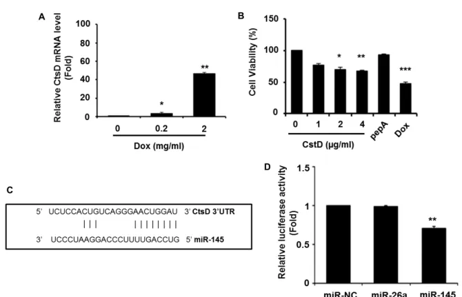
Fig. 1. Suppression of cellular viability in CtsD over-expressed cells. (A) Doxorubicin (Dox) was dose-dependently treated to 3T3-L1 cells. The CtsD level was determined by RT-qPCR after normalization with GAPDH. Values are means ± SEM of triplicate.
*
p
<0.05 versus control (0 mg/ml of Dox). **p
<0.01 versus control (0 mg/ml of Dox). (B) 3T3-L1 cells were transfected with the CtsD expression vector by dose-dependent manner. As control, CtsD inhibitor pestatinA (pepA) or Dox was treated into cells. At 24 hr post transfection, cell viability was measured using a WST-1 assay. *p
<0.05 versus control (0 μg/ml of CtsD). **p
<0.01 versus control (0 μg/ml of CtsD). ***p
<0.001 versus control (0 μg/ml of CtsD). (C) As a CtsD target microRNA, miR-145 was predicted by miRanda algorithm program. The miR-145 binding sites are resided at nucleotides 22-31 of CtsD 3’UTR. (D) For luciferase reporter assays, psiCHECK2 vector containing the full length CtsD 3’UTR was trans- fected with miR-145 mimic, miR-negative control (miR-NC) or miR-26a mimic in COS7 cells. Therenilla
luciferase activities were normalized with firefly luciferase activity. Data was expressed as a relative ratio to the miR-NC mimic. Values are shown as means ± SEM, n =3. **p
<0.01 versus miR-NC.Dox 약물을 처리한 세포의 생존율 감소 보다는 못했지만, 세 포내 CtsD plasmid의 농도가 증가할 수록 3T3-L1 세포주의 생존율도 점진적으로 줄어들었다 (Fig. 1B). CtsD inhibitor인 pepstatin A (pepA)가 첨가된 세포에서는 생존율에 큰 변화가 없었다 . 이러한 결과는 CtsD가 지방전구세포의 생존율에 직 접 영향을 미치고 있음을 나타낸다 .
다음으로 세포생존과 관련있는 CtsD 유전자의 발현을 제어 하는 microRNA을 발굴하기 위해 miRanda program을 활용 하여 CtsD 3’ UTR에 결합하는 잠정적인 microRNA을 조사하 였다 . 그 결과 2종의 microRNA (miR-22와 miR-145)가 micro- RNA support vector regression (mirSVR) 알고리즘에 의해 탐색되었다 . 이 중 mirSVR score가 보다 높은 miR-145 (mirSVR score: -0.1846)을 선발하였다(Fig. 1C). 이를 토대로 CtsD 3’
UTR 부위를 함유하는 psiCHECK reporter vector (psiCHECK- CtsD)를 클로닝하여 miR-145와의 결합 여부를 luciferase re- porter assay로 조사하였다. COS7 세포주에 psiCHECK-CtsD
벡터와 miR-145 mimic을 도입하여 luciferase 활성을 측정한 결과 microRNA negative control (miR-NC)에 비해 miR-145 mimic이 도입된 세포에서 luciferase 활성이 유의하게 감소하 였다 (Fig. 1D). CtsD 유전자를 표적으로 사용하지 않는 miR- 26a mimic은 luciferase 활성에 큰 영향을 미치지 못했다. 따라 서 , miR-145가 CtsD 유전자의 전사후 발현 조절에 직접 관여 하고 있음을 알 수 있다 .
MiR-145에 의한 CtsD mRNA 발현 제어 조사
3T3-L1 세포주에서 miR-145에 의한 내인성 CtsD 유전자의
발현 변화를 조사하기 위해 RT-qPCR을 수행하였다. 지방전구
세포에 miR-145 mimic을 transfection한 후 miR-145의 세포내
도입여부를 확인하기 위해 miR-145 발현을 조사하였다. 그 결
과 miR-145 발현이 대조군(miR-NC)에 비해 7,000배 증가하여
세포내로 miR-145 mimic이 도입 되었음을 확인하였다. 도입
된 miR-145에 의한 CtsD mRNA 발현을 조사 결과 대조군
A
B
C
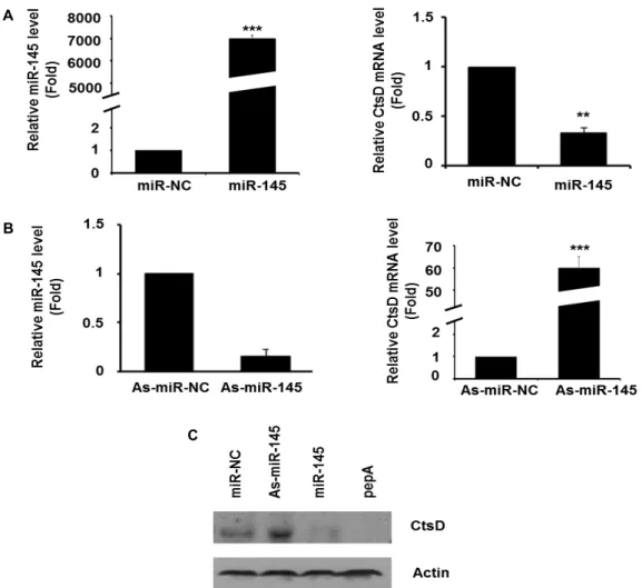
Fig. 2. The expression of CtsD in 3T3-L1 cells by miR-145. (A) 3T3-L1 cells were transfected with 50nM miR-NC or miR-145 mimic respectively. The levels of miR-145 and CtsD were determined by RT-qPCR. Values are means ± SEM of triplicate. **
p
<0.01 versus miR-NC. ***
p
<0.001 versus miR-NC. (B) 3T3-L1 cells were transfected with As-miR-NC or As-miR-145 mimic for 24 hr. The expressions of miR-145 and CtsD were determined by RT-qPCR. Values are means ± SEM of triplicate. ***p
<0.001 versus miR-NC. (C) After transfection with miR-145 mimic, As-miR-145 mimic or miR-NC, cells were lysed and Western blot analysis was conducted with antibodies against CtsD and actin. Data are a representative example of 3 in- dependent experiments.(miR-NC)에 비해 발현이 75% 감소하였다(Fig. 2A). 이외에도, miR-145 발현을 억제하는 miR-145 inhibitor (As-miR-145)을 세포주에 도입한 다음 발현을 조사한 결과 대조군 (As-miR- NC)에 비해 CtsD mRNA 발현이 대략 60배 이상 증가하였다 (Fig. 2B). 이러한 miR-145에 의한 CtsD mRNA 발현 변화가 CtsD 단백질에도 영향을 미치는지 조사하기 위해 Western blot을 실시하였다. 3T3-L1 세포주에 miR-NC가 도입된 경우 CtsD 단백질이 약하게 발현되고 있었으나, miR-145 inhibitor (As-miR-145)을 함유한 세포에서는 CtsD 단백질 발현이 증가 하였다 . miR-145 mimic이 도입된 세포에서는 CtsD inhibitor 인 pepA가 처리된 세포와 유사하게 CtsD 단백질이 거의 감지 되지 않았다 (Fig. 2C). 이러한 결과는 세포에서 miR-145가 CtsD 유전자의 발현을 직접 제어하고 있음을 나타낸다.
MiR-145에 의한 세포생존율 조사
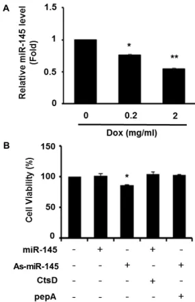
3T3-L1 세포주에서 Dox에 의한 CtsD 발현 증가를 관찰하 였기 때문에 이번에는 CtsD 유전자 발현을 억제하는 miR-145 의 발현을 조사하였다 . 농도가 다르게 Dox을 지방전구세포에 처리하고 miR-145 발현을 관찰한 결과 CtsD mRNA 발현과 반대로 miR-145 발현이 농도 의존적으로 감소하였다(Fig.
3A). 또한 세포생존율도 miR-145 inhibitor (As-miR-145)가 도 입된 세포에서 대략 20% 감소하였다(Fig. 3B). MiR-145 mimic 이 도입된 세포에서는 CtsD 발현여부와 관계없이 생존율에 변화가 없었으며 , As-miR-145을 함유한 세포에서도 CtsD in- hibitor인 pepA가 처리된 세포에서는 생존율에 차이가 없었 다 .
이러한 결과를 종합하면 지방전구세포에서 miR-145가
CtsD 발현을 제어하여 세포 사멸을 억제할 수 있음을 알 수
A
B
As-miR-145
Fig. 3. Cell viability of 3T3-L1 cells exposed to miR-145. (A) The expression of miR-145 in cells treated with Dox was determined by RT-qPCR. Data are means ± SEM of triplicate. *
p
<0.05 versus control (0 mg/ml of Dox). **p
<0.01 versus control (0 mg/ml of Dox). (B) Cellular viability in 3T3-L1 cells was determined by WST-1 assay. Cells were transfected with the combination of miR-145 mimic, As-miR-145 mimic, CtsD vector, or pepA. Values are shown as means ± SEM, n =3. *p
<0.05 versus control (no treatment).있다 . 따라서 지방전구세포의 사멸을 유도하기 위해서는 miR- 145 발현 제어가 주요한 표적이 될 수 있을 것으로 생각된다.
기존 연구에 의하면 지방분화단계에서 miR-145 발현이 증가 하여 KLF4, KLF5, IRS1 등의 표적유전자 발현을 제어하는 것 으로 알려져 있다 [2, 9, 16]. KLF4와 KLF5는 zinc-finger pro- tein으로 PPARγ와 C/EBP 유전자를 활성화시켜 지방분화를 촉진시키고 , IRS1은 인슐린에 의해 유도되는 지방분화과정의 중요한 매개자로 작용한다 [1]. 즉, 지방분화시 miR-145는 지방 분화 제어를 통해 균형 있는 분화과정을 유도하기 위한 항상 성 유지에 중요한 역할을 하는 것으로 생각된다 . 이외에도, human adipocyte에서 ADAM17은 miR-145의 표적유전자로 작용하여 세포내 TNF-α 경로를 활성화시켜 지방분해과정 (lipolysis)을 촉진시킨다[14]. TNF-α는 지방전구세포의 분화 를 억제하고 , 지방세포의 apoptosis을 유도한다[17, 19]. 이러 한 지방분화과정에서와 달리 지방전구단계에서 miR-145 역할
은 아직 보고된 것이 없다 . 따라서, 본 연구에서CtsD 유전자의 발현을 miR-145가 제어하여 지방전구세포의 세포사멸을 억제 시킬 수 있음을 보고하였다 . 이러한 연구는 향후 비만 예방 및 치료를 위한 지방세포 사멸 기전을 규명하는데 중요한 실 마리를 제공할 수 있을 것으로 기대한다 .
감사의 글
본 논문은 농촌진흥청 차세대 바이오그린 21사업(과제번호 PJ008116)의 지원에 의해 이루어진 것임.
References
1. Ali, A. T., Hochfeld, W. E., Myburgh, R. and Pepper, M.
S. 2013. Adipocytea and adipogenesis.
Eur J Cell Biol
92, 229-236.2. Birsoy, K., Chen, Z. and Friedman, J. 2008. Transcriptional regulation of adipogenesis by KLF4.
Cell Metab
7, 339-347.3. Chiellini, C., Costa, M., Novelli, S. E., Amri, E. Z., Benzi, L., Bertacca, A., Cohen, P., Del Prato, S., Friedman, J. M.
and Maffei, M. 2003. Identification of cathepsin K as a novel marker of adiposity in white adipose tissue.
J Cell Physiol
195, 309-321.4. Conus, S., Perozzo, R., Reinheckel, T., Peters, C., Scapozza, L., Yousefi, S. and Simon, H. U. 2008. Caspase-8 is activated by cathepsin D initiating neutrophil apoptosis during the resolution of inflammation.
J Exp Med
205, 685-698.5. Eguchi, A. and Feldstein, A. E. 2013. Lysosomal Cathepsin D contributes to cell death during adipocyte hypertrophy.
Adipocyte
2, 170-175.6. Emert-Sedlak, L., Shangary, S., Rabinovitz, A., Miranda, M.
B., Delach, S. M. and Johnson, D. E. 2005. Involvement of cathepsin D in chemotherapy-induced cytochrome c release, caspase activation, and cell death.
Mol Cancer Ther
4, 733- 742.7. Cornicka, A., Fettig, J., Eguchi, A., Berk, M. P., Thapaliya, S., Dixon, L. J. and Feldstein, A. E. 2012. Adipocyte hyper- trophy is associated with lysosomal permeability both in vivo and in vitro: role in adipose tissue inflammation.
Am J Physol Endocrinol Metab
303, E597-E606.8. Gregersen, L. H., Jacobsen, A. B., Frankel, L. B., Wen, J., Krogh, A. and Lund, A. H. 2010. MicroRNA-145 targets YES and STAT1 in colon cancer cells.
PLoS One
5, e8836.9. Guo, Y., Chen, Y., Zhang, Y., Zhang, Y., Chen, L. and Mo, D. 2012. Up-regulated miR-145 expression inhibits porcine preadipocytes differentiation by targeting IRS1.
Int J Biol Sci
8, 1408-1417.10. Hajer, G. R., van Haeften, T. W. and Visseren, F. L. 2008.
Adipose tissue dysfunction in obesity, diabetes, and vas- cular diseases.
Eur Heart J
29, 2959-2971.11. He, L. and Hannon, G. J. 2004. MicroRNAs: small RNAs with a big role in gene regulation.
Nat Rev Genet
5, 522-531.12. Im, E. and Kazlauskas, A. 2007. The role of cathepsins in
초록:지방세포에서 microRNA-145에 의한 Cathepsin D의 발현 제어 김현지
1․배인선
1․서강석
2*․김상훈
1*
(
1경희대학교 생물학과 ,
2순천대학교 동물자원과학과 )
Cathepsin D (CtsD)는 아스파르트산 단백질 분해효소로서 cytochrome C의 방출을 유도하여 apoptosis 기전을 활성화시킨다 . 본 연구에서는 3T3-L1 지방전구세포에서 CtsD 발현 조절에 관여하는 microRNA에 대해 조사하였 다 . 먼저 지방전구세포 사멸시 CtsD 발현 변화를 관찰하기 위하여 DNA damage agent인 doxorubicin을 3T3-L1 세포주에 노출시켜 CtsD 발현이 증가함을 확인하였다. 또한 지방전구세포주에서 CtsD가 과발현되면 세포 생존율 이 감소하였다 . miRanda program을 이용하여 CtsD 유전자를 표적으로 하는 microRNA를 탐색하여 miR-145를 선발하였다 . Luciferase reporter assay에 의해 miR-145가 CtsD 유전자의 3’ UTR 부위에 결합하여 luciferase 활성 을 감소시킴을 관찰하였다 . 3T3-L1 세포주에 miR-145 mimic을 도입한 결과 CtsD mRNA 발현과 단백질 수준이 감소하였다 . 또한 세포주에 doxorubicin을 처리한 결과 CtsD 유전자 발현 증가와 상반되게 miR-145 발현이 감소 하였다 . 이외에도 miR-145 inhibitor을 세포에 도입하면 세포 생존율이 감소하였다. 이러한 결과는 지방전구세포 의 세포사멸에 CtsD가 관여할 수 있으며, miR-145에 의해 CtsD 발현이 직접 조절되고 있음을 나타낸다. 따라서, 지방전구세포의 사멸을 유도하기 위해서는 miR-145 발현 제어가 주요한 표적이 될 수 있을 것으로 생각된다.
본 연구결과는 향후 비만 예방 및 치료를 위한 지방세포 사멸기전 규명에 중요한 기초 자료를 제공할 수 있을 것으로 기대한다 .
ocular physiology and pathology.
Exp Eye Res
84, 383-388.13. Kim, V. N. 2005. MicroRNA biogenesis: coordinated crop- ping and dicing.
Nat Rev Mol Cell Biol
6, 376-385.14. Lorente-Cebrian, S., Mejhert, N., Kulyte, A., Laurencikiene, J., Astrom, G., Heden, P., Ryden, M. and Arner, P. 2014.
MicroRNAs regulates human adipocyte lipolysis: effects of miR-145 are linked to TNF-alpha.
PLOS One
9, e86800.15. Minarowska, A., Minarowski, L., Karwowska, A. and Gacko, M. 2007. Regulatory role of cathepsin D in apoptosis.
Folia Histochem Cytobiol
45, 159-163.16. Oishi, Y., Manabe, I., Tobe, K., Tsushima, K., Shindo, T., Fujiu, K. et al. 2005. Kruppel-like transcription fator KLF5 is a key reguator of adipocyte differentiation.
Cell Metab
1, 27-39.17. Petruschke, T. and Hauner, H. 1993. Tumor necrosis fac- tor-alpha prevents the differentiation of human adipocyte precursor cells and causes delipidtaion of newly developed fat cells.
J Clin Endocrinol Metab
76, 742-747.18. Poy, M. N., Spranger, M. and Stoffel, M. 2007. microRNAs and the regulation of glucose and lipid metabolism.
Diabetes Obes Metab
9, 67-73.19. Prins, J. B., Niesler, C. U., Winterfor, C .M., Bright, N. A., Siddle, K., O'Rahilly, S., Walker, N. I. and Cameron, D. P.
1997. Tumor necrosis factor-alpha induces apoptosis of hu- man adipose cells.
Diabetes
46, 1939-1944.20. Prins, J. B. and O'Rahilly, S. 1997. Regulation of adipose cell number in man.
Clin Sci (Lond)
92, 3-11.21. Taleb, S., Cancello, R., Clement, K. and Lacasa, D. 2006.
Cathepsin s promotes human preadipocyte differentiation:
possible involvement of fibronectin degradation.
Endocrinol- ogy
147, 4950-4959.22. Trajkovski, M., Hausser, J., Soutschek, J., Bhat, B., Akin, A., Zavolan, M., Heim, M. H. and Stoffel, M. 2011. MicroRNAs 103 and 107 regulate insulin sensitivity.
Nature
474, 649-653.23. Xie, H., Lim, B. and Lodish, H. F. 2009. MicroRNAs induced during adipogenesis that accelerate fat cell development are downregulated in obesity.
Diabetes
58, 1050-1057.24. Yang, M., Zhang, Y., Pan, J., Sun, J., Liu, J., Libby, P., Sukhova, G. K., Doria, A., Katunuma, N., Peroni, O. D., Guerre-Millo, M., Kahn, B. B., Clement, K. and Shi, G. P.
2007. Cathepsin L activity controls adipogenesis and glucose tolerance.
Nat Cell Biol
9, 970-977.25. Zhang, Y. and Huang, C. 2012. Targeting adipocyte apopto- sis: a novel strategy for obesity therapy.