천연보존료 복합 황금추출물이 유산균발효 마늘추출물의 저장성 및 기능성에 미치는 영향
이희섭1․이성진2․손요한2․유희종2․조홍연1
1고려대학교 식품생명공학과
2에스케이 바이오랜드(주) 기술연구원 식품연구소
Effects of Mixed Scutellaria baicalensis Extracts as Natural Preservative on Efficacy and Storage of Lactic Acid-Fermented Garlic Extract
Hee-Seop Lee1, Sun-Jin Lee2, Johann Sohn2, Heui-Jong Yu2, and Hong-Yon Cho1
1Department of Food and Biotechnology, Korea University
2Food R&D Center, SKbioland Co., Ltd.
ABSTRACT The natural preservative “complex Scutellaria baicalensis extract (BHC)” contains Scutellaria baica- lensis, Glycyrrhiza uralensis (liquorice), Zizyphus jujube (jujube), and Astragalus propinquus (milk vetch root). BHC has been used as a natural preservative for more than 10 years to increase storage duration and quality of food with strong antibacterial activity. BHC has been added into functional foods as a subsidiary ingredient. However, no studies have been performed to test whether or not BHC affects the activity of main functional ingredients. In this study, we tested whether or not BHC has any effect on the hepatoprotective activity of lactic acid-fermented garlic extract (LAFGE) when formulated in a clinical test supplement. H2O2-induced oxidative damage in HepG2 cells was not attenuated by BHC, indicating that BHC had no influence on the protective effect of LAFGE against oxidative damage.
Furthermore, BHC had no effect on the hepatoprotective effect of LAFGE against acetaminophen-induced acute liver injury in rats, as indicated by no changes in alanine transaminase and aspartate transaminase levels. In conclusion, BHC, formulated in the clinical test supplement with LAFGE, had no effect on hepatoprotective activity, indicating BHC could be considered as a suitable natural preservative for liquefied functional food materials.
Key words: lactic acid-fermented garlic, mixed Scutellaria baicalensis extracts, liver-protective, efficacy, storage
Received 6 September 2016; Accepted 10 September 2016 Corresponding author: Hong-Yon Cho, Department of Food and Biotechnology, Korea University, Sejong 30019, Korea
E-mail: [email protected], Phone: +82-44-860-1433
서 론
본 연구의 저자들은 마늘의 기능성 강화를 목적으로 유산 균 발효를 적용하여 유산균발효 마늘추출물을 개발하였고, 알코올성 지방간 예방 효능에 관한 연구(1,2), 비알코올성 지방간 예방 효능에 관한 연구(3), 급성 간 손상 예방 효능에 관한 연구(4)를 보고한 바 있다. 유산균발효 마늘추출물은 발효 후 S-allyl cysteine(SAC), S-ethyl cysteine(SEC), S-methyl cysteine(SMC) 등의 유기황화합물들이 증가하 는 것을 확인하였으며, 이로 인해 증가한 항산화 활성이 산 화 스트레스로부터 간 손상을 예방하고 간 기능을 강화하는 것을 확인하였다. 유산균발효 마늘추출물은 마늘의 높은 당 함유량으로 인해 분말화가 어렵고, 분말화를 위해서는 고형 분 대비 2~3배에 해당하는 부형제 첨가가 요구되기 때문에 최종 제품의 제한된 크기와 중량을 고려할 때 분말 형태보다
는 농축액 형태가 적합하다. 그러나 액상제형의 건강기능성 식품은 유통기한 설정 한계가 액상제형의 일반식품보다 길 기 때문에 보존료 첨가가 불가피하다. 또한, 합성보존제보다 천연보존제를 선호하는 구매자들의 성향을 반영하여 액상 제형의 시료 제조 시 천연보존제 사용이 요구된다. 그러나 식물자원을 주원료로 사용하고 있는 건강기능식품에 있어 천연물 유래의 첨가물, 부원료 등의 사용은 주원료의 효능에 영향을 미칠 가능성을 배제할 수 없다. 특히 액상제형의 원 료에 포함되는 천연보존제가 주원료의 효능에 어떠한 영향 을 미치는지에 대한 연구는 현재까지 수행된 바가 없으며, 이에 따라 nutrigenomics를 활용한 정량적 실험과 고찰이 요구되고 있다.
황금추출물은 Bacillus cereus, Listeria monocyto- genes, Escherichia coli, Vibrio parahaemolyticus의 생 육 저해 효능(5)과 항생제 내성균인 Salmonella enter- itidis, Streptococcus aureus,enteroaggregative E. coli 등에 대한 항균력(6)이 보고되어 있고, 본 시험에 사용된 복 합 황금추출물은 주원료인 황금을 비롯해 감초, 대추, 황기 등으로부터 추출한 복합 추출물로, 시중에 사용되고 있는
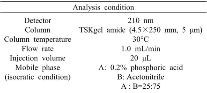
Table 1. Condition for analysis of cycloalliin Analysis condition Detector
Column Column temperature
Flow rate Injection volume
Mobile phase (isocratic condition)
210 nm
TSKgel amide (4.5×250 mm, 5 μm) 30°C
1.0 mL/min 20 μL
A: 0.2% phosphoric acid B: Acetonitrile
A : B=25:75 천연보존료이다. 하지만 천연보존료인 복합 황금추출물이
건강기능식품 주원료의 기능성에 미치는 영향에 대한 연구 는 보고된 바 없다.
따라서 본 연구는 간 기능 개선용 건강기능식품 원료로 개발 중인 유산균발효 마늘추출물과 이에 일정 비율로 첨가 된 천연보존료 복합 황금추출물이 유산균발효 마늘추출물 의 간 보호 효능에 미치는 영향을 검토하기 위해 수행되었 다.
재료 및 방법
재료 및 시약
Thiobarbituric acid(TBA), 환원형/산화형 글루타치온 (GSH), 1,1,3,3-tetraethoxypropane, 5,5’-dithiobis-(2- nitrobenzoic acid)(DTNB), hematoxylin, eosin 등은 Sigma-Aldrich Co.(St. Louis, MO, USA)에서 구입하였다.
혈청 분석기용 시약은 Roche(Basel, Switzerland)에서 구 입하였다. c-Jun N-terminal kinase(JNK), B-cell lym- phoma 2(Bcl-2), Bcl-2 associated X protein(Bax), p53 등의 항체는 Cell Signaling사(Danvers, MA, USA)로부터 구입하였다.
유산균발효 마늘추출물의 제조 및 복합 황금추출물 본 실험에서 사용한 유산균발효 마늘추출물은 경남 가월 영농조합에서 생산한 창녕산 마늘을 구입하여 다음과 같이 조제하였다. 마늘 5 kg과 증류수 10 kg을 발효조(MJS U3, Marubishi, Tokyo, Japan)에서 혼합, 교반하면서 121°C에 서 1시간 동안 멸균하였다. 37°C로 냉각한 후 Lactobacillus plantarum BL2 종균배양액 200 mL를 접종, 2일간 배양하 였다. 배양 종료 후 50°C에서 24시간 동안 숙성 과정을 거친 다음, 멸균, 여과 및 농축하여 유산균발효 마늘추출물로 사 용하였다. 복합 황금추출물은 (주)테코스사(Gangwon, Ko- rea)에서 제조한 제품을 구매하여 사용하였다.
유산균발효 마늘추출물을 이용한 액상제형 시료 제조 유산균발효 마늘추출물 농축액(고형분 60% 이상) 750 mg, 천연보존료로써 복합 황금추출물 10 mg, 액상과당 1,000 mg을 정제수 18,240 mg에 용해하여 총 20,000 mg 중량으로 제조하여 파우치 포장 후 105°C에서 30분간 살균 하였다.
유산균발효 마늘추출물을 이용한 액상제형 시료의 경시 안 정성 확인
제조된 유산균발효 마늘추출물 액상제형 시료의 경시 안 정성을 확인하기 위하여 포장된 파우치 액상 복합물을 4, 25, 45°C에서 4개월간 보관하며 1개월마다 유산균발효 마 늘추출물의 지표성분인 cycloalliin의 함량을 HPLC(Alli- ance HPLC, Waters Co., Milford, MA, USA)를 이용하여
Table 1의 조건으로 분석하였다. 또한, 1개월마다 식품공전 에 명시된 방법에 따라 일반세균수를 측정하였다.
동물실험
7주령 수컷 Wistar rat은 (주)오리엔트바이오(Seong- nam, Korea)에서 구입하였고, 온도 22±3°C, 상대습도 50
±10%, 조도 200~300 lux, 12시간 간격의 명암 주기 환경 에서 1주일간 적응시킨 후 실험에 사용하였다.
대조군(vehicle), acetaminophen(APAP) 처리군(APAP), 유산균발효 마늘추출물 투여군(APAP+G), 복합 황금추출 물 투여군(APAP+B), 복합 황금추출물이 포함된 유산균발 효 마늘추출물 투여군(APAP+G+B)의 5그룹으로 분류하 였다. APAP+G 그룹은 유산균발효 마늘추출물 500 mg/
kg, APAP+B 그룹은 복합 황금추출물 6.7 mg/kg, APAP+
G+B 그룹은 유산균발효 마늘추출물 500 mg/kg 및 복합 황금추출물 6.7 mg/kg을 하루 한 번, 7일간 경구투여 하였 고, vehicle 그룹은 생리식염수를 경구투여 하였다. 복합 황 금추출물의 투여 용량은 유산균발효 마늘추출물 액상 파우 치 제조 시 포함된 복합 황금추출물이 유산균발효 마늘추출 물의 중량 대비 1/75이기 때문에 유산균발효 마늘추출물의 투여 용량 대비 1/75 중량으로 투여하였다. 시료의 마지막 경구투여 2시간 후, vehicle 그룹을 제외한 모든 그룹은 2%
Tween 80에 현탁한 APAP 1.5 g/kg을 경구투여 하였고, vehicle 그룹은 2% Tween 80을 경구투여 하였다. 24시간 후 마취하여 심장으로부터 채혈한 다음 혈장을 분리하였고, 혈장과 간 조직은 -80°C에 보관하며 분석에 사용하였다.
본 실험은 고려대학교 실험동물윤리위원회의 승인을 받았 으며, 규정에 따라 실시하였다(KUIACUC-2016-149).
세포 내 reactive oxygen species(ROS) 소거 활성 평가 Wang과 Joseph(7)의 방법에 따라 세포 내 ROS를 측정하 였다. HepG2 cell은 5×10⁵ cells/well 농도로 96-well plate에 접종하여 24시간 배양하였다. 80 μL의 DMEM 배지 로 교체한 후, 250 μL의 DCFH-DA 10 μL, test sample 10 μL를 첨가한 다음 1시간 배양하였다. 배지를 제거하고 PBS로 2회 세척한 후, warm PBS에 희석한 500 μM의 H2O2를 처리한 다음 1시간 후에 VICTOR3(Perkin Elmer, Waltham, MA, USA)를 사용하여 excitation 485 nm, emis-
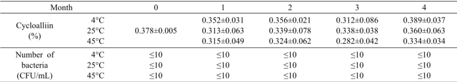
Table 2. Stability of lactic acid-fermented garlic extract included complex Scutellaria baicalensis extract during accelerated experiment
Month 0 1 2 3 4
Cycloalliin (%)
4°C 25°C 45°C
0.378±0.005
0.352±0.031 0.313±0.063 0.315±0.049
0.356±0.021 0.339±0.078 0.324±0.062
0.312±0.086 0.338±0.038 0.282±0.042
0.389±0.037 0.360±0.063 0.334±0.034 Number of
bacteria (CFU/mL)
4°C 25°C 45°C
≤10
≤10
≤10
≤10
≤10
≤10
≤10
≤10
≤10
≤10
≤10
≤10
≤10
≤10
≤10 The accelerated experiment was performed under various conditions. The index compound (cycloalliin) and the number of bacteria were analyzed monthly for 4 months during the lactic acid-fermented garlic extract samples have been stored at 4, 25, and 45°C.
sion 535 nm에서 형광도를 측정하였다.
글루타치온 함량 분석
Rahman 등(8)의 방법에 의해 글루타치온 함량을 측정하 였다. HepG2 cell은 6×10⁶ cells/well 농도로 6-well plate 에 접종하여 24시간 배양한 후 test sample을 첨가하여 24 시간 배양하였다. 4 mM의 H2O2를 처리하고 24시간 후, GSH extraction buffer[0.1% triton X-100, 0.6% sulfo- salicylic acid in 0.1 M potassium phosphate buffer(pH 7.5)]를 1 mL 첨가하여 homogenization 후 13,000 rpm에 서 5분간 원심분리 하여 상등액을 분석에 사용하였다. 간 조직의 경우 50 mg의 조직에 GSH extraction buffer 1 mL 를 첨가한 후, 상기 기술한 방법에 의해 GSH를 추출하였다.
Total glutathione을 측정하기 위해 상등액 20 μL, DTNB 60 μL, GSH reductase 60 μL를 혼합하여 1분간 반응시킨 다음 NADPH 60 μL를 첨가하고 5분 후 412 nm에서 흡광도 를 측정하였다. 산화형 GSH(GSSG)를 측정하기 위해 상등 액 100 μL에 2-vinylpyridine 2 μL를 첨가하여 1시간 동안 GSH를 masking 한 후, 6 μL의 triethanolamine을 첨가하 여 10분간 중성화하였다. 그다음 total GSH 측정 방법과 같은 방법으로 GSSG를 측정하였다.
혈청 분석
혈청분석기(Cobas c-111, Roche, Basel, Switzerland) 를 이용하여 혈중 ALT, AST 수치를 측정하였다.
Hematoxylin & eosin staining
간 조직은 10% formalin에 고정한 후 paraffin으로 em- bedding 한 다음, microtome을 이용하여 5 μm 두께로 세절 하여 slide glass에 부착시켰다. 간 조직 slide는 hematox- ylin과 eosin으로 염색한 후 광학현미경으로 조직학적 변화 를 관찰하였다.
지질과산화물 함량 측정
Fraga 등(9)의 방법에 따라 지질과산화물의 함량을 측정 하였다. 간 조직 100 mg을 1.15% KCl solution에 균질화한 후, 13,000 rpm에서 5분간 원심분리 하여 상등액을 TBARS assay에 사용하였다. 상등액 200 μL, 2% phosphoric acid
100 μL, 0.6% thiobarbituric acid solution 200 μL를 혼합 한 후, 100°C에서 20분간 반응시켰다. 13,000 rpm에서 5 분간 원심분리 한 후 상등액을 취하여 540 nm에서 흡광도 를 측정하였다. 표준물질은 1,1,3,3-tetraethoxypropane 을 사용하여 같은 방법으로 표준곡선을 작성하였고, 이로부 터 간 조직 내의 지질과산화물의 함량을 산출하였다.
Western blot
HepG2 cell은 6×10⁶ cells/well 농도로 6-well plate에 접종하여 24시간 배양한 후 test sample을 첨가하여 24시 간 배양하였다. 4 mM의 H2O2를 처리하고 24시간 후 RIPA buffer를 첨가하여 homogenization 한 다음, 13,000 rpm 에서 20분간 원심분리 하여 상등액을 취하였다. 간 조직의 경우 조직 100 mg에 RIPA buffer 1 mL를 첨가한 후 상기 방법에 의해 단백질을 추출하였다. 상등액은 Bradford as- say를 통해 protein 함량을 측정한 후 같은 양의 protein을 SDS-PAGE로 분리한 다음 PVDF membrane에 transfer 하고 5% BSA로 blocking 후, 1차 antibody를 4°C에서 overnight 반응시키고 3회 washing 한 후 2차 antibody를 상온에서 1시간 반응시킨 다음 ECL solution을 이용하여 발광시킨 뒤 LAS imagequant 2000을 이용하여 촬영하였 다.
통계처리
통계처리는 Statistical Analysis System(SAS) pro- gram(SAS 8.2, SAS Institute Inc., Cary, NC, USA)을 이 용하여 one-way analysis of variance(ANOVA) test와 Duncan’s multiple range test를 실시하여 P<0.05에서 통 계적 유의성을 검정하였다.
결과 및 고찰
복합 황금추출물이 포함된 유산균발효 마늘추출물 액상제 형의 경시 안정성
복합 황금추출물이 첨가된 유산균발효 마늘추출물 액상 제형을 4, 25, 45°C에서 4개월간 보관하며 지표성분의 함량 과 일반세균수를 측정하여 안정성을 확인하였다. 그 결과 유산균발효 마늘추출물의 지표성분인 cycloalliin의 함량은
0 50 100 150 200 250 300
Relative DCF fluorescence . intensity (%) .
Vehicle H2O2 H2O2+G H2O2+B H2O2+G+B
***
*** ***
N/A
Fig. 1. Effect of complex Scutellaria baicalensis extract as a natural preservative to intracellular reactive oxygen species (ROS) scavenging activity of lactic acid-fermented garlic extract in HepG2 cells. HepG2 cells were incubated with test sample and DCFH-DA as a fluorescence probe to ROS for 1 h, and then incubated with 500 μM of H2O2. After 1 h, fluorescence intensity was measured with VICTOR3 fluorometer at 485 nm (excitation) and 535 nm (emission). G, lactic acid-fermented gar- lic extract; B, complex Scutellaria baicalensis extract; G+B, lac- tic acid-fermented garlic extract included complex Scutellaria baicalensis extract. Data represent mean±SD (n=3). Asterisks (*) above the error bar indicate significant differences versus H2O2
group. ***P<0.001.
0.0 0.1 0.2 0.3 0.4 0.5 0.6 0.7 0.8 0.9 1.0
GSH (μM/mg protein) .
Vehicle H2O2 H2O2+G H2O2+B H2O2 +G+B
** *** ***
A N/A
0.0 0.1 0.1 0.2 0.2 0.3 0.3
GSSG (μM/mg protein) .
Vehicle H2O2 H2O2+G H2O2+B H2O2
+G+B
**
*
B N/A
0.0 1.0 2.0 3.0 4.0 5.0
GSH/GSSG ratio .
Vehicle H2O2 H2O2+G H2O2+B H2O2
+G+B
**
** **
C N/A
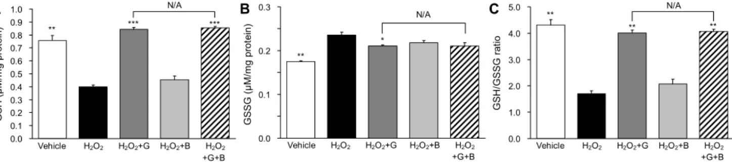
Fig. 2. Influence of complex Scutellaria baicalensis extract as a natural preservative to glutathione restoring effect of lactic acid-fer- mented garlic extract in H2O2-treated HepG2 cells. HepG2 cells were incubated with test sample for 24 h and further incubated with 4 mM of H2O2 for 24 h. Cellular glutathione was extracted, and detected by as described in Materials and Methods. Data represent mean±SD (n=3). G, lactic acid-fermented garlic extract; B, complex Scutellaria baicalensis extract; G+B, lactic acid-fer- mented garlic extract included complex Scutellaria baicalensis extract. Asterisks (*) above the error bar indicate significant differences versus H2O2 group. *P<0.05, **P<0.01, ***P<0.001.
4개월 후에도 초기 수치와 유의적 차이 없이 안정적으로 유 지되는 것을 확인하였으며, 일반세균의 증식이 억제되었다 (Table 2). 유산균발효 마늘추출물을 이용한 액상제형 시료 에 함유된 0.05%의 복합 황금추출물은 유산균발효 마늘추 출물의 저장성을 유지하기에 적합한 것으로 확인되었다.
H2O2-유도 산화적 손상에 대한 발효 마늘추출물의 간 보호 효능에 미치는 복합 황금추출물의 영향
유산균발효 마늘추출물에 포함된 복합 황금추출물이 유 산균발효 마늘추출물의 세포 내 ROS 소거 활성에 미치는 영향을 검토하였다. 유산균발효 마늘추출물(H2O2+G)은 4 mg/mL, 복합 황금추출물(H2O2+B)은 유산균발효 마늘추출 물 대비 1/75 중량으로 약 0.05 mg/mL로 처리하였고, 이들 의 혼합물(H2O2+G+B) 또한 비교 평가하였다. 그 결과 H2O2 를 처리한 그룹은 대조군 대비 세포 내 ROS가 255%로 증가 하였고, 유산균발효 마늘추출물 처리군, 복합 황금추출물 처 리군, 유산균발효 마늘/복합 황금추출물 처리군은 각각 191
%, 254%, 191%로, 복합 황금추출물 단독 처리군은 세포 내 ROS에 대해 소거 활성이 나타나지 않았으며, 또한 유산 균발효 마늘추출물의 항산화 효능에도 영향이 없음을 확인 하였다(Fig. 1).
또한, 세포 내 총 GSH, 환원형 GSH, 산화형 GSH(GSSG) 측정 결과 H2O2를 처리한 그룹은 세포 내 GSH가 0.4 μM로 감소하였고, 유산균발효 마늘추출물 처리군, 복합 황금추출 물 처리군, 유산균발효 마늘추출물/복합 황금추출물 처리군 은 각각 0.8, 0.5, 0.9 μM로 증가하였다(Fig. 2A). GSSG는 대조군, H2O2 처리군, 유산균발효 마늘추출물 처리군, 복합 황금추출물 처리군, 유산균발효 마늘추출물/복합 황금추출 물 처리군에서 각각 0.18, 0.24, 0.21, 0.22, 0.21 μM로, H2O2 처리군에 대해서 유산균발효 마늘추출물 처리군에서 만 유의적인 GSSG 감소 효과를 나타내었고, 유산균발효 마 늘추출물 처리군과 유산균발효 마늘추출물/복합 황금추출 물 혼합 처리군 사이의 유의차는 나타나지 않았다(Fig. 2B).
GSSG에 대한 GSH 함량의 비율을 비교한 결과 대조군,
H2O2 처리군, 유산균발효 마늘추출물 처리군, 복합 황금추 출물 처리군, 혼합처리군에서 각각 4.3배, 1.7배, 4.0배, 2.1 배, 4.1배로 나타났다(Fig. 2C). 유산균발효 마늘추출물은 H2O2에 대해 glutathione depletion을 효과적으로 억제해 간 손상을 예방한 것으로 생각되며, 이에 대해 유산균발효 마늘추출물 대비 1/75 중량비로 혼합된 복합 황금추출물은 단독 투여 시 항산화 효능이 나타나지 않았으며, 유산균발효 마늘추출물에 대한 영향은 나타나지 않았다.
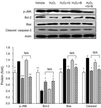
복합 황금추출물이 유산균발효 마늘추출물의 산화적 간 손상 보호 효능에 미치는 영향을 protein level에서 검토하 였다. DNA damage, 산화 스트레스 등에 반응하여 세포자 멸을 유도하는 mitogen-activated protein kinase(MAPK) family 중 하나인 JNK, anti-apoptotic protein으로 알려진
p-JNK Bcl-2 Bax Cleaved caspase-3 Actin
Vehicle H2O2 H2O2+G H2O2+B H2O2
+G+B
0.0 0.2 0.4 0.6 0.8 1.0 1.2 1.4 1.6 1.8
Protein (fold) .
p-JNK Bcl-2 Bax Cleaved caspase-3
* * * *
** **
**
* *
**
*** **
N/A N/A N/A
N/A
Fig. 3. Influence of complex Scutellaria baicalensis extract as a natural preservative to effect of lactic acid-fermented garlic extract on apoptosis-related protein expression in H2O2-treated HepG2 cells. HepG2 cells were incubated with test sample for 24 h and further incubated with 4 mM of H2O2 for 24 h. Proteins were extracted and quantificated by Bradford’s assay, and sepa- rated by SDS-PAGE. Transferred proteins to PVDF membrane were detected by p-JNK, Bcl-2, Bax, caspase-3 antibodies. Data represent mean±SD (n=3). G, lactic acid-fermented garlic ex- tract; B, complex Scutellaria baicalensis extract; G+B, lactic acid-fermented garlic extract included complex Scutellaria bai- calensis extract. Asterisks (*) above the error bar indicate sig- nificant differences versus H2O2 group. *P<0.05, **P<0.01, ***P<
0.001.
Bcl-2, pro-apoptotic protein으로 알려진 Bax 등을 검토 하였다. 산화스트레스는 JNK의 인산화를 유도하며, 인산화 된 JNK는 Bcl-2의 작용을 저해하고, Bax의 작용을 활성화 하여 apoptosis signal을 증폭시킨다. Cleaved caspase-3 는 procaspase-3의 활성형으로, apoptosis pathway의 end point effector로써 세포골격을 분해하는 역할을 수행 한다(10,11). 유산균발효 마늘추출물은 산화스트레스에 대 해 Bcl-2는 upregulation, apoptosis 관련 인자들은 down- regulation 함으로써 간세포 보호 효능을 나타내었으며, 복 합 황금추출물은 유산균발효 마늘추출물의 산화스트레스 완화 효능에 대해 영향이 나타나지 않음을 확인하였다(Fig.
3).
Acetaminophen에 의한 급성 간 손상 동물 모델에서 발효 마늘추출물의 간 보호 효능에 미치는 복합 황금추출물의 영향
복합 황금추출물이 유산균발효 마늘추출물의 간 보호 효 능에 미치는 영향을 확인하기 위해 acetaminophen(APAP) 으로 간 손상을 유도한 rat 모델에서 혈중 ALT(GOT) 및
AST(GPT) 수치를 확인하였다. 그 결과 vehicle 그룹은 ALT 30.2 U/L, AST 74.6 U/L, APAP 그룹은 각각 69.3 U/L, 210 U/L로 많이 증가하였고, APAP+B 그룹은 각각 73.3 U/L, 254 U/L로 APAP 그룹과 통계적 유의성이 나타나지 않았다. APAP+G 그룹은 ALT 44 U/L, AST 140.3 U/L APAP+G+B 그룹은 ALT 49.1 U/L와 137.2 U/L로 모두 APAP 그룹보다 유의적으로 감소하였으며, 두 그룹 간의 유 의적 차이는 나타나지 않았다. 혈중 간 손상 수치를 나타내 는 지표인 AST, ALT의 결과로 인체적용 시험용 시료에 포 함된 복합 황금추출물은 유산균발효 마늘추출물과의 해당 배합비에서 유산균발효 마늘추출물의 간 보호 효능에 대해 상승 효과(synergistic effect) 또는 상가 효과(additive ef- fect) 등의 추가적인 영향을 미치지 않음을 확인하였다(Fig.
4).
지질과산화물(MDA) 측정 결과 vehicle 그룹은 179 nM/
mg tissue, APAP 그룹은 275 nM/mg tissue로 증가하였 고, APAP+B 그룹은 276 nM로 APAP 그룹과 통계적인 유 의차가 없었다. APAP+G 그룹과 APAP+G+B 그룹은 각각 250, 248 nM로 두 그룹 간의 유의차는 없었으며, APAP에 대해 유의적으로 MDA 수치를 감소시켰다. 또한, 간 조직 내 글루타치온 함량은 vehicle, APAP, APAP+G, APAP+B, APAP+G+B 그룹 순으로 각각 0.93, 0.7, 0.97, 0.76, 0.92 μm/mg tissue로 나타났고, 복합 황금추출물 단독처리군과 유산균발효 마늘/복합 황금추출물 혼합 처리군 사이의 유의 차는 없었다. 따라서 복합 황금추출물은 유산균발효 마늘추 출물의 지질과산화물 생성 및 글루타치온 고갈 방지 효능에 대해 영향을 미치지 않았음을 확인하였다(Fig. 5).
조직학적 분석 결과 APAP는 조직 괴사와 울혈 증상 및 혈관 주변부에 경미한 지방증(steatosis)이 관찰되었다. 유 산균발효 마늘추출물을 투여한 쥐의 간 조직에서는 괴사, 울혈, 지방증 등의 이상 병변이 관찰되지 않았고, 정상대조 군과 유사하였다. 복합 황금추출물 투여군은 APAP 그룹과 유사한 증상이 관찰되었다. 복합 황금추출물을 포함하는 유 산균발효 마늘추출물을 투여한 쥐의 간 조직은 정상군과 유 사하였고, 이는 복합 황금추출물이 유산균발효 마늘추출물 의 간 보호 효능에 간섭하지 않았음을 의미한다(Fig. 6).
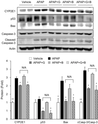
유산균발효 마늘추출물의 간 보호 효능에 대한 복합 황금 추출물의 간섭 여부를 protein level에서 검토하였다. Ace- taminophen의 biotransformation에 관여하는 CYP2E1은 대사 과정 중 친핵성 대사산물인 N-acetylbenzoquinonei- mine(NAPQI)이 일부 생성되며, acetaminophen 과량 복용 시 NAPQI의 과량 축적으로 인해 DNA damage 및 gluta- thione depletion, oxidative stress 등에 의한 간 손상으로 이어진다(12). CYP2E1은 APAP 그룹에서 vehicle 그룹 대 비 2.5배 증가하였고, APAP+G 그룹은 2.0배, APAP+B 그룹은 2.6배로 APAP 그룹과 유의적 차이를 나타내지 않았 고, APAP+G+B 그룹은 2.1배로 유산균발효 마늘추출물 단독 투여군과 유의적 차이가 없었다. Tumor suppressor
0 10 20 30 40 50 60 70 80 90 100
Vehicle APAP APAP+G APAP+B APAP+G+B
ALT (U/L) .
***
**
***
N/A
0 50 100 150 200 250 300
Vehicle APAP APAP+G APAP+B APAP+G+B
AST (U/L) .
N/A
***
** **
Fig. 4. Influence of complex Scutellaria baicalensis extract as a natural preservative to hepatoprotective effect of lactic acid-fermented garlic extract against acetaminophen-induced acute liver injury in rats. Rats were administrated with lactic acid-fermented garlic extract (APAP+G), complex Scutellaria baicalensis extract (APAP+B), and mixture of G and B (APAP+G+B) for 7 days. 2 h later after final administration, APAP administrated at dose of 1.5 g/kg except vehicle group. After 24 h, serum and the liver tissues were collected and analysed. Serum ALT and AST were measured with serum analyzer. Data represent mean±SD. Asterisks (*) above the error bar indicate significant differences versus APAP group. **P<0.01, ***P<0.001.
0 50 100 150 200 250 300 350
Vehicle APAP APAP+G APAP+B APAP+G+B
Hepatic MDA (nM/mg tissue) .
***
N/A
*
*
0.0 0.2 0.4 0.6 0.8 1.0 1.2
Vehicle APAP APAP+G APAP+B APAP+G+B
Hepatic GSH (μM/mg tissue) .
N/A
*** **
**
Fig. 5. Influence of complex Scutellaria baicalensis extract as a natural preservative to effect of lactic acid-fermented garlic extract on hepatic lipid peroxidation and glutathione depletion induced by acetaminophen. Rats were administrated with lactic acid-fermented garlic extract (APAP+G), complex Scutellaria baicalensis extract (APAP+B), and mixture of G and B (APAP+G+B) for 7 days.
2 h later after final administration, APAP administrated at dose of 1.5 g/kg except vehicle group. After 24 h, serum and the liver tissues were collected and analysed. Hepatic MDA and glutathione were detected by thiobarbituric acid reactive substance assay and DTNB method, respectively. Data represent mean±SD. Asterisks (*) above the error bar indicate significantly differences versus APAP group. *P<0.05, **P<0.01, ***P<0.001.
Vehicle APAP APAP+G APAP+B APAP+G+B
x40
x400
Fig. 6. Influence of complex Scutellaria baicalensis extract as a natural preservative to effect of lactic acid-fermented garlic extract in histopathologic changes against acetaminophen-induced acute liver injury in rats. Rats were administrated with lactic acid-fermented garlic extract (APAP+G), complex Scutellaria baicalensis extract (APAP+B), and mixture of G and B (APAP+G+B) for 7 days.
2 h later after final administration, APAP administrated at dose of 1.5 g/kg except vehicle group. After 24 h, the liver sections stained with hematoxylin and eosin.
Vehicle APAP APAP+G APAP+B APAP+G+B
CYP2E1 p53 Bax
Caspase-3 Cleaved Caspase-3 Actin
0 1 1 2 2 3 3 4
Protein (Fold) .
CYP2E1 p53 Bax cCasp-3/Casp-3
N/A N/A
N/A
N/A
** **
* * *
* *
*
*
*
*
□ Vehicle ■ APAP
■ APAP+G ■ APAP+B ▨ APAP+G+B
Fig. 7. Influence of complex Scutellaria baicalensis extract as a natural preservative to effect of lactic acid-fermented garlic extract on liver injury-related proteins expression in APAP-chal- lenged rat livers. Rats were administrated with lactic acid-fer- mented garlic extract (APAP+G), complex Scutellaria baica- lensis extract (APAP+B), and mixture of G and B (APAP+G+
B) for 7 days. Proteins were extracted from rat livers and quanti- ficated by Bradford’s assay, and separated by SDS-PAGE.
Transferred proteins to PVDF membrane were detected by CYP 2E1, p53, Bax, cleaved and procaspase-3 antibodies. cCasp-3/
Casp-3 means the ratio of cleaved caspase-3 (cCasp-3) to pro- caspase-3 (Casp-3). Data represent mean±SD. Asterisks (*) above the error bar indicate significant differences versus APAP group. *P<0.05, **P<0.01.
p53은 DNA가 손상되었을 때 세포 자멸을 일으키는 pro- tein으로(13), vehicle 대비 APAP, APAP+G, APAP+B, APAP+G+B 그룹 순으로 각각 1.2배, 0.5배, 1.0배, 0.4배 발현되었고, APAP 그룹에서 vehicle 그룹 대비 소폭 증가 하였으나 유의적인 차이는 없었고, APAP+B 그룹 또한 ve- hicle 그룹과 유의차는 없었다. 그러나 APAP+G 그룹과 APAP+G+B 그룹은 두 그룹 간의 유의차는 없었으나, APAP 그룹에 대해 p53 발현량이 유의적으로 감소하였다.
Proapoptotic protein인 Bax는 mitochondria membrane 에 pore를 형성하여 mitochondria 내 cytochrome c를 세 포질로 방출하는 역할을 수행하며, cytochrome c는 apop- tosis signal pathway를 촉진하는 중요한 mediator이다.
Vehicle 대비 APAP, APAP+G, APAP+B, APAP+G+B 순으로 각각 2.3배, 1.1배, 1.8배, 1.1배 발현도를 나타내었 고, APAP+G 그룹과 APAP+G+B 그룹에서 APAP 그룹에 대해 유의적으로 감소하였고, APAP+B 그룹은 APAP 그룹
과 유의차가 없었다. Caspase-3는 apoptosis의 endpoint effector로써 세포 골격을 이루는 분자들의 분해를 담당한 다. 비활성형 procaspase-3에 대한 활성형 cleaved cas- pase-3의 비율은 vehicle 그룹 대비 APAP 그룹에서 2.9배 로 증가하였고, APAP+G 그룹과 APAP+G+B 그룹에서 각 각 1.8배, 1.8배로 감소하였고 두 그룹 간의 유의적 차이는 없었다. APAP+B 그룹은 2.7배로 APAP 그룹과 유의적 차 이를 보이지 않았다(Fig. 7).
결과적으로 복합 황금추출물에 포함된 천연물 유래의 성 분들이 유산균발효 마늘추출물의 간 보호 효능에 상승적 또 는 추가적인 영향을 미치지 않는 것을 세포실험과 동물실험 을 통하여 확인하였다. 이는 천연보존제를 비롯해 건강기능 식품의 주원료 이외 첨가되는 천연부원료, 천연첨가물 등이 첨가 농도와 관련하여 주원료의 효능에 영향을 줄 수 있는지 를 검토한 것으로, 향후 천연첨가물이 첨가되어 제조된 건강 기능식품의 기능성 인정에 있어 심사의 중요한 자료를 제공 할 수 있을 것으로 생각한다.
요 약
본 연구는 유산균발효 마늘추출물의 간 보호 효능에 대해 천연보존료인 복합 황금추출물이 유산균발효 마늘추출물의 간 보호 효능에 미치는 영향 여부를 확인하기 위해 수행되었 다. 복합 황금추출물은 H2O2에 의한 산화적 손상에 대한 유 산균발효 마늘추출물의 간 보호 효능에 있어서 유의적인 영 향을 미치지 않았으며, acetaminophen에 의한 급성 간 손 상 동물 모델에서 시료에 첨가된 복합 황금추출물은 유산균 발효 마늘추출물의 간 손상 억제 효능에 있어 유의적인 영향 을 미치지 않았다. 이상의 결과들을 종합 고찰할 때 복합 황금추출물은 기능성 원료의 유효성에 영향을 미치지 않으 면서 액상제형 건강기능성식품의 안전성을 확보할 수 있는 천연보존제로 판단되었다.
REFERENCES
1. Jung EB, Choi JH, Yu HJ, Kim KH, Lee SK, Hwang YI, Lee SH. 2013. Organosulfur compounds in fermented garlic extracts and the effects on alcohol induced cytotoxicity in CYP2E1-transfected HepG2 cells. J Korean Soc Food Sci Nutr 42: 342-347.
2. Choi JH, Kim MS, Yu HJ, Kim KH, Lee HS, Cho HY, Lee SH. 2014. Hepatoprotective effects of lactic acid-fermented garlic extracts on fatty liver-induced mouse by alcohol. J Korean Soc Food Sci Nutr 43: 1642-1647.
3. Lee HS, Lim WC, Choi JH, Yu HJ, Kim KH, Lee SH, Cho HY. 2014. Ameliorating effects of lactic acid-fermented gar- lic extracts on oleic acid-induced hepatic steatosis. Korean J Food Sci Technol 46: 762-768.
4. Lee HS, Lim WC, Lee SJ, Lee SH, Yu HJ, Lee JH, Cho HY. 2016. Hepatoprotective effects of lactic acid-fermented garlic extract against acetaminophen-induced acute liver in- jury in rats. Food Sci Biotechnol 25: 867-873.
5. Cho SH, Kim YR. 2001. Antimicrobial characteristics of
Scutellariae Radix extract. J Korean Soc Food Sci Nutr 30:
964-968.
6. Jang JS, Kim JH, Kwon MJ. 2011. Antibacterial activity of Scutellaria baicalensis extract against antibiotic resistant bacteria. Korean J Food Nutr 24: 708-712.
7. Wang H, Joseph JA. 1999. Quantifying cellular oxidative stress by dichlorofluorescein assay using microplate reader.
Free Radic Biol Med 27: 612-616.
8. Rahman I, Kode A, Biswas SK. 2006. Assay for quantitative determination of glutathione and glutathione disulfide levels using enzymatic recycling method. Nat Protoc 1: 3159- 3165.
9. Fraga CG, Leibovitz BE, Tappel AL. 1988. Lipid peroxi- dation measured as thiobarbituric acid-reactive substances in tissue slices: characterization and comparison with homo- genates and microsomes. Free Radic Biol Med 4: 155-161.
10. El-Hassan H, Anwar K, Macanas-Pirard P, Crabtree M, Chow SC, Johnson VL, Lee PC, Hinton RH, Price SC, Kass GE. 2003. Involvement of mitochondria in acetaminophen- induced apoptosis and hepatic injury: roles of cytochrome c, Bax, Bid, and caspases. Toxicol Appl Pharmacol 191:
118-129.
11. Jaeschke H, Bajt ML. 2006. Intracellular signaling mecha- nisms of acetaminophen-induced liver cell death. Toxicol Sci 89: 31-41.
12. Lee SS, Buters JT, Pineau T, Fernandez-Salguero P, Gonza- lez FJ. 1996. Role of CYP2E1 in the hepatotoxicity of acetaminophen. J Biol Chem 271: 12063-12067.
13. Schäfer T, Scheuer C, Roemer K, Menger MD, Vollmar B. 2003. Inhibition of p53 protects liver tissue against endo- toxin-induced apoptotic and necrotic cell death. FASEB J 17: 660-667.