Ⅰ. 서 론
신체 전반에 걸쳐 병발하는 악성 흑색종은 종종 치험 례가 보고되고 있으나, 특히 구강 악안면 영역의 악성 흑색종은 보 고가 부족한 편이며 그 예후가 불량하고 치료방식에서도 아직 많은 이견이 있는 편이다. 따라서 진단 시 주의가 필요하고 치 료방법도 신중히 선택할 필요성이 있다. 본 연구에서는 최근 7 년간 부산대학교병원 구강악안면외과에 내원하여 악성 흑색 종으로 진단받고 치료받은 환자들의 증례들을 후향적 연구로 서 분석하여 비교하였다. 또한 5년 생존율이 4-20%로 보고1-3)되 고 있는 구강 악안면 영역의 악성 흑색종에 대한 문헌들과 비 교 하여 본원의 임상증례들과의 진단법, 치료방식, 예후 등을 비교, 검토하고자 한다.
Ⅱ. 연구대상과 방법
2001년부터 2007년까지 7년간 부산대학교병원 구강 악안면
외과에 내원하여 치료받은 환자 중 악성 흑색종으로 진단받은 환자들 중 Follow-up이 가능한 환자들을 선택하여 각 환자들에 대한 임상기록 및 이학적 검사기록, 인터뷰를 통해 얻어진 후 향적 자료를 비교, 검토 및 분석하여 각 병소의 육안적 소견, 조 직 및 조직면역화학적소견, 치료방법 및 예후 등을 비교, 분석 하였다.증 증례례 11
: 70세 남성으로서
2005년 4월14일 초진 시 상악 구개부 전방 치조골 1군데에서
흑색 병소 (size: 0.8 cm×0.8 cm× 0.5 cm)를 관찰하고 일차 생검 후 악성흑색종(T1 N0 M0)으로 진단하였다. 치료로는 일차수 술로 상악 부분골 절제술- segmental maxillectomy (splitted ant.palate, alveolar portion)을 시행하여 병소부를 광범위하게 제거하
였다. 이후 수술부위에 복부에서 채취한 피부를 즉시 이식을 시행하였다.이후 경과관찰 1개월 째 부가적인 항암요법을 시행하였다.
항암제 투여방식으로는 Interferon 300 FU ×15 days (one time)
for 10 times 으로 일년 간 시행하였다. 환자에 대한 정확한 진단
구강내 악성흑색종에 대한 임상연구
김욱규*∙허진호∙황대석∙김용덕∙신상훈∙김종렬∙정인교 부산대학교 치의학전문대학원 구강악안면외과학교실
Abstract (J. Kor. Oral Maxillofac. Surg. 2008;34:611-615)
CLINICAL STUDY ON MALIGNANT MELANOMA IN ORAL CAVITY
Uk-Kyu Kim*, Jin-Ho Heo, Dae-Seok Hwang, Yong-Deok Kim, Sang-Hun Shin, Jong-Ryoul Kim, In-Kyo Chung
Department of Oral and Maxillofacial Surgery, School of Dentistry, Pusan National University
The prognosis of oral malignant melanoma is poor compared with cutaneous melanoma. It may be related to the difficulty of wide enough resection, the early hematogenous matastases, higher stage at initial diagnosis, and tendency to growth vertically. In the view of histological differences between oral mucosa and skin, it is impossible use Clark’s and Breslow’s classifications for prognosis. The great problem is that there is still no consensus on the treatment due to rarity .
Because data collection from case reports is considered to be the best source of information and should be pooled to analyze key determinants of outcome, We analysed 6 cases of primary malignant melanoma of the oral cavity which were diagnosed and treated in Pusan National University Hospital on recent 7 years and reviewed the literatures. Immunohistochemical study on S 100 Protein, GP 100 (HMB-45) with biopsy was usable to confirm the melanoma. Three patients who were treated by surgery, chemotherapy are alive, but a patients who couldn’t received benefit care surgi- cally due to poor condition was died of distant metastasis, and two patients who refused to surgery are still alive.
Neck dissection including wide excision is recommended if lymph node involvement is suspected. Additionally, adjuvant chemotherapy could be considered as supporting therapy for malignant melanoma.
Key words: Melanoma, S 100 protein, HMB-45, Clark’s classification
*본 연구는 부산대학교 자유연구과제 학술연구비 지원에 의해 이루어 졌음 김 욱 규
602-739 부산시 서구 아미동1-10
부산대학교 치의학전문대학원 구강악안면외과학교실 Uk-Kyu Kim
Dept. of OMFS, Pusan National University, School of Dentistry, 1-10, Ami-dong, Seo-gu, Busan, 602-739, Republic of Korea Tel: 82-51-240-7803
E-mail: kuksjs@pusan.ac.kr
을 위해서 생검 및 방사선 소견외 조직면역화학법인 S100 pro-
tein, HMB-45를 써서 진단하여 양성반응을 얻었다. 수술 후 2년 6개월까지 구강 및 경부, 신체타부위에서의 재발 및 전이 소견
없이 건강은 양호한 상태로 유지되었으나 그후 추적 진단용PET-CT상에서 우측 폐에서 약 직경 2cm 크기의 대상성이 높은
단일 병소가 발견되어 부분 폐절제술을 시행 후 생검을 통해 전이된 악성 흑색종으로 진단하였다. 술 후 6개월째인 현재 환 자건강은 양호한 편이다(그림 1~5).
시행한 진단법 고찰해 보면
1. histology:
제거한 종물의 각질층에서 점막세포의 멜라닌화를 관찰함.
2. radiographic findings: 상악전치부 치조골부의 병소이환이
의심이 되었음.3. 추천되는 immunohistochemisty
- S100 protein (시행 및 양성반응), GP 100 (HMB-45)(시행 및
양성반응), Micropthalmia transcription factor (MART1)(미시 행), Melano-A immunostain (미시행)증 증례례 22
: 63세 여성으로서
2005년 11월 8일 초진 시 상악구개부의 3군데에서 흑색병소
들을 관찰하였다. 크기는 각각 0.5 cm ×0.7×05 cm, 0.9 cm ×0.6×0.5 cm, 0.4×0.5×0.5 cm이었다(stage T1 N0 M0). 3군데 병
소들을 일과성으로 제거하려고 일차수술시 구개부 점막은 모 두 박리, 제거 한 후 복부피부이식술을 시행하였다. 환자의 경 제적 사정상 부가적인 항암치료는 시행되지 못하였다.Immunohistochemical test로서 S100 protein(+), GP 100(+) (HMB- 45)
항원-항체검사를 시행하여 악성 흑색종 병소임을 재확인 하였다.술 후 6개월째인 2006년 4월 19일 수술부위가 아닌 타부위인 상악우측치조골의 설측 치은점막부에서 재차 흑색병소가 관 찰되어 조직검사결과 재발성 미세침윤성 악성 흑색종으로 진 단되어 이차 절제수술을 계획하였다 (size: 0.5 cm×0.5 cm×0.4
cm /focal microinvasive melanoma/ T1 Nx M0). 2006년 5월 11일 이
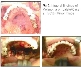
차수술은 상악 구치부 치조골 포함한 부분 상악골 절제술, 우 측 설골상부 경부곽청술을 적용하였다. 단 면역조직화학적 검 사 상 S100 protein(+), (HMB-45)(-)로 나타남.현재는 술후 2년째로서 구강내 및 구강외에서 특이소견이 관찰되지 않고 환자의 건강은 양호한 편이다 (Fig. 6 - Fig. 13).
Fig. 1.Intraoral findings (Case 1, M/70)
Fig. 6.Intraoral findings of Melanoma on palate(Case 2, F/65)- Mirror Image
Fig. 7.Intraoperative findings-removal of the lesion (Left)
Total skin graft(Right) Fig. 2.CT findings-
Arrow indicates the lesion site
Fig. 3.Segmental maxillectomy (Left)
Skin grafting (Right)
Fig. 4.Histologic findings - melanotic pigmentation in dermis layer on palate(Left)
Melanin on gingiva Right)
Fig. 5.Healed state on 15 months after operation - bare condition (Left)
Prosthesis restoration (Right)
Ⅲ. 결 과
총 6명의 환자 중 5명의 환자가 남성이었다. 연령은 60세부 터 87세까지의 분포를 보였다. 발생부위로는 경구개에서 발생 한 환자가 5명으로 가장 많았으며, 상악 전치부 치조부에한정 되어 병발한 증례는 1건이었다. 병기의 분류는 일반적인 흑색 종의 분류법을 따랐으며, 4명의 환자에서 림프절의 전이를 보 이지 않는 Stage I의 양상을 나타내었고, 다른 한명의 환자에서 는 경부림프절의 전이 (Stage II)를 보여 경부 곽청술을 시행하 였으나 재발하여 다발성 경부전이(Stage III)가 나타나 2차 경부 곽청술을 시행한 증례 (증례5)도 있었다 (표1).
림프절 전이는 2명의 환자에서 관찰되었고, 원격전이는 2명 의 환자에서 발견되었다. 원격전이의 장소는 췌장과 폐 부위 였다. 6명의 환자 중 3명의 환자만 악성 흑색종의 치료(증례1,
2, 5)에 동의하였는데 치료에 동의하지 않은 사유로는 고령(2
명), 전신 건강(간경화)으로 나타났다. 치료를 받지 않은 경우 에서 stage III로 판정 되었된 환자는 1년 이내에 흑색종으로 인 해 사망한 것으로 조사되었다. 치료에 동의하였던 3명의 환자 는 각각 수술, 수술+항암요법, 수술+면역항암치료를 시행 받 았으며 그중 1명에서 술 후 2년 6개월째 폐전이가 발견되어 폐부분절제 수술하였고(증례1) 현재 수술한 환자 모두는 생존중 이다.
Ⅳ. 토 의
Workshop on Oral Malignant Melanomas (WOMM, 1997)에서는
구강내 흑색종을 피부흑색종과는 다르게 고려해야 한다고 주 장하였다. ‘in situ OMM’,
‘invasive OMM’‘invasive with in situcomponent OMM’
‘atypical melanocytic proliferation’,
의 용어가 보 다 나은 용어임을 보고하였다. 비 전형 멜라닌세포의 증식증(Atypical melanocytic proliferation)은 구강 내 점막의 멜라닌화가
진행되는 것으로 과염색체, 불규칙성 유사분열상을 가진 각진 핵 (hyperchromatic and angulated nuceli with infrequent mitotic activi-ty)을 동반함을 의미하였다
2-4). 본 증례1, 2에서의 조직 소견 상
에서도 비슷한 병리조직학적 소견을 관찰할 수 있었다.
대부분의 구강 악성흑색종(OMM)은 경구개및 상악 치조골 부위에서 발생(50 - 77% 차지)한다고 알려져 있고 흑색종 확진 전에 40%환자에서 멜라닌색소침착(melanotic pigmentation)이 일어난다고 하고 이러한 흑색반점은 10 - 15개월에 걸쳐 급격 한 성장이 된다고 보고되고 있다. 본 증례들에서도 환자가 멜 라닉 색소침착을 인지하여 흑색반점으로 진행되기 까지 5-10
Fig. 8. Melanotic pigmen- tation in dermis layer, H-E staining ×100(left)
S-100 positive reaction × 200 (Right)
Fig. 9.Recurred lesion on right maxilla
Fig. 10.Resection of the lesion (Con’t)
Fig. 11.Buccal fat pad grafting (Con’t)
Fig. 13.HMB-45 negative results from 2nd operation specimen
Fig. 12.Healed state of palate (Con’t)
Table 1. Case Analysis
No. Age/Sex Duration(till Dx) Site Stage
1(증례1) 72/M 12 Mo. Hard palate Stage I
2(증례2) 63/F 10 Mo. Hard palate Stage I
3 70/M Unknown Hard palate Stage I
4 87/M 5 Hard palate Stage I
5 60/M 3 Hard palate Stage II -> III
6 65/M Unknown Mx. ant.(alveolus) Stage III
개월 정도 걸리는 것으로 판정되었다.
진단으로는 생검(biopsy)이 필수적이고 세포막간 이상
(Junctional anomalies) 증가된 멜라닌세포, 비전형세포(atypical cells)의 출현 등이 흑색종 질환임을 의심할 수 있고 암종세포
는 방추형(spindle shape), 상피성(plasmocytoid or epithelioid)이고 멜라닌화(amelanotic)를 띤다. 감별진단으로서 미분화성악성 종양과 anaplastic lymphoma와 구분해야하고 이를 위해 면역화 학적 처리가 요구 된다5-8).
S-100 protein과 homatropine methylbromide antigen(HMB-45)에
의 양성반응이 대부분의 흑색종 병소에서 나타난다. S-100 pro-tein은 방추형, 신경세포형의 멜라닌세포(spindled, more neural- appearing melanocytes)에 반응하고 HMB-45는 구형세포(round cells)를 확인하는데 사용된다. 상피세포기원병소와 달리 흑색
종은 또한 간엽세포(mesenchymal cells)의 marker인 vimentin에도 양성반응을 나타낸다. 최근에는 microphthalmic transcription fac-tor, tyrosinase, melano-A immunostains을 써서 melanocytes를 구분
해 내는데 쓰이고 있다. 1994년 Gazit와 Daniels5)은 S-100(50증례 중 46개 증례에서)와 HMB-45(epithelioid variations대부분)를 써 서 흑색종구분에 유용했음을 보고 하였다. 본 증례1, 2에서도 2 개의 면역화학요법을 적용한 항체인자들을 써서 악성 흑색종 을 확진하였다.구강점막과 피부에서의 흑색종들의 예후를 Clark씨와
Breslow씨 분 류 법 으 로 판 단 하 기 에 는 어 려 움 이 있 다 . Batsakis(1982)와 Patel(2002)는 구강내 점막 흑색종 또한 0.5mm
이상 병소침범이 있을시 예후가 좋지 않음을 보고하였다9). Rapini(1985)는 5년 생존율을 15-38%정도로 보고하였다
10). 이러
한 예후불량은 세포형(histotype)과 연관될 뿐 아니라 충분히 제 거가 힘든 부위에 위치한 점(palate, alveolar crest)과 조기 혈행성 전이, 환자의 노령화 등이 원인임이 보고되고 있다11).
본 증례 6 례 중 수술을 시행한 3 례 중 2 례 (T1N0M0)에서는 주위 골까지 함께 절제하였고 예후는 양호하였고 다른 1례에 서는 T2N1M0로서 이미 경부로의 전이가 진행된 경우에서는 일차절제술 및 경부 곽청술을 시행하였고 환자는 생존하였다.
구강 내 악성흑색종의 예후는 피부흑색종의 예후와 비교해서 명확한 예후를 추정하기는 어려웠다.
치료방식으로 수술 외에 radiation therapy도 시도되고 있고 항 암요법도 제시되고 있다.
Umeda와 Shimada(1994)는 치료방식으로 (1) 건강한 주위조직 1.5cm을 포함한 일차 병소부 절제술 (2) 임파절 전이부 절제술-
경부 곽청술 (Stage II) (3) DTIC-ACNU-VCR을 이용한 항암요법 을 주장하였다12). 면역요법은 보조치료로 권장하고 있고 IFN alpha-2b therapy, bacille Calmette-Guerin (BCG), recominant inter- leukin-2(rIL-2)가 현재 임상 시험 중에 있다
13-16). 본 증례 1에서는
수술 후 인터페론 항암치료를 부가적으로 시행하였고 치료 중 부작용은 없었으며 환자의 예후는 양호하였다. 수술 후 치료 의 효과증진 및 정신 안정 적 측면에서는 권장 할 만 하였다.병소부위의 절제 수술이 가장 이상적인 치료법이며 lymph
node involvement가 어느 정도까지 진행되었느냐에 따라 악성
흑생종 환자의 예후가 달라지는 지에 대한 명확한 지침은 없 으나 흑색종 병소의 invasion정도와 임파절 이환과의 연관성은 분명 상관이 있음이 알려져 있다. Patel(2002)등은 구강 흑색종 은 다른 두개및 경부 흑색종(sinonasal type, 6%)보다 임파절 로 의 전이가 26%로서 높다고 보고하였다9)
. 본 6 례 중 수술 3 증례
에서 CT상 전이가 의심된 2례에서는 임파절 절제술(경부곽청 술)을 시행하여 임파절 전이여부를 확인하였고 1례에서 전이 가 level 2로 관찰되었다. 환자에서 임파절 전이가 의심되는 큰 크기의 병소는 일차 절제술 외에 임파절 절제술을 함께 시행 함이 구강내 악성흑색종 치료의 원칙으로 추천된다.Ⅴ. 결 론
본원에서 추적 관찰한 구강내 악성흑색종의 치료 예후 등을 관찰한 결과 다음의 결과를 얻었다.
1. 진단을 위해 생검법외 면역조직화학검사법으로서 S100 protein, HMB-45을 이용하여 확진하였고 면역화학검사를
시행한 환자들 모두에서 양성반응을 얻었다.2. 수술시 점막 조직절제 외 부가적인 골절제술 및 경부전이
가 의심된 경우는 경부 임파절 에 대한 예방적 경부 곽청술 이 추천되었다.3. 환자는 수술 후 인터페론 등의 부가적인 항암요법이 적극
권장되었다.4. 수술 후 6개월마다의 정기적인 검진을 시행하여 재발 및
전이위험을 관찰해 나간다면 구강 내 악성 흑색종 환자에 대한 예후도 양호하게 예측될 수 있을 것으로 생각되었다.참고문헌
1. Garzino-Demo P, Fasolis M, Maggiore LT: Oral mucosal melanoma: a series of casse reports, J Cranio-facial Surg 32, 251- 257, 2004
2. Mendenhall WM, Amdur RJ, Hinerman RW, et al: Head and Neck Mucosal Melanoma, Am J Clin Oncol 28:626-630, 2005 3. Greene GW, Haynes JW, Dozier M, et al: Primary malignant
melanoma of the oral mucosa, Oral Surg Oral Med Oral Pathol 68:1435-1441, 1953 4. Hicks MJ, Flaitz CM: Oral mucosal melanoma:epidemiology and pathobiology, Oral Oncol 36:152- 169, 2000
5. Gazit D, Daniels TE: Oral melanocytic lesions:differneces in expression of HMB-45 and S-100 antigens in round and spindle cells of malignant arid benign lesions, J Oral Pathol Med 23(2):
60-64, 1994
6. Manolidis S, Donald PJ: Malignant mucosal melanoma of the head and neck: Review of the literature and report of 14 patients, Cancer 80:1373-1386, 1997
7. Molife R, Hancock BW: Adjuvant therapy of malignant melanoma, Crit Rev Oncol Hematol 44:81-102, 2002
8. Pandey M, Mathew A, Iype EM, et al: Primary malignant mucosal melanoma of the head and neck region: pooled analysis of 60 pub- lished cases from India and review of literature, Eur J Cancer Prev 11:3-10, 2002
9. Patel SG, Prasad ML, Escrig M, et al: Primary mucosal malignant melanoma of the head and neck, Head Neck 24:247-257, 2002 10. Rapini RP, Golitz LE, Greer Jr RO, et al: Primary malignant
melanoma of the oral cavity, A review of 177 cases, Cancer
55:1543-1551, 1985
11. Regezi JA, Hayward JR, Pickens TN: Superficial melanoma of oral mucous membranes, Oral Surg Oral Med Oral Pathol 45:
730-740, 1978
12. Umeda M, Mishima Y, Teranobu O et al: Heterogeneity of pri- mary malignant melanomas in oral mucosa: an analysis of 43 cases in Japan, Pathology 20:234-241, 1988
13. Goerres GW, Stoeckli SJ, Schulthess GK et al: FDG PET for mu- cosal malignant melanoma of the head and neck, Laryngoscope, 112:381-385, 2002
14. Yii NW, Eisen T, Nicolson M, et al: Mucosal malignant melanoma of the head and neck: the Marsden experience over half a century, Clin Oncol 15: 199-204, 2003
15. Stern SJ, Guillamondegui OM: Mucosal melanoma of the head and neck, Head Neck 132:22-27, 1991
16. Prasad ML, Patel SG, Huvos AG, et al: Primary mucosal melanoma of the head and neck. A proposal for microstaging lo- calized, stage I (lymph node-negative) tumors, Cancer 100: 1657- 1664, 2004