424
Copyright © 2019 The Korean Society of Fisheries and Aquatic Science pISSN:0374-8111, eISSN:2287-8815
서 론
샛비늘치목(Myctophiformes) 샛비늘치과(Myctophidae) 어 류는전세계에 33속 248종(Eschmeyer et al., 2019), 일본에 21 속 83종(Okiyama, 2014), 우리나라에 2속 3종[깃비늘치(Ben- thosema pterotum), 샛비늘치(Myctophum affine), 얼비늘치 (Myctophum asperum)]이보고되어있다(MABIK, 2018). 샛 비늘치과어류는점심해성(Bathypelagic) 어류로 100 m에서 1000 m 사이의깊은곳에서발견되며, 표층(10-100 m)과중 층(200-2000 m) 사이를수직 회유한다(Kozlov, 1995; FAO fisheries, 1997; Karuppasamy et al., 2007, 2011; Cherel et al., 2010; Venecia, 2010). 몸의측면을따라다양한위치에발광 포와발광기관이있으며(Beebe, 1934; Haygood et al., 1994), 발광포의위치는샛비늘치과어류의종특이적분류키로이용
된다(Paxton, 1972). 또한, 성별에따라발광기관의위치와크 기가광범위하게변하기도한다(Herring, 2007; Martin et al., 2018).
2017년 2월우리나라부산주변해역과제주도주변해역에 서샛비늘치과치어 2개체가처음으로채집되었다. 이를형태 적으로동정한결과, Notoscopelus japonicus로추정되었고일 본에서대여한 N. japonicus 성어와 mtDNA 16S rRNA 염기 서열을비교하여분자적으로검증하였다. N. japonicus는북서 태평양에서가장우세한샛비늘치과어류중하나이지만(Fujii and Uyeno, 1976; Willis et al., 1988; Watanabe et al., 1999;
Kazuhisa et al., 2001), 아직국내에는보고된바가없다. 본연 구에서는우리나라에서처음보고되는이종에게새로운국명 으로 “등점샛비늘치”를제안한다.
한국 해역에서 출현한 샛비늘치과(Myctophidae) 어류 1미기록종
Notoscopelus japonicus (Tanaka, 1908) 치어의 분자동정 및 형태기재
박경현·김진구*·지환성
1·최정화
1부경대학교 자원생물학과, 1국립수산과학원 수산자원연구센터
Molecular Identification and Morphological Description of Juveniles of the Previously Unrecorded Species Notoscopelus japonicus (Tanaka, 1908) (Myctophidae) in Korean Waters
Gyeong-Hyun Park, Jin-Koo Kim*, Hwan-Sung Ji1 and Jung Hwa Choi1
Department of Marine Biology, Pukyong National University, Busan 48513, Korea
1Fisheries Resources Research Center, National Institute of Fisheries Science, Tongyeong 53064, Korea
Two juvenile (9.48 and 10.11 mm in standard length) specimens of the lanternfish
Notoscopelus japonicus
, belong- ing to the family Myctophidae, were collected from the waters around Busan and Jeju Island in February 2017. This species is characterized by having melanophores along the dorsal side of the body and one photophore (PO5) in front of the pelvic fin. It is distinguished fromBenthosema pterotum
in having many melanophores on the dorsal side and a single photophore (PO5) on the trunk.N
.japonicus
is also distinguished from its congenericN
.resplendens
andN
.caudispinosus
in having no melanophores at the base of the anal fin. Analyzing 521 base pairs of the 16S rRNA sequence, the specimens were identical toN
.japonicus
adults from Japan (K2P distance <0.004). This is the first record ofN
.japonicus
in Korea, and we propose a new Korean name “Deung-jum-saet-bi-neul-chi” for this species.Key words:
Notoscopelus japonicus
, Juvenile, Unrecorded species, Myctophidae*Corresponding author: Tel: +82. 51. 629. 5927 Fax: +82. 51. 629. 5931 E-mail address: [email protected]
This is an Open Access article distributed under the terms of the Creative Commons Attribution Non-Commercial Licens (http://creativecommons.org/licenses/by-nc/3.0/) which permits unrestricted non-commercial use, distribution, and reproduction in any medium, provided the original work is properly cited.
Received 29 May 2019; Revised 19 June 2019; Accepted 10 July 2019
저자 직위: 박경현(학부생), 김진구(교수), 지환성(연구사), 최정화(연구관) https://doi.org/10.5657/KFAS.2019.0424
Korean J Fish Aquat Sci 52(4), 424-429, August 2019
재료 및 방법
채집
2017년 2월 9일에우리나라부산주변해역(34˚N, 129˚E) 에서샛비늘치과치어 1개체, 2017년 2월 13일에제주도주변 해역(32˚N, 127˚E)에서샛비늘치과치어 1개체가국립수산과 학원시험조사선에의해봉고네트(망구 80 cm, 망목 500 µm) 로채집되었다(Fig. 1). 채집된표본은즉시 5% 중성해수-포르 말린수용액에고정되었으며, 1시간후세척하여 99% 알코올 에고정하였다. 이후, 표본은부경대학교어류학실험실어류플 랑크톤표본실(Pukyong National University, Ichthyoplankton Laboratory, PKUI)에등록및보관하였다.
형태분석
치어의부위별용어와명칭은 Nakabo (2002)와 Okiyama (2014)를참고하였고, 계수형질 3개[Dorsal fin rays (D), anal fin rays (A), pectoral fin rays (P1)], 계측형질 5개[Standard length (SL), preanal length (PAL), head length (HL), orbit di- ameter (OD), snout length (SNL)]는 Okiyama (2014)를참고 하여계수및계측하였다. 외부형태는입체해부현미경(SZH- 16, Olympus, Japan)을이용하여관찰하고, 현미경용사진촬 영장치(Active measure program, Mosaic 2.0; Fuzhou Tucsen photonics, Fuzhou, China)를이용하여 0.01 mm까지측정한 뒤스케치하였다.
분자분석
채집된치어 2개체의우측눈알에서 accuprep genomic DNA extraction kit (Bioneer, Republic of Korea)를사용하여제조 업자의 protocol에따라 total DNA를추출하였다. 추출한 total DNA는 PCR (polymerase chain reaction) 실험전까지 5°C 냉 장고에서보관하였다. Mitochondrial DNA (mtDNA)의 16S rRNA 영역을증폭시키기위해 16Sar-5’ (5’-CGC CTG TTT ATC AAA AAC AT-3’)와 16Sbr-3’ (5’-CCG GTC TGA ACT CAG ATC AGG T-3’) primer (Ivanova et al., 2007)를이용하 였다. Polymerase chain reaction (PCR)은 10X PCR buffer 2 µL, 2.5 mM dNTP 1.6 µL, 16Sar-L primer 1 µL, 16Sbr-H primer 1 µL, Takara Taq polymerase 0.1 µL를섞은혼합물에 total DNA 2 µL를첨가한후, 총 20 µL가될때까지 3차증류 수를넣고 thermal cycler (Bio-rad MJ mini PCT-1148, USA) 를이용하여다음과같은조건에서 PCR을수행하였다[Initial denaturation 95°C에서 11분; PCR reaction 35 cycles (denatur- ation 94°C에서 1분, annealing 52°C에서 1분, extension 72°C 에서 1분), final extension 72°C에서 5분]. 염기서열은 ABI PRISM 3730XL analyzer (96 capillary type)에서 BigDye (R) Terminator v3.1 cycle sequencing kits (Applied Biosys- tems, Foster City, CA, USA)를이용하여얻었다. MtDNA 16S
rRNA 염기서열은 BioEdit version 7 (Hall, 1999)의 clustarW multiple alignment (Thompson et al., 1994)를이용하여정렬 하였다. 유전거리는 mega X 프로그램(Tamura et al., 2018)을 이용하여 kimura 2-parameter model (Kimura, 1980)로계산하 고 1,000번의 bootstrap을수행하여작성하였다. 염기서열을비 교하기위하여 national center for biotechnology information (NCBI)에 등록된 샛비늘치과 어류 Notoscopelus japonicus (AP012252), 깃비늘치(Benthosema pterotum, KR231722), Notoscopelus resplendens (AB042171), Notoscopelus cau- dispinosus (AP012256) 4종의염기서열을이용하였다. 또한, 일본국립과학박물관에 등록된일본산 N. japonicus 성어표 본(NSMT-P 92385)을대여받아위와같은방법으로염기서열 을얻어비교하였다. 본연구에서사용된한국산샛비늘치과치 어 2개체와일본산 N. japonicus 성어의 16S rRNA 염기서열은 NCBI에서다음과같은등록번호를부여받았다(등록번호, MN 148516-MN 148518).
결 과
Notoscopelus japonicus Tanaka, 1908 (Table 1, Figs. 2-3) (Korean name : Deung-jum-saet-bi-neul-chi)
Notoscopelus japonicus Tanaka, 1908: 5 (type locality: Off Fig. 1. Map showing the sampling area of Notoscopelus japonicus juvenile in the southern sea of Korea (=Korea Strait) and around Jeju Island.
Table 1. Measurements and counts of Notoscopelus japonicus juveniles and three Myctophidae species Notoscopelus japonicus Benthosema
pterotum Notoscopelus
resplendens Notoscopelus caudispinosus Present study Okiyama
(2014) PKUI 614 Okiyama
(2014) Okiyama
(2014) PKUI 612 PKUI 613
Standard length (SL, mm) 9.48 10.11 9.7 10.1 11.0 7.6
Measurements (% of SL)
Preanal length 60.4 54.9 58.7 53.3 60.2 65.8
Head length 30.8 29.4 36.0 31.1 30.7 39.5
Orbit diameter 9.9 9.8 12.0 8.9 10.2 17.1
Snout length 7.7 6.9 8.0 5.6 6.8 7.9
Counts
Dorsal-fin rays 21 21 22 ? 21-24 26
Anal-fin rays 18 20 18 18 19-20 20
Pectoral-fin rays 10 10 10 11 11-12 13
Misaki, Sagami Sea, Japan); Fujii and Uyeno, 1976: 229 (Ja- pan); Paxton, 1979 (U.S.A.): 16; Masuda et al., 1984 (Japan):
75; Peden et al., 1985: 7(Canada); Peden and Hughes, 1986:
3(Canada); McAllister, 1990: 110 (Canada); Shinohara et al., 1996: 167 (Japan); Nakabo, 2000: 385 (Japan); Shinohara et al., 2001: 301(Japan); Nakabo, 2002: 385 (Japan); Mundy, 2005: 232 (Hawaii); Okiyama, 2014: 231 (Japan); Parin et al., 2014: 159 (Russia); Shinohara et al., 2014: 239 (Japan).
관찰표본
PKUI 612, 1개체, 체장 9.48 mm, 부산 주변 해역(34˚N, 129˚E), 2017년 2월 9일, 봉고네트; PKUI 613, 1개체, 체장 10.11 mm, 제주도주변해역(32˚N, 127˚E), 2017년 2월 13일, 봉고네트.
형태 기재
채집된치어 2개체의계수및계측자료는 Table 1에나타내 었다. Notoscopelus japonicus 치어는방추형의긴체형을보 인다. 두부에서꼬리부분으로갈수록체고가낮아진다. 머리 크기는보통이며눈은동그랗다. 눈의직경(9.8-9.9%)은문장 (6.9-7.7%)보다길다. 입은머리에비해큰편이며위턱이아래 턱보다크다. 가슴지느러미는타원형에가까운형태이고몸의 크기에비해작은편이다. 배지느러미는가슴지느러미보다크 다. 항문은몸의중앙에있고뒷지느러미의기부에가깝게위 치한다. 뒷지느러미는배지느러미보다길고꼬리쪽으로갈수 록길이가짧아진다. 등지느러미는뒷지느러미보다약간길고 꼬리쪽으로갈수록길이가짧아진다. 등지느러미뒤쪽으로작 은기름지느러미(adipose fin)가있다. 꼬리지느러미는가랑이 형이다. 흑색소포는시엽과소뇌경계부에 1개, 등지느러미기 저를따라 10개, 체측중앙에척추골을따라 5개존재한다. 발
Fig. 2. Photos showing diagnostic characters distinguishing the Notoscopelus japonicus juvenile (A, PKUI 612, 9.48 mm SL; B, PKUI 613, 10.11 mm SL) and Benthosema pterotum juvenile (C, PKUI 614, 10.1 mm SL). N. japonicus juvenile has ten melano- phores on the dorsal side and one photophore on the trunk, but B.
pterotum juvenile has no melanophores and nine photophores on the trunk. Scale bars=1.0 mm. SL, Standard length.
광포는배지느러미기저바로앞쪽에한개(PO5) 관찰되었다 (Fig. 2A, 2B, Fig. 3A, 3B).
분자동정
샛비늘치과(Myctophidae) 치어 2개체의 mtDNA 16S rRNA 영역 521 base-pair의염기서열을 NCBI에등록된 N. japonicus 의염기서열과비교하였다. 그결과, 샛비늘치과치어 2개체는 N. japonicus 성어와유전거리 0.000-0.004로가장가깝게유집 되었으며, 그다음으로 Notoscopelus caudispinosus 과유전거
리 0.026-0.030, Notoscopelus resplendens과유전거리 0.081- 0.085, Benthosema pterotum과는유전거리 0.139-0.144로유 집되었다. 또한, 일본산 N. japonicus 성어(NSMT-P 92385)의 염기서열과도비교하였는데, 채집된샛비늘치과치어 2개체는 일본산 N. japonicus 성어와유전거리 0.004로유집되었다(Fig.
4).
고 찰
본연구는 2017년 2월부산주변해역과제주도주변해역 에서채집된샛비늘치과(Myctophidae) 치어 2개체를대상으 로형태및분자동정을실시한결과 Notoscopelus japonicus 으로확인되었다. 본종의상위분류군인샛비늘치과어류는국 내에깃비늘치(Benthosema pterotum), 샛비늘치(Myctophum affine), 얼비늘치(Myctophum asperum) 3종만알려져있고이 번에새롭게보고되는 Notoscopelus속은아직국내에보고된 바없다(MABIK, 2018).
채집된 N. japonicus 개체와깃비늘치(PKUI 614)의계수및 계측형질을비교한결과는 Table 1에나타내었다. N. japonicus 2개체는체측등쪽등지느러미기저부분에흑색소포가 10개 존재하지만 깃비늘치 개체의등지느러미 기저에는 흑색소포 가없다. N. japonicus 2개체에서는배지느러미기저앞에 1개
의발광포(PO5)가관찰되었으나깃비늘치에서는몸통에다수
의발광포(PVO1, PO1, PO3, PO4, PO5, VO1, VO2, AOa1, AOa2)가관찰되었다(Fig. 2).
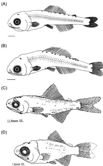
Fig. 3. Illustrations showing diagnostic characters distinguishing the Notoscopelus japonicus juvenile (A, PKUI 612, 9.48 mm SL;
B, PKUI 613, 10.11 mm SL), Notoscopelus resplendens juvenile [C, Okiyama (2014), 11.0 mm SL] and Notoscopelus caudispi- nosus juvenile [D, Okiyama (2014), 7.6 mm SL]. N. resplendens juvenile and N. caudispinosus juvenile have several melanophores on the base of anal fin, but N. japonicus juvenile has no melano- phores on the base of anal fin. Scale bars=1.0 mm. SL, Standard length.
Fig. 4. Neighbor joining tree based on partial mitochondrial DNA 16S rRNA sequences, showing the relationships among two juve- niles (PKUI 612, 613) of N. japonicus, one adult species (NSMT-P 92385) of N. japonicus, two species of Notoscopelus (N. caudispi- nosus, N. resplendens), and one outgroup (Benthosema pterotum).
The tree was constructed using the kimura 2-parameter model and 1,000 bootstrap replications. The letters in parentheses and super- scripts indicate NCBI registration number and voucher specimen number, respectively. The bottom bar indicates a genetic distance of 0.020. NCBI, National center for biotechnology information.
본종과같은속에속하는 N. resplendens및 N. caudispinosus 의계수및계측형질을비교한결과는 Table 1에나타내었다. 유 사한크기에서뒷지느러미수(anal fin rays)는 N. japonicus 2 개체(PKUI 612, PKUI 613)와 Okiyama (2014)의 N. resplen- dens, N. caudispinosus가일치하지만등지느러미수(dorsal fin rays)는 N. resplendens와 N. caudispinosus가 N. japonicus보 다많고가슴지느러미수(pectoral fin rays) 또한 N. resplen- dens와 N. caudispinosus가 N. japonicus보다많았다. 또한, N.
japonicus 2개체에는 뒷지느러미기저에흑색소포가없는반
면, N. resplendens와 N. caudispinosus에는뒷지느러미기저에 흑색소포가다수존재하여잘구분되었다(Fig. 3).
Okiyama (2014)에의하면, 일본산 N. japonicus 자치어(체장 2.3-9.8 mm)는 PO5 발광포를가지며시엽과소뇌경계부측 면, 소뇌밑, 아가미뚜껑밑, 척추중앙선양측에흑색소포가있 다고언급되어있다. 또한, 등지느러미기저시작부분에서꼬 리자루부분앞까지흑색소포가줄지어있고, 뒷지느러미기저 에흑색소포가없다고하였다. 본연구에서관찰한N. japonicus 치어또한 Okiyama (2014)와대부분잘일치하였다. 나아가, mtDNA 16S rRNA 염기서열분석결과에서도 N. japonicus 성 어와잘일치하여형태동정결과를지지해주었다. 본종의새로 운국명으로등지느러미기저에흑색소포가줄지어있는특징 에따라 “등점샛비늘치”를제안한다.
사 사
비교표본및조직시료를제공해주신 Nakae박사(일본국립 과학박물관)와논문을세심하게검토해주신심사위원께감사
드립니다. 이논문은 2019년도국립수산과학원수산과학연구
사업(근해어업자원조사, R2019019)과 2019 년도해양수산과
학기술진흥원해양수산생명공학기술개발사업의지원을받아
수행된연구입니다(No. 20170431).
References
Beebe W. 1934. Half Mile Down. Harcourt, Brace and Com- pany, NY, U.S.A., 321-322.
Cherel Y, Fontaine C, Richard P and Labat JP. 2010. Isotopic niches and trophic levels of myctophid fishes and their pred- ators in the Southern Ocean. Limnol Oceanogr 55, 315-323.
Eschmeyer WN, Fricke R and van der Laan R. 2019. Catalog of Fishes: Genera, Species, References. Retrieved from http://
researcharchive.calacademy.org/research/ichthyology/cata- log/fishcatmain.asp on Apr 22, 2019.
FAO (Food and Agriculture Organization of the UN) fisheries.
1997. Lanternfishes: a potential fishery in the northern Ara- bian Sea. Retrieved from http://www.fao.org/3/W4248E/
w4248e34.htm on Apr 22, 2019.
Fujii E and Uyeno T. 1976. On three species of the myctophid genus Notoscopelus found in western North Pacific. Japa-
nese J Ichthyol 22, 227-233.
Hall TA. 1999. BioEdit: A user-friendly biological sequence alignment editor and analysis program for windows 95/98/
NT. Nucleic Acids Symp 41, 95-98.
Haygood MG, Edwards DB, Mowlds G and Rosenblatt RH.
1994. Bioluminescence of myctophid and stomiiform fishes is not due to bacterial luciferase. J Exp Zool 270, 225-231.
https://doi.org/10.5657/KFAS.2019.0067.
Herring PJ. 2007. Sex with the lights on? A review of biolumi- nescent sexual dimorphism in the sea. J Mar Biol Assoc UK 87, 829-842.
Ivanova NV, Zemlak TS, Hanner RH and Hebert PDN. 2007.
Universal primer cocktails for fish DNA barcoding. Mol Ecol Notes 7, 544-548. http://dx.doi.org/10.1111/j.1471- 8286.2007.01748.x.
Karuppasamy PK, Balachandran K, George S, Balu S, Persis V and Menon NG. 2007. A check list of fishes collected by IKMT from the DSL survey in the Indian EEZ of Arabian Sea. J Ind Hydro 9, 311-316.
Karuppasamy PK, Lalu RCM, Muraleedharan KR and Nair M.
2011. Myctophid and pelagic shrimp assemblages in the ox- ygen minimum zone of the Andaman Sea during the winter monsoon. Ind J Geo-Mar Sci 40, 535-541.
Kazuhisa U, Orio Y, Daiji K and Yasunori S. 2001. Diet of the mesopelagic fish Notoscopelus japonicus (Family: Mycto- phidae) associated with the continental slope off the Pacific coast of Honshu, Japan. Fish Sci 2002, 1034-1040.
Kimura M. 1980. A simple method for estimating evolutionary rates of base substitutions through comparative studies of nucleotide sequences. J Mol Evol 16, 111-120.
Kozlov AN. 1995. A review of the trophic role of mesopelagic fish of the family Myctophidae in the Southern Ocean eco- system. CCAMLR Sci 2, 71-77.
MABIK (Marine Biodiversity Institute of Korea). 2018. Nation- al List of Marine Species. Namu Press, Seocheon, Korea, Martin RP, Olson EE, Girard MG, Smith WL and Davis MP. 25.
2018. Light in the darkness: New perspective on lanternfish relationships and classification using genomic and morpho- logical data. Mol Phylogenet Evol 121, 71-85. https://doi.
org/10.1016/j.ympev.2017.12.029.
Masuda H, Amaoka K, Araga C, Uyeno T and Yoshino T. 1984.
The fishes of the Japanese Archipelago. Tokai Univ Press, Tokyo, Japan.
McAllister DE. 1990. A list of the fishes of Canada. Syllogeus 64, 1-310.
Mundy BC. 2005. Checklist of the fishes of the Hawaiian Archi- pelago. Bishop Mus Press, Honolulu, Hawaii, U.S.A.
Nakabo T. 2000. Fishes of Japan with pictorial keys to the spe- cies, Second ed. Tokai Univ Press, Tokyo, Japan.
Nakabo T. 2002. Fishes of Japan with pictorial keys to the spe- cies, English ed. Tokai Univ Press, Tokyo, Japan.
Okiyama M. 2014. An atlas of the early stage fishes in Japan.
Tokai Univ Press, Tokyo, Japan.
Parin NV, Evseenko SA and Vasil'eva ED. 2014. Fishes of Rus- sian seas: Annotated catalogue. KMK Scientific Press, Mos- cow, Russia.
Paxton JR. 1972. Osteology and relationships of the lanternfish- es (family Myctophidae). Bull Nat’l His Mus Los Angeles Country Sci 13, 1-78.
Paxton JR. 1979. Nominal genera and species of lanternfishes (family Myctophidae). Contrib in Sci 322, 1-28.
Peden AE and Hughes GW. 1986. First records, confirmatory records, and range extensions of marine fishes off Canada's west coast. Canadian Field-Naturalist 100, 1-9.
Peden AE, Ostermann W and Pozar LJ. 1985. Fishes observed at Canadian weathership ocean station Papa [50°N, 145°W]
with notes on the trans-Pacific cruise of the CSS Endeavor.
Heritage record 18, British Columbia Provincial Mus, Vic- toria, Canada.
Shinohara G, Endo H and Matsuura K. 1996. Deep-water fishes collected from the Pacific coast of northern Honshu, Japan.
Mem Nat’l Sci Mus Tokyo 29, 153-185.
Shinohara G, Endo H, Matsuura K, Machida Y and Honda H.
2001. Annotated checklist of the deepwater fishes from Tosa Bay, Japan. Monographs Nat’l Sci Mus Tokyo 20, 283-343.
Shinohara G, Nakae M, Ueda Y, Kojima S and Matsuura K.
2014. Annotated checklist of deep-sea fishes of the Sea of Japan. Nat’l Mus Nat Sci Monographs 44, 225-291.
Tanaka S. 1908. On some rare fishes of Japan, with descriptions of two new genera and six new species. J Coll Sci Imperial Univ Tokyo 23, 1-24.
Tamura K, Stecher G, Peterson D, Filipski A and Kumar S. 2013.
MEGA6: Molecular evolutionary genetics analysis version 6.0. Mol Biol Evol 30, 2725-2729. https://doi.org/10.1093/
molbev/mst197.
Thompson JD, Higgins DG and Gibson TJ. 1994. CLUSTAL W: improving the sensitivity of progressive multiple se- quence alignment through sequence weighting, position- specific gap penalties and weight matrix choice. Nucl Acids Res 22, 4673-4680.
Venecia C, Manguesh G and Karuppasamy PK. 2010. A review on mesopelagic fishes belonging to family Myctophidae.
Rev Fish Biol Fisheries 21, 339-354.
Watanabe H, Moku M, Kawaguchi K, Ishimaru K and Ohno A.
1999. Diel vertical migration of myctophid fishes (Family Myctophidae) in the transitional waters of the western North Pacific. Fish Oceanogr 8, 115-127.
Willis JM, Pearcy WG and Parin NV. 1988. Zoogeography of midwater fishes in the Subarctic Pacific. Bull Ocean Res Inst Univ Tokyo 26, 79-142.