Ⅰ. 서 론
유전질환의 하나인 Prader-Labhart-Willi 또는 Prader-Willi syndrome (프라더윌리증후군, PWS)는 복 잡한 신경행동적인 장애로 환자의 70%에서 15번 염색체 장완(15q11-q13)의 결실를 보인다1). 나머지 25%에서는 materal disomy 15(UPD)이 관찰되며 2 - 5%는 im- printing 결함을 보이고2,3) 10,000명에서 15,000명당 1명 에서 발견된다4). 환자들은 키가 작고 지적장애 혹은 학습장
애, 성기발육부전, 낮은근긴장, 수면장애, 높은 통증역치와 같은 특성을 보인다5). 비만이 관찰되는 가장 흔한 유전 질환 이고 PWS 환자의 15%가 갑상선저하증을 보인다6,7). 외관 상으로는 좁은 이마, 아몬드 형태의 눈, 구각부가 아래로 쳐 져 있고 얇은 상순을 지니고 있다8). 구강 내 특성은 다발성 치아우식증, 법랑질결함, 치아 맹출지연, 우상치, 불량한 구 강위생, 치은염, biofilm 축적, 지도혀를 보인다9,10). 점진적 인 치아의 마모10)와 위식도 역류11)도 보고되고 있다. 타액의 점도가 진하고 끈적이는 양상을 지니고 있고 신생아에서 진 단지표로 사용하기도 한다12). 타액분비의 감소는 내분비 이 상과 행동장애의 결과로 생기는 현상이기 때문에 구강 내 감염에 대한 감수성이 높다9).
단계별로 증상을 나눌 수도 있다. 신생아기에서는 심각한
◆ 증 례
프라더윌리증후군 환자에서의 치과 치료 : 증례보고
이수정∙백광우*
아주대학교병원 소아치과학교실
DENTAL TREATMENT IN A PRADER-WILLI SYNDROME PATIENT : A CASE REPORT
Soo Jeong Lee, Kwangwoo Baek*
Division of Pediatric Dentistry, Department of Dentistry, Ajou University School of Medicine
Prader-will syndrome (PWS) is a genetic disorder displayed with short stature and behavioral problems.
The major manifestation is hyperphagia and obesity but there is no known effective drug that can solve this problem. This article presents a case report of a 3-year 9-month old girl affected with PWS syndrome treated under general anesthesia due to lack of cooperation and multiple caries.
The dentist should work as a member of the multidisciplinary medical team. Careful long term follow up is required to prevent dental caries and periodontal diseases as well as maintain good oral hygiene. A strict diet control should also be encouraged. [J Korean Dis Oral Health Vol.11, No.2: 67-71, December 2015] Key words :Prader-willi syndrome, Dental caries, General anesthesia
Abstract
Corresponding author : Kwangwoo Baek
164 Worldcup-ro, Youngtong-gu, Suwon, 16499, Korea Division of Pediatric Dentistry, Department of Dentistry, Ajou University School of Medicine
Tel: +82-31-219-5869, Fax: +82-31-219-5868
성장하면서 빠는 힘은 개선된다13). 환자의 주요 특징 중 하 나는 저신장이다. 평균 성인 남자 신장은 155 cm, 여자는 148 cm이며 성장호르몬 분비가 부족하기 때문에 나타나는 현상이다14). 따라서 성장호르몬대체치료(growth hormone replacement)가 진행되며 치료를 통해서 환자의 근육량을 높이고 지방량을 낮추며 신체적 활동을 증대시키고 호흡기 능을 향상시킬 수 있다15). 시상하부의 기능적인 장애 때문에 환자는 포만감을 느끼지 못하고 식욕항진을 보인다16). 적절 한 식사조절이 이루어지지 않는다면 낮은 대사능력 및 활동 력 때문에 동일 체중의 정상인보다 칼로리 요구량이 약 60% 적고 식사량은 증가되면서 비만이 된다17). 청소년기에 서는 성적발달이 미숙하고 반항심이 크고 고집이 세다6). 경 도의 지적장애를 보이고12)25%는 자폐를 동반한다18). 주로 3세에서 30세까지 사망하며 가장 흔한 사망의 원인은 비만 으로 인한 합병증인 것으로 알려져 있다19). 급하게 식사하는 과정 중에 환자가 숨이 막히거나(choking), 위파열, 조직괴 사 때문에 사망하기도 한다20,21). 비만의 합병증으로는 심폐 부전, 수면무호흡증, 혈전정맥염, 만성 다리 부종이 있다6). 성인 비만환자 중 25%가 제2형 당뇨를 경험하며 평균 발병 시기는 만20세이다22). 식욕항진에 대한 메커니즘에 관해서 는 명확하게 밝혀진 바가 없으며 음식 섭취 후에도 높은 ghrelin 수준 때문이라고 추측하지만 PWS의 식욕을 억제 하는 약물은 아직 없다13). 따라서 PWS의 건강한 삶을 위해 서는 의료전문가와 보호자의 긴밀한 협조가 필요하다.
Ⅱ. 증례 보고
만 3세 9개월 여자 환자가 우식을 주소로 아주대학교 치 과병원 소아치과에 내원하였다(Fig. 1). 환자는 출생 시 2.7 kg이었고, 신생아 때 근긴장저하(hypotonia) 때문에
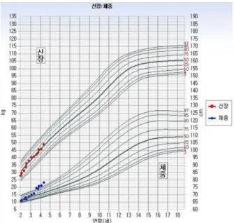
본원에서 산부인과와 소아청소년과에서 치료를 받았다. 환 자는 발달력상 걸음마는 20개월에 시작하였고 30개월에 대 소변을 가리게 되었다. 환자는 본원 유전학클리닉과 언어발 달치료를 받고 있었고 Bayley 영유아 발달검사 결과 정신 연령이 23 - 26개월정도로 추정되었다. 갑상선기능저하증 으로 씬지로이드정(Synthyroid, Bukwang Pharm., Seoul, Korea) 약물을 복용하고 있었고 지노트로핀 (Genotropin, Pfizer, Inc., New York, NY, USA) 성장 호르몬주사를 맞고 있었다. 보호자는 환자가 식탐이 많고 최근에는 냉장고를 뒤지는 행동을 보인다고 진술하였다 (Fig. 2).
Fig. 1. Extra-oral examination.
Fig. 2. Growth pattern according to weight and height.
Fig. 4. Post-operative intraoral photo.
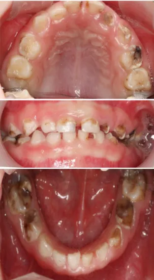
환자의 협조도가 부족하고 구강 내 다수의 우식이 관찰되 어 2015년 5월에 전신마취 하 치과치료를 진행하기로 결정 하였다. 수술 당일 검사 구강위생상태가 불량하였고, 끈적 이는 점성의 타액분비가 관찰되었다(Fig. 3). 다수의 우식 이 관찰되어 치료는 다음과 같이 진행하였다. 상악우측유측 절치, 상악우측유견치, 하악좌측유중절치, 하악좌측유측절 치는 레진수복하였고 상악우측제2유구치는 아말감으로 치 료하였다. 상악우측유중철치와 상악좌측유중절치는 치수절 제술 후 레진수복하였다. 상악좌측제1유구치와 하악우측제 2유구치는 치수절단술 후 기성금속관치료를 하였다. 상악좌 측유견치는 치수절제술 후 기성금속관치료를 하였다. 상악 우측제1유구치와 상악좌측제2유구치, 하악좌측유견치와 하 악좌측제2유구치는 기성금속관 치료를 하였다(Fig. 4). 상 악좌측유측절치, 하악좌측제1유구치와 하악우측제1유구치 는 우식이 심하고 예후가 불량할 것으로 판단되어 발치하였 다(Fig. 5). 현재는 3개월 간격으로 정기적인 검진을 시행 하고 있다.
Fig. 5. Extracted teeth (Mandibular left and right primary first
Ⅲ. 고 찰
프라더윌리증후군 환자의 식이조절은 유아기 때에 시작 하는 것을 추천한다. 환자는 극단적인 불안, 공격성 성향을 음식과 관련하여 보이는 경향이 있고13)식이조절을 하지 못 한 PWS 환자들은 비만이 생후 12 - 36개월부터 나타나기 때문이다6). 통증에 대한 역치가 높기 때문에 위장장애를 느 끼지 못하고 구토능력이 떨어진다23). 만 2 - 6세까지는 2 - 3개월마다 몸무게를 확인하여 비만의 초기증상이 발견되면 즉시 관리하는 노력이 필요하다6).
본 증례에서는 치과치료의 필요성을 보여주는 예이다. 법 랑질저형성증과 초기우식은 PWS 환자의 40%에서 발견된 다24). 신생아 및 유아기 때 영양결핍이 법랑질저형성증과 관 련되어 있는 것으로 나타났지만 최근에는 PWS 환자의 60%가 만 1세 이전에 진단을 받게 됨으로써 적절한 영양공 급이 가능해졌고 법랑질저형성증이 나타날 위험은 감소하 였다25). 치아우식과 캔디다감염은 과도한 설탕섭취, 법랑질 저형성증, 끈적이고 점성이 높은 타액, 불량한 구강위생상 태의 결과 발생한다26). PWS 환자의 불량한 구강위생상태 는 환자의 전신상태와도 연관되어 있고 환자는 기회 감염의 위험에 노출된다. 낮은 타액유량과 높은 치은염의 위험은 주기적인 스케일링을 통해서 관리가 필요하며 클로르헥시 딘과 같은 구강청결제는 구강병소를 예방할 수 있다27). 영구 치열기에서는 치아 부식과 교모로 인한 치아 마모 문제가 크며 보철 수복 시 고려해야 한다7). 최근에는 PWS 환자의 식습관 개선되고 구강관리 중요성에 대한 인식이 향상되었 다. 또한 치아마모로 인해 수복치료를 받으면서 낮은 타액 분비에도 불구하고 우식률(DMFS)이 정상인 대조군보다 같거나 낮은 것으로 보고되기도 하였다7).
전신마취 하에서 치과치료를 시행할 때 예후가 불량할 것 으로 예상되는 치아는 보존하기 위한 노력을 기울이는 대신 발치를 하는 것이 추천된다28). 수복 재료의 선택에 있어서는 구강 위생 관리가 부족한 환경에서도 예상수명이 긴 재료를 선택해야 한다28). 본 증례에서는 전신마취 하에서 환자에게 기성금속관과 같은 적극적인 수복을 하였고 유치열기의 보 존적인 치료를 통해서 영구치 맹출까지 정상적인 저작을 가 능하게 하는 것이 목표였다. 보호자에게 식이조절에 관한 교육을 진행하였고 3개월마다 정기적인 검진을 시행하고 있 다.
치과치료 시 PWS 환자의 지적장애 또는 학습장애 때문 에 어려움을 겪을 수 있다. 지적장애는 주로 경도의 수준에 서 나타나기 때문에 지속적인 관심을 통해 예방적인 접근이 무엇보다 필요하다고 사료된다.
Ⅳ. 요 약
본 논문에서 소개된 환자는 전형적인 PWS 증상인 섭식 장애, 타액분비감소, 지적장애, 비만과 같은 특성이 관찰되 었다. 전신마취 하에서 적극적인 치과 치료를 시행하였고 보호자에게 비만 및 치아 우식을 예방하기 위해 탄수화물과 당분 섭취를 제한하는 식습관에 대한 교육을 시행하였다.
환자의 내원 횟수를 늘려 치과 치료를 위한 전신마취까지 발전하지 않도록 정기적인 관리가 필요하다.
REFERENCES
1. Cataletto M, Angulo M, Hertz G, Whitman B : Prader-Willi syndrome: A primer for clinicians.
Int J Pediatr Endocrinol, 1:12, 2011.
2. Cassidy SB, Forsythe M, Heeger S et al. : Comparison of phenotype between patients with Prader-Willi syndrome due to deletion 15q and uniparental disomy 15. Am J Med Genet, 68:
433-440, 1997.
3. Nicholls RD, Knepper JLn : Genome organiza- tion, function, and imprinting in Prader-Willi and Angelman syndromes. Annu Rev Genomics Hum Genet 2:153-175, 2001.
4. Wattendorf DJ, Muenk M : Prader-Willi Syndrome. Am Fam Physician, 72:827-830, 2005.
5. Scardina GA, Fuca G, Messina P : Oral diseases in a patient affected with Prader-Willi syndrome.
Eur J Paediatr Dent, 8:96-99, 2007.
6. Cassidy SB, Driscoll DJ : Prader-Willi syndrome.
Eur J Hum Genet, 17:3-13, 2009.
7. Miller J, Silverstein J, Shuster J, Driscoll DJ, Wagner M : Short-term effects of growth hor- mone on sleep abnormalities in Prader-Willi syn- drome. J Clin Endocrinol Metab, 91:413-417, 2006.
8. Saeves R, Espelid I, Nordgarden H, et al. : Severe tooth wear in Prader-Willi syndrome. A case-control study. BMC Oral Health, 12:12, 2012.
9. Olczak-Kowalczyk D, Witt A, Gozdowski D, Ginalska-Malinowska M : Oral mucosa in chil- dren with Prader-Willi syndrome. J Oral Pathol Med, 40:778-784, 2011.
10. Bailleul-Forestier I, Verhaehe V, Voels A, et al. :
The oro-dental phenotype in Prader-Willi syn- drome: a survey of 15 patients. Int J Paediatr Dent, 18:40-47, 2008.
11. Bots CP, Schueler YT, Brand HS, van Nieuw AA : A patient with Prader-Willi syndrome.
Characteristics, oral consequences and treatment options. Ned Tijdschr Tandheelkd, 111:55-58, 2004.
12. Stephenson JB : Neonatal presentation of Prader-Willi syndrome. Am J Dis Child, 146:
151-152, 1992.
13. Miller JL, Strong TV, Heinemann J : Medication Trials for Hyperphagia and Food-Related Behaviors in Prader-Willi Syndrome. Diseases, 3:78-85, 2015.
14. Butler MG, Meaney FJ : Standards for selected anthropometric measurements in Prader-Willi syndrome. Pediatr, 88:853-860, 1991.
15. Carrel AL, Myers SE, Whitman BY, Allen DB : Growth hormone improves body composition, fat utilization, physical strength and agility, and growth in Prader-Willi syndrome: A controlled study. J Pediatr, 134:215-21, 1999.
16. Swaab DF, Purba JS, Hofman MA : Alterations in the hypothalamic paraventricular nucleus and its oxytocin neurons (putative satiety cells) in Prader-Willi syndrome: a study of five cases. J Clin Endocrinol Metab, 80:573-579, 1995.
17. Holm VA, Pipes PL : Food and children with Prader-Willi syndrome. Am J Dis Child, 130:1063-1067, 1976.
18. Veltman MW, Craig EE, Bolton PF : Autism spectrum disorders in Prader-Willi and Angelman syndromes: a systematic review. Psychiatr Genet, 15:243-254, 2005.
19. Vogels A, Van Den Ende J, Fryns JP, et al. : Minimum prevalence, birth incidence and cause of death for Prader-Willi syndrome in Flanders.
Eur J Hum Genet, 12:238-40, 2004.
20. Stevenson DA, Heinemann, J, Scheimann AO, et al. : Gastric rupture and necrosis in Prader-Willi syndrome. J Pediatr Gastroenterol Nutr, 45:272- 274, 2007.
21. Stevenson DA, Heinemann J, Scheimann AO, et al. : Deaths due to choking in Prader-Willi syn- drome. Am J Med Genet A, 143:484-7, 2007.
22. Butler JV, Whittington JE, Webb T, et al. : Prevalence of, and risk factors for, physical ill- health in people with Prader-Willi syndrome: a population-based study. Dev Med Child Neurol, 44:248-255, 2002.
23. Davies PSW, Knight B : Need to know nutrition for children with Prader Willi syndrome : a guide for parents and carers, Brisbane University of Queensland, 2012.
24. Goldstone AP : Prader-Willi syndrome: advances in genetics, pathophysiology, and treatment.
Trends Endocrinol Metab, 15:12-20, 2004.
25. Saeves R, Nordgarden H, Espelid I, et al. : Salivary flow rate and oral findings in Prader- Willi syndrome: a case-control study. Int J Paediatr Dent, 22:27-36, 2012.
26. Friedlander AH, Yagiela JA, Paterno VI, Mahler ME : The pathophysiology, medical management and dental implications of fragile X, Rett, and Prader-Willi syndromes. J Calif Dent Assoc, 31:693-702, 2003.
27. Yanagita M, Hirano H, Murakami S, et al. : Periodontal disease in a patient with Prader-Willi syndrome: a case report. J Med Case Reports, 5:329, 2011.
28. Kong EK, Jung YJ, Baek KW : Dental Treatment for a Patient with Seizure History and Intellectual Disability under General Anesthesia:
A Case Report. J Dent Anesth Pain Med, 8: 35- 39, 2008.