Morbidity of Solid Cancer in Behçet’s Disease:
Analysis of 11 Cases in a Series of 506 Patients
So Young Na, Jaeyoung Shin, and Eun-So Lee
Department of Dermatology, Ajou University School of Medicine, Suwon, Korea.
Received: May 15, 2012 Revised: September 7, 2012 Accepted: September 10, 2012 Corresponding author: Dr. Eun-So Lee, Department of Dermatology, Ajou University School of Medicine, 164 World cup-ro, Yeongtong-gu, Suwon 443-721, Korea.
Tel: 82-31-219-5190, Fax: 82-31-219-5189 E-mail: [email protected]
∙ The authors have no financial conflicts of interest.
© Copyright:
Yonsei University College of Medicine 2013 This is an Open Access article distributed under the terms of the Creative Commons Attribution Non- Commercial License (http://creativecommons.org/
licenses/by-nc/3.0) which permits unrestricted non- commercial use, distribution, and reproduction in any medium, provided the original work is properly cited.
Purpose: Behçet’s disease (BD) is rarely reported to be associated with malignan-
cies in the literature. However, the frequency of cancer in BD patients remains un- known. This study evaluated cancer morbidity in BD patients compared with that in the general population of Korea. Materials and Methods: A retrospective chart review was performed on 506 patients visiting our hospital from 1994 to 2011 for BD. We analyzed the standardized morbidity rate (SMR), which is the ratio of ob- served to expected malignancies. Furthermore, we reviewed cases of solid cancer in BD patients in the literature. Results: Of the 506 patients with BD, 11 (2.17%) developed cancer. We found a variety of solid cancers without predominance and no hematologic malignancies. The total number of cancers observed was less than expected, which was determined from the statistical data of the National Cancer Information Center of Korea, with an SMR of 0.023 (95% confidence interval, 0.012-0.039).
Conclusion: BD may be associated with a lower cancer-relatedmorbidity compared with the general population of Korea.
Key Words:
Behçet’s disease, malignancy, morbidity, solid cancer
INTRODUCTION
Behçet’s disease (BD) is a chronic relapsing systemic vasculitis, characterized by diverse manifestations including recurrent orogenital ulcers, uveitis, skin lesions, and arthritis, as well as the involvement of the gastrointestinal tract, central ner- vous system and blood vessels.
1The pathogenesis of BD is regarded to be partially associated with autoimmunity on the basis of the evidence of effective immuno- suppressive treatment and detected auto-antibodies.
2The risk of malignancy is higher in patients with antineutrophil cytoplasmic antibody-associated vasculitis and Henoch-Schönlein purpura than in controls.
3A high risk of malignancy is also reported in autoimmune rheumatic diseases such as systemic lupus erythematosus, systemic sclerosis, and dermatomyositis.
4-7These characteristics are assumed to be possible causes for the development of malignancies in patients with BD.
However, there are only a few case reports and case series regarding the associa-
tion between malignant diseases and BD in the literature.
8Due to the scant evi-
dence of the relationship between BD and malignancy, the prevalence of malignan-
cy in BD patients remains unclear. Here we report 11 cases of BD patients associated
Cancer Information Center (http://www.cancer.go.kr/ncic/
cics_f/04/041/index.html) in 2008. The results are expressed as SMRs and 95% confidence intervals.
RESULTS
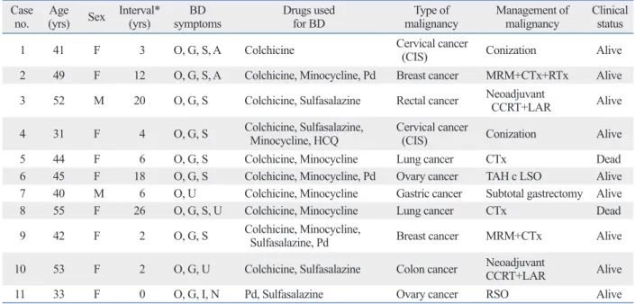
Of the 506 BD patients from our hospital, 11 (2.17%) devel- oped malignancies. The clinical findings and other character- istics of these 11 patients are summarized in Table 1. Nine patients were women, and 2 patients were men. The mean±
standard deviation (SD) ages at diagnosis of BD and malig- nancy were 35.4±7.4 years and 44.2±7.7 years, respectively.
The mean±SD follow-up duration for our patients was 76.4±53.2 months (range, 12-174 months). BD preceded the malignancy in 9 patients (81.8%), concurrently devel- oped in 1 patient (9.1%), and developed after malignancy in 1 patient (9.1%). The median duration between BD and malignancy was 6 years (range, 0-26 years).
Solid cancers were diagnosed in all patients without he- matologic disease. The various types of solid cancers iden- tified included lung, breast, ovary, cervical, and colon can- cer in 2 patients each, and gastric cancer in 1 patient.
Among the BD-related symptoms, all 11 patients pre- sented with oral ulcers, followed by genital ulcers (90.9%), with malignancy and analyzed the cancer morbidity of BD
compared to that of the general population of Korea.
MATERIALS AND METHODS
A total of 506 BD patients, who visited the Department of Dermatology of Ajou University Hospital between 1994 and 2011, were included in this study. All patients fulfilled either the International criteria for the diagnosis of BD
9or the revised criteria of the Behçet’s Disease Research Com- mittee of Japan.
10To identify which BD patients were asso- ciated with malignancy, the medical charts were reviewed retrospectively. The following data were collected: the age at diagnosis of BD and malignancy, type of malignancy, sex, duration of disease, clinical features of BD, and treat- ment regimens for BD and the malignancy. The Institution- al Review Board approved this study (IRB number: AJIRB- MDB-12-007).
The standardized morbidity rate (SMR) was used to com- pare the observed and expected morbidities. In this study, the SMR was the ratio of the observed morbidity in BD pa- tients to the morbidity in the total age- and sex-matched Korean population. The expected number of malignancies was determined from the statistical data of the National
Table 1. Characteristics of the 11 Behçet’s Disease Patients Who Developed Solid Cancer Case
no. Age
(yrs) Sex Interval* (yrs) BD
symptoms Drugs used
for BD Type of
malignancy Management of
malignancy Clinical status
1 41 F 3 O, G, S, A Colchicine Cervical cancer
(CIS) Conization Alive
2 49 F 12 O, G, S, A Colchicine, Minocycline, Pd Breast cancer MRM+CTx+RTx Alive
3 52 M 20 O, G, S Colchicine, Sulfasalazine Rectal cancer Neoadjuvant
CCRT+LAR Alive
4 31 F 4 O, G, S Colchicine, Sulfasalazine,
Minocycline, HCQ Cervical cancer
(CIS) Conization Alive
5 44 F 6 O, G, S Colchicine, Minocycline Lung cancer CTx Dead
6 45 F 18 O, G, S Colchicine, Minocycline, Pd Ovary cancer TAH c LSO Alive
7 40 M 6 O, U Colchicine, Minocycline Gastric cancer Subtotal gastrectomy Alive
8 55 F 26 O, G, S, U Colchicine, Minocycline Lung cancer CTx Dead
9 42 F 2 O, G, S Colchicine, Minocycline,
Sulfasalazine, Pd Breast cancer MRM+CTx Alive
10 53 F 2 O, G, U Colchicine, Sulfasalazine Colon cancer Neoadjuvant
CCRT+LAR Alive
11 33 F 0 O, G, I, N Pd, Sulfasalazine Ovary cancer RSO Alive
BD, Behçet’s disease; O, oral ulcer; G, genital ulcer; S, skin lesions; A, arthritis; U, uveitis; I, intestinal involvement; N, neurologic involvement; Pd, pred- nisolone; HCQ, hydroxychloroquine; CIS, carcinoma in situ; MRM, modified radical mastectomy; CTx, chemotherapy; RTx, radiation therapy; CCRT, con- current chemoradiotherapy; LAR, lower anterior resection; TAH, total abdominal hysterectomy; LSO, left salphingo-oophorectomy; RSO, right salphingo- oophorectomy.
*Interval between Behçet’s disease and cancer.
The expected number of BD patients associated with can- cers was 485.12, which was calculated by the indirect stan- dardized method from Korean data of 10-year prevalence rate of malignancy in 2008. This expected number was 44.10 times more than the observed number of patients-11 in our study. According to SMR analysis, BD patients exhibited a lower malignancy-related morbidity (SMR 0.023, 95% CI 0.012-0.039), than the general population of Korea. Male (SMR 0.024, 95% CI 0.002-0.048) and female (SMR 0.014, 95% CI 0.011-0.044) patients showed similar results.
DISCUSSION
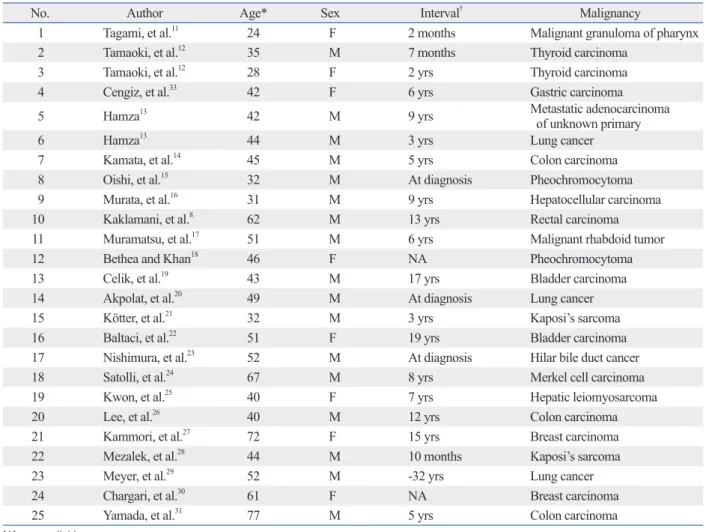
The frequency of malignancy, especially solid cancers asso- ciated with BD, was low compared with those of other au- toimmune diseases with less than 100 cases in the world lit- erature.
8,11-36We summarized the case reports for only solid skin lesions (72.7%), and uveitis (27.3%). A young female
patient (case, 11) experienced severe BD symptoms with the simultaneous involvement of the nervous and gastroin- testinal systems. No patient was treated with immunosup- pressive agents except corticosteroids before the diagnosis of the malignancy. Colchicine was used as the main treat- ment in most patients, and corticosteroids, sulfasalazine or antibiotics were administered in some cases.
Surgery was the preferred treatment option in 9 BD pa- tients although surgical procedures were different. Among them, only 1 patient (case, 6) developed wound dehiscence as a postoperative complication, which was well controlled by secondary suture. She was negative for the pathergy re- action. Chemotherapy was used in 6 patients and radiation therapy was performed in 3 patients without complication.
During the follow-up period, 2 patients (case, 5 and 8) died from tumor progression and the others are still alive with- out recurrence of cancer.
Table 2. Case Reports of Solid Cancer Associated with Behçet’s Disease in the Literature
No. Author Age* Sex Interval† Malignancy
1 Tagami, et al.11 24 F 2 months Malignant granuloma of pharynx
2 Tamaoki, et al.12 35 M 7 months Thyroid carcinoma
3 Tamaoki, et al.12 28 F 2 yrs Thyroid carcinoma
4 Cengiz, et al.33 42 F 6 yrs Gastric carcinoma
5 Hamza13 42 M 9 yrs Metastatic adenocarcinoma
of unknown primary
6 Hamza13 44 M 3 yrs Lung cancer
7 Kamata, et al.14 45 M 5 yrs Colon carcinoma
8 Oishi, et al.15 32 M At diagnosis Pheochromocytoma
9 Murata, et al.16 31 M 9 yrs Hepatocellular carcinoma
10 Kaklamani, et al.8 62 M 13 yrs Rectal carcinoma
11 Muramatsu, et al.17 51 M 6 yrs Malignant rhabdoid tumor
12 Bethea and Khan18 46 F NA Pheochromocytoma
13 Celik, et al.19 43 M 17 yrs Bladder carcinoma
14 Akpolat, et al.20 49 M At diagnosis Lung cancer
15 Kötter, et al.21 32 M 3 yrs Kaposi’s sarcoma
16 Baltaci, et al.22 51 F 19 yrs Bladder carcinoma
17 Nishimura, et al.23 52 M At diagnosis Hilar bile duct cancer
18 Satolli, et al.24 67 M 8 yrs Merkel cell carcinoma
19 Kwon, et al.25 40 F 7 yrs Hepatic leiomyosarcoma
20 Lee, et al.26 40 M 12 yrs Colon carcinoma
21 Kammori, et al.27 72 F 15 yrs Breast carcinoma
22 Mezalek, et al.28 44 M 10 months Kaposi’s sarcoma
23 Meyer, et al.29 52 M -32 yrs Lung cancer
24 Chargari, et al.30 61 F NA Breast carcinoma
25 Yamada, et al.31 77 M 5 yrs Colon carcinoma
NA, not available.
*Age at diagnosis of malignancy.
†Interval between Behçet’s disease and cancer.
ation between BD and malignancy has been performed.
Among 1769 BD patients, 32 (1.8%) developed cancer in a 12-year period.
32However, the incidence rate was not com- pared to that of the general population in that study. In the present study, we analyzed the SMR to compare the cancer morbidity in BD patients with that of the general popula- tion of Korea. The morbidity of malignancies was signifi- cantly lower in BD patients after adjusting for age and sex.
According to a literature review, BD is predominantly as- sociated with hematologic malignancies, especially myelo- dysplastic syndrome.
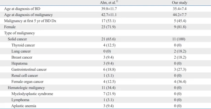
32,36In their case series in Korea, Ahn, et al.
32also reported that myelodysplastic syndrome is the most common associated disease (21.9%), followed by thy- roid cancer (12.5%). They stated that the types of solid can- cers in BD patients are presumed to be similar to those of the general population on the basis of the 2002 annual report of the Korean Central Cancer Registry program.
32However, we did not find any hematologic malignancies among the present 506 BD patients, and no particular type of solid cancer was predominant (Table 3). The difference in fre- quencies of hematologic malignancies between the study by Ahn, et al.
32and our study may result from the difference of recruitment group for BD patients. They recruited the BD patients from the department of rheumatology, whose pa- tients usually present more internal involvement such as in- testinal or vascular manifestations, and have tendency to cancers associated with BD in Table 2. In the literature, the
average age of malignancy diagnosis was 46.48±13.47 years, and the median duration between BD and malignan- cy was 6 years; these values are similar to those of the pres- ent study despite the male predominance. Malignancy was diagnosed after BD was diagnosed in most patients in both our study and the literature.
Only a few clinical researchers have made efforts to de- termine the incidence rate of malignancy in BD. Cengiz, et al.
33reported 13 cases of malignancies in 400 BD patients with a median follow-up of 10 years; however, they did not find any significant difference from the incidence of malig- nancies in the general population of Turkey. In another study from Turkey, they observed 8 patients with cancer among 387 BD patients with a 20-year follow-up.
35The estimated annual incidence rate of malignancies in BD patients is 103 in 100000, which is similar to the crude yearly cancer inci- dence of 90 in 100000 among the general population of Turkey in 1995.
35In 2005, Kaklamani, et al.
8found that among 128 BD patients, 2 developed malignancies since 1990. They also calculated the age-standardized rate (ASR) for cancer cases in their population as 1600 per 100000 in 10 years. This rate is lower, although not significantly, than the ASR of 2725 per 100000 in 10 years in the general pop- ulation of Greece. In Korea, a country known for having a high prevalence of BD, a single-center study on the associ-
Table 3. Comparison of Malignancy in Behçet’s Disease Reported as Case Series in Korea with Our Study
Ahn, et al.32 Our study
Age at diagnosis of BD 39.8±11.7 35.4±7.4
Age at diagnosis of malignancy 42.7±11.1 44.2±7.7
Malignancy at first 5 yr of BD Dx 17 (53.1) 5 (45.4)
Female 23 (71.9) 9 (81.8)
Type of malignancy
Solid cancer 21 (65.6) 11 (100)
Thyroid cancer 4 (12.5) 0 (0)
Lung cancer 0 (0) 2 (18.2)
Breast cancer 3 (9.4) 2 (18.2)
Hepatoma 3 (9.4) 0 (0)
Gastrointestinal cancer 6 (18.8) 3 (27.3)
Renal cell cancer 1 (3.1) 0 (0)
Female organ cancer 4 (12.5) 4 (36.4)
Hematologic maligancy 11 (34.4) 0 (0)
Myelodysplastic syndrome 7 (21.9) 0 (0)
Lymphoma 1 (3.1) 0 (0)
Aplastic anemia 3 (9.4) 0 (0)
BD, Behçet’s disease.
Unless otherwise indicated, values are frequency (percentage) or mean±standard deviation.
relationships between solid cancers, excluding the afore- mentioned cancers, and BD seem to be incidental. Although most authors consider autoimmune-related or immunosup- pressive drugs as risk factors for carcinogenesis in BD, there is no clear evidence that these factors induce carcino- ma or sarcoma.
33Similarly, a study from Korea, compared the characteristics of treatment including immunosuppres- sants but found no significant difference between BD pa- tients with or without malignancy.
32In addition, we did not find any BD cases treated with immunosuppressants before the development of cancer. Therefore, we think the inci- dence of cancer was coincidental.
Studies on the possible genetic mechanism of solid cancer in BD patients are rare. We found only one article mention- ing transforming growth factor-β (TGF-β), which is a potent cell growth inhibitor. Kaklamani, et al.
42showed that not only is the risk of malignancy in BD patients lower (albeit not significantly) than that of the general population, but also that TFGBR1*6A, a variant of the type I receptor of TGF-β, is implicated in breast, ovarian, and colon cancers. Based on these findings, they found that the allelic frequency of TGFBR1*6A is lower than that of the general population;
possibly indicating a protective mechanism against the devel- opment of malignancy in BD patients.
Our study has several limitations. Since it was conducted in a single center in Korea, the sample size was relatively small. The data in our study were obtained only from the De- partment of Dermatology despite the fact that BD patients usually visit several departments due to their various symp- toms. This could lead to possible recruitment bias. Moreover, the selection bias could have been amplified by the fact that the comparison was not performed on a case-to-case basis between BD patients and the corresponding normal popula- tion in terms of the period that the malignancy developed.
Although 1 patient had severe BD with symptoms including gastrointestinal and neurological involvement, the symptoms of other patients were mild and did not require immunosup- pressive therapy. Furthermore, only 27% of our BD patients associated with malignancy had ocular involvement, which is reported in approximately 70% of BD patients.
1The meth- odology of our study is limited by its retrospective nature and lack of a genetic analysis. Further studies about other candi- date genetic polymorphisms may be required to clarify the mechanism of carcinogenesis in BD.
In conclusion, cancer morbidity is significantly lower in BD patients than the general Korean population. However, further investigation, particularly multicenter surveys, are nec- administer more immunosuppressive agents including cy-
closporine and/or azathioprine.
32These drugs have been implicated in the development of hematologic malignancies by the direct effect of the drugs on DNA replication and in- direct effect on cellular regulation.
8Previous research has tried to determine the cause of sol- id cancers in BD cases in the literature. Colon carcinoma confined to the ileocecal region with histopathological evi- dence of transmural ulcer scarring has been reported; in ad- dition, the ileocecal region is the most commonly involved region in cases of BD with gastrointestinal involvement.
31Recently, the biology of chronic inflammation was deter- mined to play a major role in cancer development. Chronic inflammation, which can induce attenuated local cell-medi- ated immunity and elevated angiogenesis, may provide an ideal environment to nurture mutated cells and help them evade immune surveillance.
26The fact that inflammatory bowel diseases share some clinical features with rheumatic diseases, and the well-known association between colorec- tal cancer and inflammatory bowel diseases, support the role of inflammation in cancer development.
37Therefore, the possibility of cancer development from an ulcer scar due to intestinal BD should be carefully considered.
31Long-term cyclophosphamide therapy is reported to be
associated with the development of anaplastic bladder car-
cinoma in BD patients.
19,33It is well known that the risk of
bladder carcinoma increases with cumulative doses of cyclo-
phosphamide and that the histology is always high grade.
19Accordingly, the possibility of bladder cancer development
should be considered in long-term cyclophosphamide treat-
ment, particularly in young patients with long life expectan-
cies. Several cases of Kaposi’s sarcoma after immunosup-
pressive therapy in BD patients have been also documented
in the literature.
21,28,34The causative immunosuppressive
drugs of this disease alone or in combination therapy in-
clude corticosteroids, azathioprine, methotrexate, cyclo-
phosphamide, and cyclosporin A. The association between
Kaposi’s sarcoma and immunodeficiency induced by cyto-
toxic drugs had been established. Use of immunosuppres-
sive agents is also associated with lymphoproliferative dis-
orders, as shown in methotrexate-related lymphoma in
patients with rheumatoid arthritis.
38Furthermore, reactiva-
tion or de novo infection of various pathogens, such as Ep-
stein-Barr virus and human T-lymphotropic virus-1, are in-
volved in not only hematological malignancies but also in
several autoimmune and rheumatic diseases,
39,40which may
be a consequence of therapeutic immunosuppression.
41The
Small-cell carcinoma as a cause of superior vena cava syndrome in a patient with Behçet’s disease. Respiration 2000;67:593.
21. Kötter I, Aepinus C, Graepler F, Gärtner V, Eckstein AK, Stübiger N, et al. HHV8 associated Kaposi’s sarcoma during triple immu- nosuppressive treatment with cyclosporin A, azathioprine, and prednisolone for ocular Behçet’s disease and complete remission of both disorders with interferon alpha. Ann Rheum Dis 2001;60:
83-6.
22. Baltaci S, Gögüs C, Karamürsel T, Tulunay O. Invasive bladder carcinoma in a patient with Behçet’s disease. Int J Urol 2003;10:
669-71.
23. Nishimura K, Fujiki R, Hirotsu J, Ueda T, Nakamura E, Yoshido- mi M, et al. A case of hilar bile duct cancer with intestinal Behçet’s disease. Kurume Med J 2004;51:169-73.
24. Satolli F, Venturi C, Vescovi V, Morrone P, De Panfilis G. Merkel- cell carcinoma in Behçet’s disease. Acta Derm Venereol 2005;
85:79.
25. Kwon KM, Jang BK, Chung WJ, Park KS, Cho KB, Hwang JS, et al. [A case of primary hepatic leiomyosarcoma with intrahepatic and abdominal subcutaneous metastasis in Behcet’s disease]. Ko- rean J Hepatol 2005;11:386-91.
26. Lee JE, Sohn JW, Lee KH, Park YS, Kim KH, Choi JW, et al. Co- lon Cancer in Behcet’s Disease. Yeungnam Univ J Med 2006;23:
124-30.
27. Kammori M, Tsuji E, Ogawa T, Takayoshi N, Kurabayashi R, Takubo K, et al. The pathological findings of vasculitis simultane- ously occurring with carcinoma, invasive breast carcinoma in a patient with Behçet’s disease. Breast Cancer 2006;13:378-81.
28. Mezalek ZT, Harmouche H, Attar NE, Serraj K, Aouni M, Ad- naoui M, et al. Kaposi’s sarcoma in association with Behcet’s dis- ease: case report and literature review. Semin Arthritis Rheum 2007;36:328-31.
29. Meyer J, Wahidi M, Shofer S, Evans J, Crawford J, Kelsey CR.
Formation of a bronchoesophageal fistula following concurrent radiation and chemotherapy for lung cancer in the setting of Be- hçet’s disease. J Thorac Oncol 2008;3:1361-2.
30. Chargari C, Kirova YM, Fourquet A, Campana F. Severe acute ra- diation-related skin toxicity in a breast cancer patient with Be- hçet’s disease. Radiother Oncol 2009;91:139.
31. Yamada M, Shiroeda H, Nomura T, Hayashi R, Sato K, Tsutsumi M, et al. Colon cancer arising in an ulcer scar due to intestinal Be- hçet’s disease. Intern Med 2011;50:429-32.
32. Ahn JK, Oh JM, Lee J, Koh EM, Cha HS. Behcet’s disease asso- ciated with malignancy in Korea: a single center experience.
Rheumatol Int 2010;30:831-5.
33. Cengiz M, Altundag MK, Zorlu AF, Güllü IH, Ozyar E, Atahan IL. Malignancy in Behçet’s disease: a report of 13 cases and a re- view of the literature. Clin Rheumatol 2001;20:239-44.
34. Shahram F, Daneshmandi S, Bayan N, Nadji A, Akbarian M, Da- vatchi F. Behcet’s disease and malignancy, report of 11 cases. In:
Bang D, Lee E-S, Lee S, editors. Behcet’s disease. Seoul: Design Mecca Publishing Co.,; 2000. p.515-9.
35. Kural-Seyahi E, Fresko I, Seyahi N, Ozyazgan Y, Mat C, Hamuryudan V, et al. The long-term mortality and morbidity of Behçet syndrome: a 2-decade outcome survey of 387 patients fol- lowed at a dedicated center. Medicine (Baltimore) 2003;82:60-76.
36. Bang D, Lee J-H, Lee E-S, Lee S. Ten Cases of Behcet’s disease associated with Tumor. In: Bang D, Lee E-S, Lee S, editors. Be- hcet’s disease. Seoul: Design Mecca Publishing Co.; 2000. p.526-8.
37. Rubin DC, Shaker A, Levin MS. Chronic intestinal inflammation:
essary to verify the correlation between BD and malignancy.
REFERENCES
1. Sakane T, Takeno M, Suzuki N, Inaba G. Behçet’s disease. N Engl J Med 1999;341:1284-91.
2. Direskeneli H. Autoimmunity vs autoinflammation in Behcet’s disease: do we oversimplify a complex disorder? Rheumatology (Oxford) 2006;45:1461-5.
3. Pankhurst T, Savage CO, Gordon C, Harper L. Malignancy is in- creased in ANCA-associated vasculitis. Rheumatology (Oxford) 2004;43:1532-5.
4. Szekanecz Z, Szekanecz E, Bakó G, Shoenfeld Y. Malignancies in autoimmune rheumatic diseases - a mini-review. Gerontology 2011;57:3-10.
5. Liang JA, Sun LM, Yeh JJ, Lin WY, Chang SN, Sung HC, et al.
Malignancies associated with systemic lupus erythematosus in Taiwan: a nationwide population-based cohort study. Rheumatol Int 2012;32:773-8.
6. Abu-Shakra M, Guillemin F, Lee P. Cancer in systemic sclerosis.
Arthritis Rheum 1993;36:460-4.
7. So MW, Koo BS, Kim YG, Lee CK, Yoo B. Idiopathic inflamma- tory myopathy associated with malignancy: a retrospective cohort of 151 Korean patients with dermatomyositis and polymyositis. J Rheumatol 2011;38:2432-5.
8. Kaklamani VG, Tzonou A, Kaklamanis PG. Behçet’s disease as- sociated with malignancies. Report of two cases and review of the literature. Clin Exp Rheumatol 2005;23(4 Suppl 38):S35-41.
9. Criteria for diagnosis of Behçet’s disease. International Study Group for Behçet’s Disease. Lancet 1990;335:1078-80.
10. Mizushima Y. [Revised diagnostic criteria for Behçet’s disease in 1987]. Ryumachi 1988;28:66-70.
11. Tagami H, Makimoto K, Yamazaki M, Kodera M. [Unusual case of malignant pharyngeal granuloma--with symptoms resembling Behcet’s syndrome]. Hifuka Kiyo 1970;65:253-61.
12. Tamaoki N, Habu S, Yoshimatsu H, Tsuchiya M, Watanabe H.
Thymic change in Behçet’s disease. Keio J Med 1972;21:201-13.
13. Hamza MH. Cancer complicating Behçet’s disease treated with chlorambucil. Ann Rheum Dis 1986;45:789.
14. Kamata T, Yamaguti A, Fusida S, Ishida T, Katou M, Sekino H, et al. A case report of the development of intestinal Behcet disease immediately after sigmoidectomy for sigmoid cancer. Jpn J Gas- troenterol Surg 1988;21:2331-4.
15. Oishi S, Koga B, Sasaki M, Umeda T, Sato T. Pheochromocytoma associated with Behçet’s disease. Jpn J Clin Oncol 1989;19:283-6.
16. Murata I, Omagari K, Nishihata S, Kamiya T, Imanishi T, Tanaka Y, et al. [A case of Behçet disease associated with hepatocellular carcinoma]. Gan No Rinsho 1989;35:625-31.
17. Muramatsu M, Kotake S, Yoshikawa K, Sasamoto Y, Matsuda H, Yamawaki S. The development of malignant rhabdoid tumor in a patient with Behçet’s disease treated with ciclosporin. Graefes Arch Clin Exp Ophthalmol 1998;236:798-9.
18. Bethea L, Khan S. Behçet’s disease and pheochromocytoma. J S C Med Assoc 1999;95:295-8.
19. Celik I, Altundağ K, Erman M, Baltali E. Cyclophosphamide-as- sociated carcinoma of the urinary bladder in Behçet’s disease.
Nephron 1999;81:239.
20. Akpolat T, Yildiz L, Akpolat I, Bekir Selcuk M, Ozbek N, Yücel I.
including primary biliary cirrhosis. Hepatol Res 2005;31:116-9.
40. Chen MR. Epstein-barr virus, the immune system, and associated diseases. Front Microbiol 2011;2:5.
41. Zandman-Goddard G, Shoenfeld Y. Infections and SLE. Autoim- munity 2005;38:473-85.
42. Kaklamani VG, Sadim M, Koumantaki Y, Kaklamanis P, Pasche B. Role of polymorphisms in Adamantiades-Behçet’s disease. J Rheumatol 2008;35:2376-8.
inflammatory bowel disease and colitis-associated colon cancer.
Front Immunol 2012;3:107.
38. Lioté F, Pertuiset E, Cochand-Priollet B, D’Agay MF, Dombret H, Numéric P, et al. Methotrexate related B lymphoproliferative dis- ease in a patient with rheumatoid arthritis. Role of Epstein-Barr virus infection. J Rheumatol 1995;22:1174-8.
39. Harada M, Kumemura H, Fujita A, Yanagimoto C, Harada R, Hashimoto O, et al. A human T-cell lymphotropic virus type-1 (HTLV-1) carrier complicated with various autoimmune diseases