미토콘드리아 DNA 돌연변이에 따른 Leigh 증후군의 임상 양상 분석
연세대학교 의과대학 소아과학교실1, 병리학교실2
지나리
1・허선미
1・김세훈
2・이민정
1・이철호
1・이영목
1Submitted: 14 September, 2017 Revised: 29 September, 2017 Accepted: 30 September, 2017
Correspondence to Young-Mock Lee, MD, PhD Department of Pediatrics, Yonsei University College of Medicine, Gangnam Severance Hospital, 211, Eonju- ro, Gangnam-gu, Seoul 06273, Korea
Tel: +82-2-2019-3354, Fax: +82-2-3461-9473 E-mail: [email protected]
Leigh Syndrome: Subgroup Aanalysis according to Mitochondrial DNA Mutation
Purpose: Leigh syndrome (LS) is a rare, progressive neurodegenerative disorder with
characteristic abnormalities in the central nervous system. Such patients present with heterogeneous clinical symptoms and genetic abnormalities; thus, prognosis is difficult to anticipate. The present study investigates whether distinct patient characteristics are associated with mitochondrial DNA (mtDNA) mutation in LS patients.
Methods: : We retrospectively analyzed data from patients diagnosed with LS at
our hospital who were assessed using genomic sequencing of mtDNA. A subgroup analysis was performed to divide patients according to the mtDNA sequencing results.
Results: Among the 85 patients enrolled, 18 had mtDNA mutations. Most patients
had lactic acidosis and a lactate/pyruvate ratio above 20, indicating respiratory chain abnormalities. In the subgroup analysis, the mutation group had a significantly higher female-to-male ratio, alanine level, ocular involvement, and midbrain and medulla abnormalities on magnetic resonance imaging (MRI).
Conclusion: The subgroup analysis indicates that mtDNA sequencing is
recommended for female patients, or those who exhibit ocular involvement, high alanine levels, or MRI findings with lesions in the midbrain and medulla.
Key Words: Mitochondria, Mitochondrial DNA, Leigh syndrome, Alanine, Brainstem
Na Lee Jee, MD
1, Sun Mi Her, MD
1, Se Hoon Kim, MD, PhD
2, Min Jung Lee, MSc
1, Chul Ho Lee, MSc
1, Young-Mock Lee, MD, PhD
11
Department of Pediatrics,
2Department of Pathology, Yonsei University College of Medicine, Seoul, Korea
Copyright © 2018 by The Korean Child Neurology Society
http://www.cns.or.kr
Introduction
Leigh syndrome (LS) is a rare, genetically heterogeneous, and progressive neurodegenerative disorder1-3). LS involves the progression of focal and necroti- zing lesions in the central nervous system, including the basal ganglia, cerebel- lum, and brainstem4). Clinical presentations include psychomotor delay, weak- ness, hypotonia, ataxia, dystonia, brainstem dysfunction, respiratory difficulty, swallowing dysfunction, and ophthalmological abnormalities such as nystagmus and optic atrophy5). The age at onset of LS is typically between 3 and 12 months.
Most patients die within a few years of being diagnosed6).
Since the identification of the first pathogenic mutation in a patient with LS in 1991, more than 75 disease-associated genes have been identified. Several stu- dies have reported mitochondrial DNA (mtDNA) mutations in approximately 10–
20% of LS cases6-8). These mutations are usually associated with defects involving
mitochondrial oxidative phosphorylation9). Mutations in 13 of 37 mtDNA-encoded genes that encode subunits of mitochondrial respiratory chain complexes have been associated with LS8).
LS is a rare and heterogeneous disease; therefore, it is difficult to obtain large amounts of data from patients. The objective of the present study is to compare differences in clinical features, laboratory findings, and imaging characteristics of LS in indivi- duals with and without mtDNA mutations.
Materials and Methods
1. Participants
This study was a retrospective analysis of patients diagnosed with LS and followed up at Gangnam Severance Hospital from March 2006 to February 2016. Patients who fulfilled the follow- ing criteria were included: (1) characteristic features of LS on neuroimaging, i.e., symmetrical hyperintense lesions in the basal ganglia and/or brainstem on T2-weighted magnetic resonance imaging (MRI); (2) abnormal energy metabolism indicated by a severe defect in oxidative phosphorylation or pyruvate dehydrogenase complex activity9), and (3) genetic analysis of whole mtDNA performed at a diagnostic workup. All procedures conducted were approved by the Institutional Review Board of Gangnam Severance Hospital in Seoul, Korea.(3-2017-0618).
2. mtDNA analysis
DNA was extracted from peripheral blood leukocytes or primary cultured fibroblast cells, using the QIAcube System and QIAamp DNA Blood Mini Extraction Kit (Qiagen, CA) and was stored in 10 mM of Tris buffer solution at -20℃. mtDNA was amplified using long-range polymerase chain reaction (PCR).
PCR conditions were as follows: 98℃ for 30 seconds, 30 cycles at 98℃ for 10 seconds, 72℃ for 8 minutes 15 seconds, and a final extension at 72℃ for 10 minutes. PCR products were run on a 1%
agarose gel, and the expected 16.5-kb fragments were excised.
DNA was purified using Agencourt AMPure XP (Beckman Coulter, IN). Quantification was performed using 4200 TapeSta- tion (Agilent, UK). We fragmented the PCR product into 150–
200-bp segments with NEBNext dsDNA Fragmentase® (New England Biolabs, MA), according to the manufacturer’s protocol.
The enzyme-fragmented PCR product was used as the input for the Accel-NGS® 2S PCR-Free DNA Library Kit following the manufacturer’s protocol. Final libraries were evaluated on the 4200 TapeStation and quantitated using Qubit (Thermo Fisher Scientific, MA). Libraries were sequenced by synthesis on MiSeq for paired 150-bp read lengths using Illumina MiSeq V3 Kits
(Illumina, CA). The sequenced reads were mapped to the human mitochondria reference (NC_012920) with the Burrows- Wheeler Aligner (BWA), and variants were identified with the Genome Analysis ToolKit (GATK). Sequence variants were filtered in accordance with various quality parameters.
3. Subgroup definitions and data analysis
Patients were divided into either a mtDNA mutation-positive or mtDNA mutation-negative group. The two groups were statistically compared and analyzed on the basis of biochemical screening tests, neuroradiological findings, and pathologic findings. Patient data included sex, age of first symptoms, organ involvement, biochemical study (i.e., serum lactate, serum pyruvate, lactate-pyruvate ratio, creatine kinase (CK), plasma amino acids, and urine organic acid), brain MRI findings, respiratory chain enzyme activities, muscle histochemistry, and mtDNA sequencing findings. Patients with known nuclear DNA (nDNA) mutations were excluded6-8).
4. Statistical analysis
Statistical analysis was performed using SPSS version 22.0 (SPSS Inc., Chicago, IL). Descriptive statistics included the mean, standard deviation (SD), median, and range. Continuous variables are presented as means and were compared between groups using two-sample t-tests. Categorical variables are presented as counts and percentages and were compared between groups using the chi-square test and Fisher’s exact test.
P values less than 0.05 were considered statistically significant.
Results
Among the 85 patients who underwent mitochondrial gene analysis, 67 (78.8%) had normal mtDNA and 18 (21.2%) had
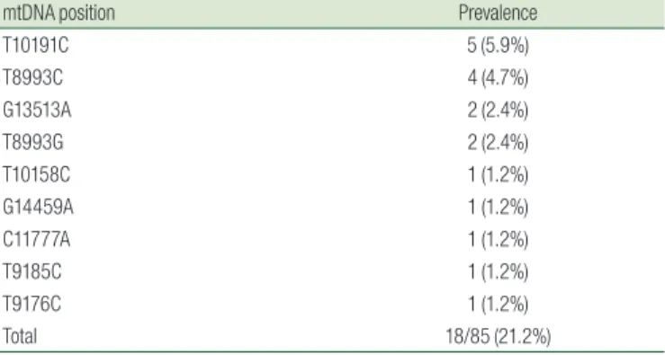
Table 1. The Position of mitochondrial DNA (mtDNA) Mutations in
Patients with Leigh Syndrome (LS) (n=18)
mtDNA position Prevalence
T10191C 5 (5.9%)
T8993C 4 (4.7%)
G13513A 2 (2.4%)
T8993G 2 (2.4%)
T10158C 1 (1.2%)
G14459A 1 (1.2%)
C11777A 1 (1.2%)
T9185C 1 (1.2%)
T9176C 1 (1.2%)
Total 18/85 (21.2%)
mtDNA, mitochondrial DNA.
mtDNA mutations. The most frequent mutation, T1019C, a mutation in the ND3 gene of the mitochondrial respiratory chain (MRC) complex I, was present in 5 patients (5.9%)10,11). The prevalence of the position of a mtDNA mutation is shown in Table 1.
Of the 85 patients with LS, 41 were male and 44 were female (Table 2). The average age of the first clinical presentation was 1.56 (SD=2.08) years old. All 85 patients (100%) had central nervous system involvement, and other organs that were affected are shown in Table 2. Subgroup analysis was performed to compare the mtDNA mutation positive and negative groups.
The proportion of females was significantly higher in the positive group than in the negative group (P=0.022). The age at onset of first symptoms was significantly greater in the positive group than in the negative group (P=0.028). The incidence of ocular involvement was also significantly higher in the mtDNA mutation positive group (P=0.009). In contrast, there was no
significant difference between the two groups in the involve- ment of other organs.
The laboratory data of all patients are summarized in Table 3.
Table 2. Comparison of Pediatric Clinical Variation in Leigh Syndrome (LS) between mitochondrial DNA (mtDNA) Mutation Positive and Negative
Groups (n=85)
Clinical feature Specification Total (n=85) mtDNA (+) (n=18) mtDNA (–) (n=67) P-value
Sex (female) 44 (40.7%) 15 (75.0%) 30 (44.8%) 0.022*
Age at onset of first symptom (years) 1.56±2.08 2.34±2.69 1.30±1.79 0.028*
Organ involvement CNS 85 (100%) 18 (100.0%) 67 (100.0%)
Muscular 43 (50.6%) 9 (50.0%) 34 (50.7%) 0.955
Respiratory 24 (28.2%) 6 (33.3%) 18 (26.9%) 0.555
GI tract 33 (38.8%) 5 (27.8%) 28(41.8%) 0.279
Ear 4 (4.7%) 0 (0%) 4 (6.0%) 0.379
Eye 8 (9.4%) 5 (27.8%) 3 (4.5%) 0.009*
Heart 6 (7.1%) 1 (5.6%) 5 (7.5%) 1.000
Hematology 1 (1.2%) 0 (0.0%) 1 (1.5%) 1.000
Nephrology 5 (5.9%) 2 (11.1%) 3 (4.5%) 0.285
*P values <0.05 were considered statistically significant.
Table 3. Comparison of Biochemical Laboratory Findings in Patients with Leigh Syndrome (LS) with or without mitochondrial DNA (mtDNA)
Mutations (n=85)
Biochemical laboratory findings Total (n=85) mtDNA (+) (n=18) mtDNA (–) (n=67) P-value Reference value
Lactate At the time of diagnosis 2.91±1.71 2.85±1.40 2.93±1.79 0.877 0.5-2.2mmol/L
Last visit 2.43±1.27 2.61±1.01 2.39±1.33 0.523
Pyruvate At the time of diagnosis 0.17±0.10 0.13±0.07 0.18±0.11 0.072 0.034-0.102 mmol/L
Last visit 0.16±0.07 0.15±0.06 0.16±0.0.07 0.941
Lactate/ pyruvate ratio At the time of diagnosis 21.89±28.37 36.53±13.52 17.96±10.18 0.189
Last visit 18.17±18.87 18.02±6.62 18.21±21.02 0.970
CK At the time of diagnosis 148.94±133.16 122.67±107.87 156.00±139.04 0.349 44-245 IU/L
Last visit 162.25±161.78 106.35±61.49 176.72±176.95 0.112
Plasma amino acid (n=72) Alanine 432.93±189.03 517.19±220.99 408.86±173.65 0.042* 143-439 nmol/mL
Glycine 329.4±920.72 705.94±1,948.72 221.82±75.08 0.336 81-436 nmol/mL
Proline 181.39±90.56 178.69±84.77 182.16±92.87 0.894 52-298 nmol/mL
Threonine 125.72±86.72 151.75±168.77 118.29±41.08 0.443 189.8 nmol/mL
Urine organic acid (n=59) Abnormal findings 28(47.5%) 6/11 (54.5%) 22/48 (45.8%) 0.602
mtDNA, mitochondrial DNA; CK, creatine kinase.
*P values <0.05 were considered statistically significant.
Table 4. Comparison of Leigh Syndrome (LS) Magnetic Resonance
Imaging (MRI) Findings between mitochondrial DNA (mtDNA) Mutation Positive and Negative Groups (n=85)
Involved lesion Total (n=85) mtDNA (+) (n=18) mtDNA (–) (n=67) P-value Basal ganglia 80 (94.1%) 17 (94.4%) 63 (94.0%) 1.000
Thalamus 28 (32.9%) 8 (44.4%) 20 (29.9%) 0.242
Brainstem 31 (28.7%) 10 (55.6%) 21 (31.3%) 0.058
Midbrain 30 (35.3%) 11 (61.1%) 19 (28.4%) 0.010
Pons 14 (16.5%) 4 (22.2%) 10 (14.9%) 0.483
Medulla 19 (22.4%) 8 (44.4%) 11 (16.4%) 0.022*
Cerebellum 23 (27.1%) 6 (33.3%) 17 (25.4%) 0.555
Atrophy 44 (51.8%) 7 (38.9%) 37 (55.2%) 0.218
Cortex 23 (27.1%) 6 (33.3%) 17 (25.4%) 0.555
White matter 35 (41.2%) 3 (16.7%) 32 (47.8%) 0.017*
mtDNA, mitochondrial DNA.
*P values <0.05 were considered statistically significant.
Biochemical laboratory measurements showed that the mean lactate and pyruvate levels were elevated, but in most patients, creatine kinase activity was not elevated relative to the reference value. In addition, no differences between the two groups were found for lactate, pyruvate, the lactate-pyruvate ratio, and CK levels and abnormal findings in urine organic acid. Plasma amino acid analysis revealed that the mean alanine level was elevated compared to the reference value and that the alanine levels of the two groups were significantly different (P=0.042).
The neuroimaging findings are summarized in Table 4. Of the 85 patients, 80 (94.1%) had basal ganglia involvement, and 31 (28.7%) had brainstem involvement, of which the midbrain was the most commonly affected site (30/31; 96.7%). In addition, 23 of the 85 patients (27.1%) showed cortex involvement, and 35 patients (41.2%) showed white matter involvement. MRI findings showed no significant differences in the involvement of the basal ganglia, thalamus, cortex, and cerebellum between the two groups. The ratio of abnormalities in the brainstem was significantly higher in the mtDNA mutation-positive group, especially in the midbrain (P=0.010) and medulla (P=0.014). The ratio of white matter involvement was lower in mtDNA mutation- positive group (P=0.017).
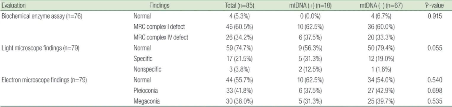
A muscle biopsy biochemical enzyme assay identified 46 patients (60.5%) with MRC 1 complex deficiency, 26 patients (34.2%) with MRC 4 deficiency, and 4 normal patients (5.3%).
Moreover, 17 patients (21.5%) showed mitochondrial disease- specific findings, such as ragged red fiber and abnormal histochemical staining. Three patients (3.8%) had nonspecific findings and 59 patients (74.7%) were normal. An electron microscope examination revealed 33 patients (41.8%) with pleioconia and 30 (38.0%) with megaconia. No differences were found between the two groups (Table 5).
Discussion
Previous studies have identified clinical differences according to the MRC complex type in LS12). More recently, however, studies have attempted to classify clinical features that are associated with genetic variation in patients with LS11-13). Phenotype analysis with the same mutation is limited because of the rarity of the disease.
Therefore, our study performed a genome-wide analysis of mtDNA rather than analyze a single mutation. We analyzed age, sex, biochemical markers, and the pathology of the patients who underwent mtDNA analysis. We diagnosed and treated patients with LS for the past 10 years using the same analytical procedures at a single institution.
LS typically presents clinical features in the first 2 years of life14). The mean age of clinical presentation in the patients in our study was 1.56 years, similar to the results of previous studies. Although previous studies of LS did not report sex differences6,15), we found that the proportion of females in the mtDNA mutation group was significantly higher than that in the non-mutation group (P=0.022).
The age of onset was significantly higher in the mtDNA mutation positive group. We performed a genome-wide analysis of mtDNA for all the patients included in our study. Therefore, the patients without a mtDNA mutation likely have a nDNA mutation. LS with a nDNA mutation often presents with more severe outcomes than LS with a mtDNA mutation4). Therefore, due to nDNA mutations, the age of onset of the mtDNA mutation-free group may be younger.
The clinical manifestation reported in our study is similar to that reported in previous studies, involving symptoms such as delayed development, seizure, motor weakness, loss of consciousness, muscle atrophy, and ataxia8). The incidence of ocular involvement in the mtDNA mutation-positive group was significantly higher;
therefore, additional mitochondrial genetic testing should be considered.
Screening tests for LS include an analysis of lactate and pyruvate
Table 5. Comparison of Diagnostic Evaluation in mitochondrial DNA (mtDNA) Mutation Positive and Negative Groups of Patients with Leigh
Syndrome (LS) (n=85)
Evaluation Findings Total (n=85) mtDNA (+) (n=18) mtDNA (–) (n=67) P-value
Biochemical enzyme assay (n=76) Normal 4 (5.3%) 0 (0.0%) 4 (6.7%) 0.915
MRC complex I defect 46 (60.5%) 10 (62.5%) 36 (60.0%)
MRC complex IV defect 26 (34.2%) 6 (37.5%) 20 (33.3%)
Light microscope findings (n=79) Normal 59 (74.7%) 9 (56.3%) 50 (79.4%) 0.055
Specific 17 (21.5%) 5 (31.3%) 12 (19.0%)
Nonspecific 3 (3.8%) 2 (12.5%) 1 (1.6%)
Electron microscope findings (n=79) Normal 44 (55.7%) 10 (62.5%) 34 (54.0%) 0.540
Pleioconia 33 (41.8%) 6 (37.5%) 27 (42.9%) 0.698
Megaconia 30 (38.0%) 5 (31.3%) 25 (39.7%) 0.535
mtDNA, mitochondrial DNA; MRC, mitochondrial respiratory chain.
*P values <0.05 were considered statistically significant.
levels, the lactate/pyruvate ratio, serum amino acid abnormalities, and urine organic acid16,17). Oxidative phosphorylation deficiency may lead to lactic acidosis. Pyruvate accumulates and is eventually metabolized into lactate by lactate dehydrogenase or transaminated into alanine by alanine aminotransferase, leading to an increase in these two substances in the blood, urine, and cerebrospinal fluid18). Therefore, assessment of alanine levels is useful in screening and diagnosing mitochondrial diseases16,19,20). In our study, the alanine level was significantly higher in the mtDNA mutation group, indicating that alanine levels may be helpful in differentiating patients with mtDNA mutations.
Bilateral, symmetrical hyperintensities in T2-weighted images are a characteristic finding in LS15). These lesions are commonly found in the basal ganglia, especially in the putamen, or in variable areas within the brainstem, such as the substantia nigra, nucleus ruber, or medulla oblongata12,21). In our study, brain involvement was present in all patients, and most of the patients had lesions in the basal ganglia or brainstem. In particular, the ratio of midbrain and medulla lesions in the brainstem was significantly higher in the group with mtDNA mutations than those in the group without mtDNA mutations (P=0.010 and 0.022, respectively). In the mtDNA mutation-positive group, the proportion of white matter involvement, a nonspecific finding in LS, was significantly lower than that in the non-mutation group (P=0.017). Previous studies that analyzed MRI features and the MRC complex reported no specific correlations between MRI findings and MRC complex type22). In contrast, a previous investigation has reported a difference in MRI findings between groups with and without nDNA mutations, such as the SURF1 mutation23). Our study found a significant difference in MRI findings between mtDNA mutation-positive and -negative groups. Thus, MRI findings correlate better with genetic findings than with pathologic findings. Imaging differences can signal differences in the natural course of the disease, suggesting that clinical differences in treating individuals with LS may depend on the presence of mtDNA mutations.
Although LS is a single syndrome, over 75 genes are known to cause this disease9). These genes are important for oxidative phosphorylation7). The gold standard of LS diagnosis is gradually progressing from neuropathologic to genetic analysis because previous analyses have not uncovered clinically relevant correla- tions24-26). Previous biochemical and clinical diagnostic methods for LS may require reanalysis and reclassification based on genotype.
Diagnosis using genotype would improve predictions of the clinical course from the time of diagnosis. However, genome-wide mtDNA and nDNA analysis are difficult and costly. Thus, identifying the characteristics of the patients with LS that have mtDNA mutations could present relevant criteria for determining whether mtDNA or
nDNA analysis should be performed. The results of our study suggest that mtDNA sequencing is recommended to evaluate patients with LS who are female or who exhibit ocular involvement, high alanine levels, or MRI findings of lesions in the midbrain and medulla.
A limitation of our study is that we could not compare the clinical features of the mtDNA mutation group and the nDNA mutation group. Further studies are required to analyze the characteristics of patients with mutations in nDNA.
요약
목적: Leigh 증후군은 진행하는 드문 신경퇴행성 질환으로 중추신 경계의 이상을 특징으로 한다. Leigh 증후군 환자들은 다양한 임상 증상 및 유전적 이상을 나타내기 때문에 예후를 예측하기 어렵다. 따 라서, 본 연구에서는 Leigh 증후군 환자들에서의 미토콘드리아 DNA 돌연변이와 환자들의 임상 양상과의 연관성에 대해 분석하였다.
방법: Leigh 증후군으로 진단받은 환자 중에서 미토콘드리아 DNA 유전자 돌연변이 검사를 시행한 85명의 환자들의 자료를 후향적으로 분석하였다. 미토콘드리아 DNA 돌연변이 결과에 따라 환자군을 나누 고, 집단 간의 임상 양상 차이를 통계 분석을 시행하였다.
결과: 연구에 참여한 85 명의 환자 중 18 명이 미토콘드리아 DNA 돌연변이를 가지고 있었다. 대부분의 환자는 젖산수치 및 젖산/피루 베이트 비율이 20 이상이었으며 이는 호흡 연쇄 반응의 이상에 부합 하는 결과였다. 미토콘드리아 DNA 돌연변이가 있는 환자군은 미토콘 드리아 DNA 돌연변이가 없는 환자군과 비교하여, 여성 비율, 알라닌 수치, 안구 침범, 자기 공명 영상(MRI)상 중뇌 및 숨뇌 이상이 통계학 적으로 유의하게 높았다.
결론: 임상적으로 Leigh 증후군이 의심될 때, 여성환자, 안구침범 을 보이는 환자, 높은 알라닌 수치를 보이는 환자, MRI상 중뇌나 숨뇌 에 이상소견이 보이는 환자에서는 미토콘드리아 DNA 돌연변이 검사 를 우선적으로 고려할 수 있다.
References
1) Leigh D. Subacute necrotizing encephalomyelopathy in an infant. J Neurol Neurosurg Psychiatry 1951;14:216-21.
2) Zhu Z, Yao J, Johns T, Fu K, De Bie I, Macmillan C, et al. SURF1, encoding a factor involved in the biogenesis of cytochrome C oxidase, is mutated in Leigh syndrome. Nat Genet 1998;20:337- 43.
3) van Erven PM, Cillessen JP, Eekhoff EM, Gabreëls FJ, Doesburg WH, Lemmens WA, et al. Leigh syndrome, a mitochondrial encephalo(myo)pathy. A review of the literature. Clin Neurol Neurosurg 1987;89:217-30.
4) Baertling F, Rodenburg RJ, Schaper J, Smeitink JA, Koopman WJ,
15) Lee HF, Tsai CR, Chi CS, Lee HJ, Chen CC. Leigh syndrome:
clinical and neuroimaging follow-up. Pediatr Neurol 2009;40:88- 93.
16) Morava E, van den Heuvel L, Hol F, de Vries MC, Hogeveen M, Rodenburg RJ, et al. Mitochondrial disease criteria: diagnostic applications in children. Neurology 2006;67:1823-6.
17) Parikh S, Goldstein A, Koenig MK, Scaglia F, Enns GM, Saneto R, et al. Diagnosis and management of mitochondrial disease: a consensus statement from the mitochondrial medicine society.
Genet Med 2015;17:689-701.
18) Janssen AJ, Smeitink JA, van den Heuvel LP. Some practical aspects of providing a diagnostic service for respiratory chain defects. Annal Clin Biochem 2003;40:3-8.
19) Stern HJ. Lactic acidosis in paediatrics: clinical and laboratory evaluation. Ann Clin Biochem 1994;31 (Pt 5):410-9.
20) Stacpoole PW. Lactic acidosis and other mitochondrial disorders. Metabolism 1997;46:306-21.
21) Valanne L, Ketonen L, Majander A, Suomalainen A, Pihko H.
Neuroradiologic findings in children with mitochondrial disorders. AJNR Am J Neuroradiol 1998;19:369-77.
22) Kim J, Lee SK, Kim EY, Kim DI, Lee YM, Lee JS, et al.
Neuroradiologic findings in children with mitochondrial disorder: correlation with mitochondrial respiratory chain defects. Eur Radiol 2008;18:1741-8.
23) Rossi A, Biancheri R, Bruno C, Di Rocco M, Calvi A, Pessagno A, et al. Leigh syndrome with COX deficiency and SURF1 gene mutations: MR imaging findings. AJNR Am J Neuroradiol 2003;24:1188-91.
24) Haack TB, Gorza M, Danhauser K, Mayr JA, Haberberger B, Wieland T, et al. Phenotypic spectrum of eleven patients and five novel MTFMT mutations identified by exome sequencing and candidate gene screening. Mol Genet Metab 2014;111:342-52.
25) Lieber DS, Calvo SE, Shanahan K, Slate NG, Liu S, Hershman SG, et al. Targeted exome sequencing of suspected mitochondrial disorders. Neurology 2013;80:1762-70.
26) Calvo SE, Compton AG, Hershman SG, Lim SC, Lieber DS, Tucker EJ. Molecular diagnosis of infantile mitochondrial disease with targeted next-generation sequencing. Sci Transl Med 2012;4:118ra10.
Mayatepek E, et al. A guide to diagnosis and treatment of Leigh syndrome. J Neurol Neurosurg Psychiatry 2014;85:257-65.
5) Thorburn DR, Rahman S. Mitochondrial DNA-associated Leigh syndrome and NARP. In: Pagon RA, Adam MP, Ardinger HH, Wallace SE, Amemiya A, Bean LJH, et al., editors. GeneReviews.
Seattle(WA): University of Washington, Seattle; 1993.
6) Sofou K, De Coo IF, Isohanni P, Ostergaard E, Naess K, De Meirleir L, et al. A multicenter study on Leigh syndrome: disease course and predictors of survival. Orphanet J Rare Dis 2014;9:52.
7) Ma YY, Wu TF, Liu YP, Wang Q, Song JQ, Li XY, et al. Genetic and biochemical findings in Chinese children with Leigh syndrome.
J Clin Neurosci 2013;20:1591-4.
8) Rahman S, Blok RB, Dahl HH, Danks DM, Kirby DM, Chow CW, et al. Leigh syndrome: clinical features and biochemical and DNA abnormalities. Ann Neurol 1996;39:343-51.
9) Lake NJ, Compton AG, Rahman S, Thorburn DR. Leigh syndrome: one disorder, more than 75 monogenic causes. Ann Neurol 2016;79:190-203.
10) Leshinsky-Silver E, Lev D, Tzofi-Berman Z, Cohen S, Saada A, Yanoov-Sharav M, et al. Fulminant neurological deterioration in a neonate with leigh syndrome due to a maternally transmitted missense mutation in the mitochondrial ND3 gene. Biochem Biophys Res Commun 2005;334:582-7.
11) Lim BC, Park JD, Hwang H, Kim KJ, Hwang YS, Chae JH, et al.
Mutations in ND subunits of complex I are an important genetic cause of childhood mitochondrial encephalopathies. J Child Neurol 2009;24:828-32.
12) Medina L, Chi TL, DeVivo DC, Hilal SK. MR findings in patients with subacute necrotizing encephalomyelopathy (Leigh syndrome): correlation with biochemical defect. AJR Am J Roentgenol 1990;154:1269-74.
13) Ruhoy IS, Saneto RP. The genetics of Leigh syndrome and its implications for clinical practice and risk management. Appl Clin Genet 2014;7:221-34.
14) Ostergaard E, Hansen FJ, Sorensen N, Duno M, Vissing J, Larsen PL, et al. Mitochondrial encephalomyopathy with elevated methylmalonic acid is caused by SUCLA2 mutations. Brain 2007;130:853-61.