ISSN 0378-6471 (Print)⋅ISSN 2092-9374 (Online)
https://doi.org/10.3341/jkos.2018.59.6.527
Original Article
근시의 정도에 따른 안압, 뇌척수압 및 사상판경유압력차의 관계
The Relationships of Intraocular Pressure, Cerebrospinal Fluid Pressure, and Trans-lamina Cribrosa Pressure Differences with Myopia
나승관1⋅온영훈1⋅김찬윤2⋅이시형1
Seung Kwan Nah, MD1, Young-Hoon Ohn, MD1, Chan Yun Kim, MD2, Si Hyung Lee, MD1
순천향대학교 의과대학 부천병원 안과학교실1, 연세대학교 의과대학 안과학교실 시기능개발연구소2 Department of Ophthalmology, Bucheon Hospital, Soonchunhyang University College of Medicine1, Bucheon, Korea The Institute of Vision Research, Department of Ophthalmology, Yonsei University College of Medicine2, Seoul, Korea
Purpose: To investigate the relationships between myopia and the three parameters of intraocular pressure (IOP), estimated cerebrospinal fluid pressure (CSFP), and the trans-lamina cribrosa pressure difference (TLCPD).
Methods: A total of 6,933 adults (≥19 years of age) who participated in the Korea National Health and Nutrition Examination Survey (2008–2012). These subjects were divided into two groups: young age group (19–49 years of age) and old age group (≥
50 years of age). The estimated CSFP was calculated as CSFP (mmHg) = 0.44 body mass index (kg/m2) + 0.16 diastolic blood pressure (mmHg) - 0.18 age (years) - 1.91. The TLCPD was calculated by subtracting the CSFP from the IOP.
Results: The mean estimated CSFP in the total population was 13.7 ± 0.1 mmHg (young, 14.2 ± 0.1 mmHg; old, 11.5 ± 0.1; p <
0.01), the mean IOP in the total population was 14.0 ± 0.1 mmHg (young, 14.0 ± 0.1 mmHg; old, 14.1 ± 0.1; p = 0.724), and the mean TLCPD in the total population was 0.7 ± 0.1 mmHg (young, 0.3 ± 0.1 mmHg; old, 3.0 ± 0.2; p < 0.001). After adjusting for confounding factors, multivariate linear regression analyses revealed significant positive associations between the degree of myopia and the estimated CSFP (p < 0.001; β, 0.12; spherical equivalent [SE], 0.03), as well as IOP (p < 0.001; β, 0.29; SE, 0.05). As a result, a higher TLCPD also showed a significant association with more myopic refractive error (p=0.002; β, 0.18; SE, 0.06). In subgroup analyses, a similar association was shown only in the young age group (estimated CSFP, p < 0.001; β, 0.12;
SE, 0.03; IOP, p < 0.001; β, 0.28; SE, 0.05; TLCPD, p = 0.005; β, 0.17; SE: 0.06), while the old age group did not show a sig- nificant association between TLCPD and the degree of myopia (p = 0.274; β, 0.18; SE, 0.16).
Conclusions: The calculated TLCPD showed an association with high myopia. It was consistent with the potential role of high myopia in the pathogenesis of open-angle glaucoma.
J Korean Ophthalmol Soc 2018;59(6):527-536
Keywords: Cerebrospinal fluid pressure, Intraocular pressure, Myopia, Open-angle glaucoma
■Received: 2017. 11. 23. ■ Revised: 2018. 2. 28.
■Accepted: 2018. 6. 4.
■Address reprint requests to Si Hyung Lee, MD
Department of Ophthalmology, Soonchunhyang University Bucheon Hospital, #170 Jomaru-ro, Bucheon 14584, Korea Tel: 82-32-621-6718, Fax: 82-32-621-5018
E-mail: [email protected]
* This work was supported by the government (the Ministry of Education) Research Fund (2017) of National Research Foundation of Korea (No. 2017R1D1A1B03029944).
* Conflicts of Interest: The authors have no conflicts to disclose.
ⓒ2018 The Korean Ophthalmological Society
This is an Open Access article distributed under the terms of the Creative Commons Attribution Non-Commercial License (http://creativecommons.org/licenses/by-nc/3.0/) which permits unrestricted non-commercial use, distribution, and reproduction in any medium, provided the original work is properly cited.
녹내장은 시신경유두테의 점진적인 소실 및 이에 동반되 는 시야이상을 유발하는 질환으로 실명에 이르게 되는 주 요 원인 중 하나이다.1 이러한 녹내장의 위험인자로는 대표 적인 고안압 이외에도 근시, 고령, 당뇨병, 인종, 가족력, 고 혈압 등이 있다.1 특히 녹내장과 최근에 유병률이 높아지는 근시와의 관계에 대한 연구는 그동안 많이 시도되었으며, 그중에 근시가 개방각녹내장의 발생에 있어 위험인자라는 점은 여러 연구에서 알려져 있다. 근시가 녹내장 병인에 영
향을 주는 기전으로는 근시안 자체의 해부학적 요인과 더 불어 유전적요인, 안압, 혈류장애도 제시되었다.2-17
녹내장에 있어서의 최근 많은 연구가 시도된 관련인자로 는 안압, 뇌척수압, 사상판경유압력차가 있다.18,19 안압의 경우 녹내장과 가장 밀접하게 연관되어 있는 인자로 알려 져 있으며, 안압을 낮추는 것은 녹내장의 발생과 진행을 막 는데 있어서 명확하게 증명된 유일한 인자 중 하나이다.20 뇌척수압과 사상판경유압력차 또한 안압과 관련하여 녹 내장에 영향을 끼치는 중요한 요인 중 하나이다. 시신경유 두는 3가지 층인 표층신경섬유층, 사상판전층, 사상판층으 로 이루어져 있으며, 사상판은 신경세포의 축삭과 망막혈 관이 지나가는 구조물이다. 시신경은 지주막하강에 둘러싸 여 있어서 안압의 영향뿐만 아니라 지주막하강내의 뇌척수 액압력의 영향도 받게 된다. 여기서 사상판은 안구내의 안 압과 안구 밖에서 작용하는 뇌척수압이 만나게 되는 해부 학적인 부분으로 이때의 두 압력의 차이는 사상판경유압력 차로 정의한다(사상판경유압력차, trans-lamina cribrosa pres- sure difference [TLCPD] = intraocular pressure [IOP] – in- tracranial pressure [ICP]).18,20 이러한 해부학적 특성 때문에 사상판 경유 압력차가 커질수록 사상판의 형태학적 변형이 일어나게 되고 기계적인 시신경 손상이 야기되면서 녹내장 으로 진행하는 것으로 알려져 있고, 이는 특히 정상안압 녹 내장 환자에서 녹내장 발생 기전과 밀접한 연관이 있는 것 으로 보인다.18,19
이러한 뇌척수압 및 사상판경유압력차와 녹내장의 연관 성에도 불구하고, 녹내장의 주요 위험인자 중 하나인 근시 에서의 사상판경유압력차에 대한 연구는 많이 이뤄지지 않 고 있다. 이에 본 연구에서는 국민건강영양조사자료를 바 탕으로 한국인에서 근시의 정도와 안압, 뇌척수압, 사상판 경유압력차 간의 연관성을 비교, 분석하여 근시에서 개방 각 녹내장 발생기전에서 사상판경유압력차가 미치는 영향 을 확인해 보고자 하였다. 뇌척수압 및 사상판경유압력차 는 Xie et al21의 체질량지수, 이완기 혈압과 나이를 이용하 여 뇌척수압을 계산할 수 있는 공식을 이용하여 추정하였 다.
대상과 방법
대상
본 연구는 2008-2012년 국민건강영양조사자료를 이용하 여 분석하였으며, 생명윤리위원회(institutional review board, IRB) 면제 대상이다. 국민건강영양조사는 국민건강증진법 을 바탕으로 국민건강과 영양에 관한 기초적인 통계를 산출 하기 위해 시행된 법정조사로 질병관리본부 연구윤리심의위원
회 승인을 받았다. 국민건강영양조사는 제1기(1998년)부터 제3 기(2005년)까지 3년 주기로 실시하였으며, 제4기(2007-2009년) 부터는 연중조사체계로 개편하여 조사를 실시하였고 제5기 (2010-2012년) 조사까지 공개되었다. 2008-2012년 국민건강 영양조사대상자 중 19세 이상 총 23,522명 중 백내장, 녹내 장, 사시, 망막, 굴절수술을 받은 대상자 7,717명, 녹내장 환 자 230명, 익상편 환자 3,718, 백내장환자 4,518명, 결측값 406명을 제외한 최종 6,933명을 대상으로 분석하였다.
Choi et al22에 따르면 국내 근시 유병률이 50대 전후로 현 저한 차이가 있음을 보고한 바 있으며, 이를 근거로 본 연 구에서는 50세를 기준으로 젊은 연령군(19-49세)을 Group 1, 고령군(50세 이상)을 Group 2로 나누어 연구를 진행하였 다.
방법
국민건강영양조사에서는 전신적인 요소를 확인하기 위 해 혈압(수축기 및 이완기 혈압)과 맥박, 체질량지수를 측 정하였고, 혈액검사를 통해 중성지방, high density lip- oprotein (HDL), 총 콜레스테롤수치를 측정하였다. 안검진 은 안과의사 또는 안과 전공의에 의하여 실시되었으며 안압 은 골드만압평안압계(Haag-Streit, Inc., Bern, Switzerland) 로 측정하였다. 굴절이상은 국민건강영양조사에서 수집된 항목 중 조절마비를 시행하지 않은 상태에서 자동굴절검사 기(Topcon. KR-8800; Topcon Inc., Tokyo, Japan)를 이용해 측정한 굴절이상 정도를 연령별로 수집하여 분석하였다.
굴절값은 각각의 눈에서 얻어진 구면렌즈대응치값(구면렌 즈값 +1/2×원주렌즈값)을 이용하였고, 근시의 정도는 경도 근시(-2.99D ≤ standard error [SE] ≤ -0.50D)와 중등도근시 (-5.99D ≤ SE ≤ -3.00D), 고도근시(SE ≤ -6.00D) 3가지의 군으로 나눠서 분석하였다.
안압과 굴절치는 우안 값을 대표 값으로 포함하였다. 이때 우안과 좌안 굴절치는 높은 상관관계를 보였으며(Pearson’s correlation = 0.91, p<0.001), 우안과 좌안 안압도 높은 상 관관계를 보였다(Pearson’s correlation = 0.82, p<0.001).
뇌척수압은 이전에 발표된 공식(추정뇌척수압=0.44×체 질량지수[kg/m2]+0.16×이완기혈압[mmHg]–0.18×나이 [years]–1.91)을 이용하여 추정뇌척수압을 구하였다. 사상판경 유압력차는 추정뇌척수압에서 안압을 뺀 값으로 하였다(사상 판경유압력차[TLCPD] = 안압[IOP]–뇌척수압[cerebrospinal fluid pressure, CSFP]).21
통계분석
통계프로그램은 SAS version 9.3 (SAS Inc., Cary, NC, USA)를 사용하였다. 각 군별 기본 특성 및 임상적 특성에
대한 통계적 차이를 검정하기 위하여 범주형 변수의 경우 Rao-Scott χ2 test를 사용하였고, 연속형 변수의 경우 Wald’s F test를 사용하였다. 굴절률 이상(디옵터)의 정도 혹은 근 시의 정도에 따른 3가지 인자(안압, 추정평균뇌척수압, 평 균사상판경유압력차) 간의 연관성을 찾기 위해 단순 및 다 중 선형 회귀분석(linear regression analysis)을 수행하였다.
다중 선형 회귀분석 시 두 가지 보정군으로 나누어서 분석 을 시행하였는데, 나이와 성별 보정군(Model 1)과 나이와 성별, 체질량지수, 고혈압, 당뇨, 고지질혈증에 따른 보정군 (Model 2)으로 나눠서 시행하였다. 회귀분석의 모든 변수 들에 대해 다중 공선성(multicollinearity)에 대한 검사를 시 행하였으며, 그중 분산팽창계수(variance inflation factor)가 5 이하인 변수들만 다중 선형 회귀분석 보정군에 포함되었 다. 모든 통계적인 검정은 양측의 검정을 기반으로 유의수 준 0.05보다 작은 유의확률을 보이는 경우 유의한 것으로 간주하였다.
결 과
전신 검사 및 안과 검사가 가능했던 19세 이상의 성인은 총 6,933명이었고, 그중 젊은 연령군(19-49세, Group 1)은 5.307명, 고령군(50세 이상, Group 2)은 1,626명이었다. 평 균 연령은 Group 1에서 32.3세, Group 2에서 56.1세였으며, 남녀 비율은 Group 1에서 여성이 49.8%, Group 2에서 53.4%였다. 도심 지역에서 Group 1은 73.4%, Group 2는 74.9%가 거주하고 있었다. 고혈압은 Group 1에서 13.3%
Group 2에서 40.3%, 당뇨는 Group 1에서 2.4%, Group 2에 서 11.9%, 고지혈증은 Group 1에서 5.5%, Group 2에서 15.7%의 유병률을 보였다.
수축기 혈압은 Group 1에서 111.7 mmHg, Group 2에서 123.2 mmHg로 두 군 간 의미있는 차이를 보였으며(p<0.001), 이완기 혈압은 Group 1에서 75.3 mmHg, Group 2에서 80.3 mmHg로 두 군 간 의미있는 차이를 보였다(p<0.001). 평균 추정뇌척수압은 Group 1에서 14.2 mmHg, Group 2에서 11.5 mmHg로 두 군 간 의미있는 차이를 보였으며(p<0.001), 평균 안압은 Group 1에서 14.0 mmHg, Group 2에서 14.1 mmHg (p<0.001), 평균사상판경유압력차는 Group 1에서는 0.3 mmHg, Group 2에서 3.0 mmHg로 두 군 간 의미있는 차 이를 보였다(p<0.001). Group 1에서 정시와 원시(spherical equivalent [SE] > -0.50D)는 30.2%, 경도근시(-2.99D ≤ SE
≤ -0.50D)는 46.2%, 중등도근시(-5.99D ≤ SE ≤ -3.00D)는 17.4%, 고도근시(SE ≤ -6.00D)는 6.1%의 구성을 보였고, Group 2에서 정시와 원시(SE > -0.50D)는 70.3%, 경도근시 (-2.99D ≤ SE ≤ -0.50D)는 23.8%, 중등도근시(-5.99D ≤ SE
≤ -3.00D)는 4.3%, 고도근시(SE ≤ -6.00D)는 1.6%의 구성 을 보였다(Table 1).
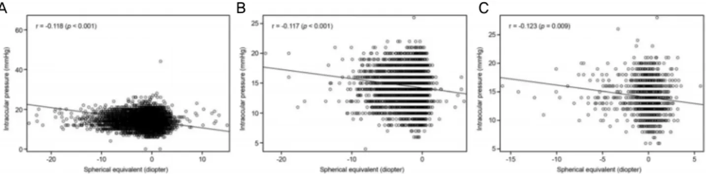
Fig. 1은 굴절률 이상에 따른 추정뇌척수압의 분포를 나 타내고 있으며, 전체 환자군(r=-0.187, p=0.030), Group 1 (r=-0.105, p<0.001), Group 2 (r=-0.187, p=0.030) 모두에서 음의 상관관계를 보이고 있다. Fig. 2는 굴절률 이상에 따 른 안압의 분포를 나타내고 있으며, 전체 환자군(r=-0.118,
p<0.001), Group 1 (r=-0.117, p<0.001), Group 2 (r=-0.123, p=0.009) 모두에서 마찬가지로 음의 상관 관계를 보이고
있다. Fig. 3는 굴절률 이상에 따른 사상판경유압력차의 분 포를 나타내고 있으며, 전체 환자군(r=-0.088, p<0.001)과 Group 1 (r=-0.096, p=0.001)에서 음의 상관 관계를 보였으 나, Group 2 (r=-0.038, p=0.591)에서는 유의한 상관관계를 보이지 않았다.굴절률 이상(디옵터)과 3가지 인자(추정뇌척수압, 안압, 사상판경유압력차) 간의 다중선형회귀분석을 나이, 성별 보정군(Model 1)과 나이, 성별, 체질량지수, 이완기 혈압, 고혈압, 당뇨, 고지질혈증에 따른 보정군(Model 2)으로 나 눠서 시행하였다(Table 2).
먼저 추정뇌척수압은 전체 환자군에서 Model 1 (β:
-0.04, SE: 0.02, p=0.033)과 Model 2 (β: -0.03, SE: 0.01,
p<0.001) 모두 굴절률과 통계적으로 유의한 상관관계를 보
였고, Group 1에서도 Model 1 (β: -0.03, SE: 0.02,p=0.046), Model 2 (β: -0.03, SE: 0.01, p<0.001)로 모두 통
계적으로 유의한 상관관계를 나타내었으나, Group 2에서는 Model 1 (β: -0.07, SE: 0.05, p = 0.135)에서는 통계적으로 유의하지 않았고, Model 2 (β: -0.05, SE: 0.02, p = 0.029) 는 통계적으로 유의하였다.굴절률과 안압의 관계는 전체 환자군에서 Model 1 (β:
-0.12, SE: 0.02, p<0.001)과 Model 2 (β: -0.11, SE: 0.02,
p<0.001) 모두 통계적으로 유의한 상관관계를 보였고,
Group 1에서 Model 1 (β: -0.12, SE: 0.02, p< 0.001), Model 2 (β: -0.11, SE: 0.02, p<0.001)로 모두 통계적으로 유의한 상관관계를 나타내었으며, Group 2에서는 Model 1 (β: -0.12, SE: 0.05, p=0.010)은 통계적으로 유의하였으나, Model 2 (β: -0.09, SE: 0.05, p=0.065)에서는 통계적으로 유의하지 않았다.사상판경유압력차는 전체 환자군은 Model 1 (β: -0.07, SE: 0.02, p=0.001)과 Model 2 (β: -0.08, SE: 0.02, p<0.001) 모두 굴절률과 통계적으로 유의한 상관관계를 보였고, Group 1에서도 Model 1 (β: -0.07, SE: 0.03, p = 0.002), Model 2 (β: -0.08, SE: 0.02, p<0.001)로 모두 통계적으로 유의한 상관관계를 나타내었으나, Group 2에서는 Model 1 (β: -0.05, SE: 0.06, p=0.468)과 Model 2 (β: -0.04, SE:
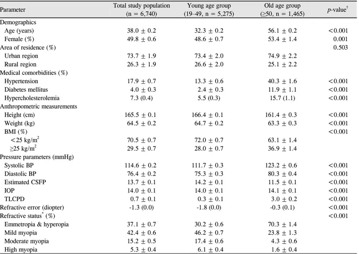
Parameter Total study population (n = 6,740)
Young age group (19–49, n = 5,275)
Old age group
(≥50, n = 1,465) p-value† Demographics
Age (years) 38.0 ± 0.2 32.3 ± 0.2 56.1 ± 0.2 <0.001
Female (%) 49.8 ± 0.6 48.6 ± 0.7 53.4 ± 1.4 0.001
Area of residence (%) 0.503
Urban region 73.7 ± 1.9 73.4 ± 2.0 74.9 ± 2.2
Rural region 26.3 ± 1.9 26.6 ± 2.0 25.1 ± 2.2
Medical comorbidities (%)
Hypertension 17.9 ± 0.7 13.3 ± 0.6 40.3 ± 1.6 <0.001
Diabetes mellitus 4.0 ± 0.3 2.4 ± 0.3 11.9 ± 1.1 <0.001
Hypercholesterolemia 7.3 (0.4) 5.5 (0.3) 15.7 (1.1) <0.001
Anthropometric measurements
Height (cm) 165.5 ± 0.1 166.4 ± 0.1 161.4 ± 0.3 <0.001
Weight (kg) 64.5 ± 0.2 64.7 ± 0.2 63.3 ± 0.3 <0.001
BMI (%) <0.001
<25 kg/m2 70.5 ± 0.7 72.0 ± 0.7 63.1 ± 1.4
≥25 kg/m2 29.5 ± 0.7 28.0 ± 0.7 36.9 ± 1.4
Pressure parameters (mmHg)
Systolic BP 114.6 ± 0.2 111.7 ± 0.3 123.2 ± 0.6 <0.001
Diastolic BP 76.4 ± 0.2 75.3 ± 0.3 80.3 ± 0.4 <0.001
Estimated CSFP 13.7 ± 0.1 14.2 ± 0.1 11.5 ± 0.1 <0.001
IOP 14.0 ± 0.1 14.0 ± 0.1 14.1 ± 0.1 <0.001
TLCPD 0.7 ± 0.1 0.3 ± 0.1 3.0 ± 0.2 <0.001
Refractive error (diopter) -1.3 (0.0) -1.8 (0.0) -0.3 (0.1) <0.001
Refractive status* (%) <0.001
Emmetropia & hyperopia 37.1 ± 0.7 30.2 ± 0.6 70.3 ± 1.4
Mild myopia 42.4 ± 0.6 46.2 ± 0.7 23.8 ± 1.3
Moderate myopia 15.2 ± 0.5 17.4 ± 0.6 4.3 ± 0.6
High myopia 5.3 ± 0.4 6.1 ± 0.4 1.6 ± 0.4
Values are presented as mean ± SD unless otherwise indicated.
BMI = body mass index; BP = blood pressure; CSFP = cerebrospinal fluid pressure; IOP = intraocular pressure; TLCPD = trans-lamina cribrosa pressure difference.
*Emmetropia & hypertropia (spherical equivalent [SE]) > -0.50 D), mild myopia (-2.99 D ≤ SE ≤ -0.50 D), moderate myopia (-5.99 D ≤ SE
≤ -3.00 D), high myopia (SE ≤ -6.00 D); †Rao-Scott χ2 test (for categorical variables) or Wald’s F tests (for continuous variables) was used.
Table 1. Demographics and general health characteristics of the study population
Figure 1. Spherical equivalent and estimated cerebrospinal fluid pressure. Association between estimated cerebrospinal fluid pressure
and spherical equivalent in the total study population (A), young (B) and old age group (C).0.05, p=0.287) 모두 통계적으로 유의하지 않았다.
근시의 정도(경도근시: [-2.99D ≤ SE ≤ -0.50D], 중등도 근시: [-5.99D ≤ SE ≤ -3.00D], 고도근시: [SE ≤ -6.00D])와 3가지 압력인자(추정뇌척수압, 안압, 사상판경유압력차)간
의 다중선형회귀분석 또한 나이 성별 보정군(Model 1)과 나이 성별, 체질량지수, 이완기 혈압, 고혈압, 당뇨, 고지질 혈증에 따른 보정군(Model 2)으로 나눠서 시행하였다 (Table 3).
A B C
Figure 2. Spherical equivalent and intraocular pressure. Association between Intraocular pressure and spherical equivalent in the total
study population (A), young (B), and old age group (C).Figure 3. Spherical equivalent and Trans-lamina cribrosa pressure difference. Association between Trans-lamina cribrosa pressure
difference and spherical equivalent in the total study population (A), young (B), and old age group (C).Parameters Model 1* Model 2†
Beta SE p-value Beta SE p-value
Estimated cerebrospinal fluid pressure
Total population -0.04 0.02 0.033‡ -0.03 0.01 <0.001‡
Young age group (19-49) -0.03 0.02 0.046‡ -0.03 0.01 <0.001‡
Old age group (≥50) -0.07 0.05 0.135 -0.05 0.02 0.029‡
Intraocular pressure
Total population -0.12 0.02 <0.001‡ -0.11 0.02 <0.001‡
Young age group (19-49) -0.12 0.02 <0.001‡ -0.11 0.02 <0.001‡
Old age group (≥50) -0.12 0.05 0.010‡ -0.09 0.05 0.065
Trans-lamina cribrosa pressure difference
Total population -0.07 0.02 0.001‡ -0.08 0.02 <0.001‡
Young age group (19-49) -0.07 0.03 0.002‡ -0.08 0.02 <0.001‡
Old age group (≥50) -0.05 0.06 0.468 -0.04 0.05 0.287
*Model 1: adjusted for age and sex; †Model 2: adjusted for age, sex, area of residence, body mass index, diastolic blood pressure, hypertension, diabetes mellitus, and hypercholesterolemia; ‡p < 0.05.
Table 2. Multivariate analysis of association between refractive error (D) and three pressure parameters (estimated cerebrospinal
pressure, intraocular pressure, and trans-lamina cribrosa pressure difference) in total, young age, and old age population추정뇌척수압에서 전체 환자군은 Model 1 (β: 0.13, SE:
0.05, p=0.006)과 Model 2 (β: 0.08, SE: 0.02, p<0.001) 모 두 통계적으로 유의하였고, Group 1에서도 Model 1 (β:
0.12, SE: 0.05, p=0.013), Model 2 (β: 0.08, SE: 0.02,
p<0.001)로 모두 통계적으로 유의하였으나, Group 2에서는
Model 1 (β: 0.28, SE: 0.13, p=0.031), Model 2 (β: 0.13,SE: 0.05, p=0.023)로 모두 통계적으로 유의하지 않았다 (Fig. 4).
안압은 전체 환자군에서 Model 1 (β: 0.30, SE: 0.05,
p<0.001)과 Model 2 (β: 0.29, SE: 0.04, p<0.001) 모두 통계
적으로 유의하였고, Group 1에서 Model 1 (β: 0.29, SE:0.05, p<0.001), Model 2 (β: 0.28, SE: 0.05, p<0.001)로 모
A B C
A B C
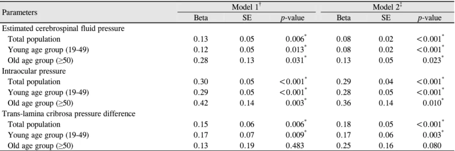
Parameters Model 1† Model 2‡
Beta SE p-value Beta SE p-value
Estimated cerebrospinal fluid pressure
Total population 0.13 0.05 0.006* 0.08 0.02 <0.001*
Young age group (19-49) 0.12 0.05 0.013* 0.08 0.02 <0.001*
Old age group (≥50) 0.28 0.13 0.031* 0.13 0.05 0.023*
Intraocular pressure
Total population 0.30 0.05 <0.001* 0.29 0.04 <0.001*
Young age group (19-49) 0.29 0.05 <0.001* 0.28 0.05 <0.001*
Old age group (≥50) 0.42 0.14 0.003* 0.36 0.14 0.010*
Trans-lamina cribrosa pressure difference
Total population 0.15 0.06 0.006* 0.18 0.05 <0.001*
Young age group (19-49) 0.17 0.07 0.009* 0.17 0.06 0.003*
Old age group (≥50) 0.13 0.19 0.483 0.25 0.16 0.080
SE = spherical equivalent.
*p < 0.05; †Model 1: adjusted for age and sex; ‡Model 2: adjusted for age, sex, area of residence, body mass index, diastolic blood pressure, hypertension, diabetes mellitus, and hypercholesterolemia.
Table 3. Multivariate analysis of association between the degree of myopia and three pressure parameters (estimated cerebrospinal
pressure, intraocular pressure, and trans-lamina cribrosa pressure difference) in total, young age, and old age populationFigure 4. Degree of myopia and estimated cerebrospinal fluid
pressure. Estimated cerebrospinal fluid pressure according to the degree of myopia in the total study population (A), young (Group 1) (B), and old (C) age group (Group 2). *0.001 ≤ p <0.05; **
p < 0.001.
A B
C
두 통계적으로 유의하였으며, Group 2에서도 Model 1 (β:
0.42, SE: 0.14, p=0.003)과 Model 2 (β: 0.36, SE: 0.14,
p=0.010) 모두 통계적으로 유의하였다(Fig. 5).
사상판경유압력차는 전체 환자군은 Model 1 (β: 0.15, SE: 0.06, p=0.006)과 Model 2 (β: 0.18, SE: 0.05, p<0.001)
모두 통계적으로 유의하였고, Group 1에서도 Model 1 (β:
0.17, SE: 0.07, p=0.009), Model 2 (β: 0.17, SE: 0.06,
p=0.003)로 모두 통계적으로 유의하였으나, Group 2에서는
Model 1 (β: 0.13, SE: 0.19, p=0.483)과 Model 2(β: 0.25, SE: 0.16, p=0.080) 모두 통계적으로 유의하지 않았다(Fig. 6).A B
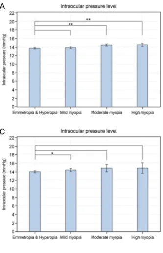
Figure 5. Degree of myopia and intraocular pressure.
Intraocular pressure according to the degree of myopia in the total study population (A), young (Group 1) (B), and old (C) age group (Group 2). *0.001 ≤ p < 0.05; **
p < 0.001.
C
C
A B
Figure 6. Degree of myopia and trans-lamina pressure difference.
Trans-lamina pressure difference according to the degree of myopia in the total study population (A), young (Group 1) (B), and old age group (Group 2) (C). *0.001 ≤ p < 0.05.
고 찰
근시와 녹내장의 밀접한 관계에 대해서 이전부터 많은 연구가 진행되었다. Mitchell et al23은 Blue Mountains Eye Study에서 개방각녹내장이 경도근시(-1.0D ≤ SE <-3.0D)에 서 4.2%, 중등도와 고도근시(≤-3.0D)에서 4.4% 유병률을 보였으며, 근시가 없는 환자에서는 1.5%의 유병률을 보여 근시에서 녹내장의 유병률이 확연히 높다는 결과를 발표하 였다. 또한 Beijing eye study에 따르면 -6.0D 이상의 고도 근시에서 녹내장성 시신경병증이 발생할 확률이 높다는 결 과를 발표하였고,24 Marcus et al25에 따르면 근시, 특히 고 도근시가 개방각녹내장의 위험요소라고 발표하였다. 또한 국민건강영양조사를 대상으로 진행된 국내 연구에서도 개 방각녹내장이 고도 근시와 연관이 있으며 특히 고도근시에 서 젊은 나이에 발병한다고 기술하고 있다.26
최근 발표된 연구들에서 낮은 뇌척수압과 높은 사상판경 유압력차가 녹내장의 주요한 위험인자로 보고된 바 있다.
Berdahl et al27,28은 척추천자를 시행한 환자군들 중 28명의 개방각 녹내장과 49명의 정상 환자들을 대상으로 시행한 후향적 연구에서 뇌척수압이 개방각 녹내장 환자에서 의미 있게 증가하였음을 보고하였고, 뇌척수압을 세 개의 군(정 상, 개방각녹내장, 정상안압녹내장)으로 나눠서 비교한 다 른 연구에서 뇌척수압이 정상안압녹내장군과 개방각녹내 장군에서 의미있게 낮았으며 이에 비해 사상판경유압력차 는 정상안압녹내장과 개방각녹내장군에서 의미있게 높았 다고 보고한 바 있다. 또한 국내에서도 국민건강영양조사 를 이용하여 시행한 연구에서 정상안압 녹내장 환자와 높 은 사상판 경유압력차가 의미있는 연관 관계가 있었다고 보고한 바 있다.29 하지만 낮은 뇌척수압과 높은 사상판경 유압력차가 녹내장과 관계가 없다는 보고들도 있어,30,31 추 후 이에 대한 추가적 연구가 필요할 것으로 사료된다.
본 연구에서 2008-2012년 시행한 국민건강영양조사를 대상으로 시행한 다중선형회귀분석에서 안압은 근시의 정 도에 따라 통계적으로 유의하게 증가하였는데 이는 Choi et al22의 연구결과와 유사하였다. 이 연구에 따르면, 2008년에 서 2011년까지 시행한 국민건강영양조사에서 젊은 연령층 (19-39세), 중년층(40-59세), 노년층(60세 이상)으로 나눠 시행한 연구결과 젊은 연령층에서 근시유병률이 높았고, 본 연구결과와 유사하게 전체 인구에서 높은 안압은 높은 근시디옵터와 연관이 있었으며 그 외에도 당뇨, 혈압 그리 고 이상지질혈증과 연관이 있었다. 세 연령층으로 나눠 시 행한 연구결과에서는 높은 안압은 젊은 연령층과 중년층에 서 근시디옵터의 정도가 큰 정도와 통계적으로 유의하게 비례하였으나, 노년층에서는 통계적으로 유의하지 않았다.
이는 본 연구에서와 같은 결과라 할 수 있다.
본 연구에서는 이전에 보고되었던 추론 공식을 통해 도 출한 추정뇌척수압 또한 근시의 정도에 따라 통계적으로 유의하게 증가하였지만 안압에 비해 적은 폭으로 증가하여 최종적으로 사상판경유압력차가 통계적으로 유의하게 증 가하는 결과를 도출하게 되었으며, 이는 연령, 성별, 거주지 역, 체질량지수, 고혈압, 당뇨 그리고 고지질혈증 등의 요인 을 보정한 이후에도 마찬가지였다. 특이적으로 연령별로 나누어 시행한 결과에서 사상판경유압력차가 젊은 연령군 (Group 1)에서 근시와 유의한 연관성을 보였으나 고령군 (Group 2)에서는 통계적으로 유의하지 않았는데, 이는 근 시의 유병률이 젊은 연령층에서 높은 것과 연관이 있을 것 으로 보인다. 본 연구를 통해 한국인에서 근시와 뇌척수압 및 사상판경유압력차의 관계를 확인할 수 있었으며, 이는 근시안에서 녹내장 발생률이 증가하는 기전과 연관성이 있 을 수 있다. 이러한 결과는 Fan et al32의 연구결과인 고도근 시에서 시신경주변 지주막하강이 확장되어 있어 녹내장에 취약할 수 있다는 내용과, Jonas and Xu33가 이전에 보고한 바 있는 고도근시에서 사상판의 두께가 감소하게 되어 안 구 내 공간과 뇌척수액공간 사이의 거리가 감소하여 결과 적으로 사상판경유압력차를 증가시키게 된다는 내용과도 어느 정도 일치한다고 볼 수 있다.
본 연구에서의 주 제한점은 뇌척수압을 직접 척추천자를 한 것이 아닌 추정공식을 통하여 구한 것이며, 이러한 추정 공식의 이용에는 논란의 여지가 있음을 이전 연구에서 보 고한 바 있다.34 하지만 본 연구에서 이용한 추정공식은 안 과적으로 대표적 population based study인 Beijing eye study 및 the central India eye and medical study에서 사용 된 바 있으며, 이를 통해 여러 인자들과의 연관성을 보고하
였다.21,35 또한 최근 보고된 국민건강영양조사자료를 이용
한 연구에서도 본 추정공식을 이용하여 추정뇌척수압 및 사상판경유압력차와 녹내장과의 연관성을 밝힌 바 있어 본 연구에서도 동일한 공식을 이용하였다.22,36 다른 제한점으 로는 국민건강영양조사의 특성상 안압을 3번에 걸쳐서 측 정하여 평균을 구하지 못하고 한 번 측정한 안압을 기록하 였기에 측정한 안압에 오차가 있을 수 있고, 본 연구가 단 면연구로 시행되었기 때문에 인과관계를 명확히 확인할 수 없다는 점이 있을 수 있다.
결론적으로 본 연구에서 근시의 정도에 따라 사상판경유 압력차가 통계적으로 유의하게 증가함을 보여주었고, 이는 근시안에서 녹내장 발생률이 증가하는 기전과 연관된다고 할 수 있다. 본 연구의 결과는 사상판경유압력차 증가로 인 해 근시가 녹내장의 발병과 진행에 영향을 미칠 수 있다는 기존 가설을 뒷받침함으로써 중등도 이상의 근시가 동반된
녹내장 환자를 치료하고 예후를 파악하는 데 있어 도움이 될 것으로 보인다.
REFERENCES
1) Crawford Downs J, Roberts MD, Sigal IA. Glaucomatous cupping of the lamina cribrosa: a review of the evidence for active pro- gressive remodeling as a mechanism. Exp Eye Res 2011;93:133-40.
2) Wu HM, Seet B, Yap EP, et al. Does education explain ethnic dif- ferences in myopia prevalence? A population-based study of young adult males in Singapore. Optom Vis Sci 2001;78:234-9.
3) Tomlinson A, Phillips CI. Ratio of optic cup to optic disc. In rela- tion to axial length of eyeball and refraction. Br J Ophthalmol 1969;53:765-8.
4) Bellezza AJ, Hart RT, Burgoyne CF. The optic nerve head as a bio- mechanical structure: initial finite element modeling. Invest Ophthalmol Vis Sci 2000;41:2991-3000.
5) Chihara E, Sawada A. Atypical nerve fiber layer defects in high myopes with high-tension glaucoma. Arch Ophthalmol 1990;108:228-32.
6) Dichtl A, Jonas JB, Naumann GO. Histomorphometry of the optic disc in highly myopic eyes with absolute secondary angle closure glaucoma. Br J Ophthalmol 1998;82:286-9.
7) Cahane M, Bartov E. Axial length and scleral thickness effect on susceptibility to glaucomatous damage: a theoretical model im- plementing Laplace's law. Ophthalmic Res 1992;24:280-4.
8) Quigley HA. Reappraisal of the mechanisms of glaucomatous op- tic nerve damage. Eye (Lond) 1987;1(Pt 2):318-22.
9) Avetisov ES, Savitskaya NF. Some features of ocular micro- circulation in myopia. Ann Ophthalmol 1977;9:1261-4.
10) Shih YF, Horng IH, Yang CH, et al. Ocular pulse amplitude in myopia. J Ocul Pharmacol 1991;7:83-7.
11) To'mey KF, Faris BM, Jalkh AE, Nasr AM. Ocular pulse in high myopia: a study of 40 eyes. Ann Ophthalmol 1981;13:569-71.
12) Perkins ES. The ocular pulse. Curr Eye Res 1981;1:19-23.
13) Lütjen-Drecoll E, Futa R, Rohen JW. Ultrahistochemical studies on tangential sections of the trabecular meshwork in normal and glaucomatous eyes. Invest Ophthalmol Vis Sci 1981;21:563-73.
14) Curtin BJ, Iwamoto T, Renaldo DP. Normal and staphylomatous sclera of high myopia. An electron microscopic study. Arch Ophthalmol 1979;97:912-5.
15) Hammond CJ, Snieder H, Gilbert CE, Spector TD. Genes and envi- ronment in refractive error: the Twin Eye Study. Invest Ophthalmol Vis Sci 2001;42:1232-6.
16) Stone EM, Fingert JH, Alward WL, et al. Identification of a gene that causes primary open angle glaucoma. Science 1997;275:668-70.
17) Mutti DO, Zadnik K, Adams AJ. Myopia. The nature versus nur- ture debate goes on. Invest Ophthalmol Vis Sci 1996;37:952-7.
18) Morgan WH, Yu DY, Cooper RL, et al. The influence of cere- brospinal fluid pressure on the lamina cribrosa tissue pressure gradient. Invest Ophthalmol Vis Sci 1995;36:1163-72.
19) Ren R, Jonas JB, Tian G, et al. Cerebrospinal fluid pressure in glau- coma: a prospective study. Ophthalmology 2010;117:259-66.
20) Marek B, Harris A, Kanakamedala P, et al. Cerebrospinal fluid pressure and glaucoma: regulation of trans-lamina cribrosa pressure. Br J Ophthalmol 2014;98:721-5.
21) Xie X, Zhang X, Fu J, et al. Noninvasive intracranial pressure esti- mation by orbital subarachnoid space measurement: the Beijing Intracranial and Intraocular Pressure (iCOP) Study. Crit Care 2013;17:R162.
22) Choi JA, Han K, Park YM, Park CK. Age-related association of re- fractive error with intraocular pressure in the Korea National Health and Nutrition Examination Survey. PLoS One 2014;9:e111879.
23) Mitchell P, Hourihan F, Sandbach J, Wang JJ. The relationship be- tween glaucoma and myopia: the Blue Mountains Eye Study.
Ophthalmology 1999;106:2010-5.
24) Xu L, Wang Y, Wang S, et al. High myopia and glaucoma suscepti- bility the Beijing Eye Study. Ophthalmology 2007;114:216-20.
25) Marcus MW, de Vries MM, Junoy Montolio FG, Jansonius NM.
Myopia as a risk factor for open-angle glaucoma: a systematic re- view and meta-analysis. Ophthalmology 2011;118:1989-94.e2.
26) Shim SH, Sung KR, Kim JM, et al. The Prevalence of Open-Angle Glaucoma by Age in Myopia: The Korea National Health and Nutrition Examination Survey. Curr Eye Res 2017;42:65-71.
27) Berdahl JP, Allingham RR, Johnson DH. Cerebrospinal fluid pres- sure is decreased in primary open-angle glaucoma. Ophthalmology 2008;115:763-8.
28) Berdahl JP, Fautsch MP, Stinnett SS, Allingham RR. Intracranial pressure in primary open angle glaucoma, normal tension glauco- ma, and ocular hypertension: a case-control study. Invest Ophthalmol Vis Sci 2008;49:5412-8.
29) Lee SH, Kwak SW, Kang EM, et al. Estimated Trans-Lamina Cribrosa Pressure Differences in Low-Teen and High-Teen Intraocular Pressure Normal Tension Glaucoma: The Korean National Health and Nutrition Examination Survey. PLoS One 2016;11:e0148412.
30) Killer HE, Miller NR, Flammer J, et al. Cerebrospinal fluid ex- change in the optic nerve in normal-tension glaucoma. Br J Ophthalmol 2012;96:544-8.
31) Linden C, Qvarlander S, Johannesson G, et al. Normal-tension glaucoma has normal intracranial pressure: a prospective study of intracranial pressure and intraocular pressure in different body positions. Ophthalmology 2018;125:361-8.
32) Fan H, Ma H, Gao R, et al. Associated factors for visibility and width of retrobulbar subarachnoid space on swept-source optical coherence tomography in high myopia. Sci Rep 2016;6:36723.
33) Jonas JB, Xu L. Histological changes of high axial myopia. Eye (Lond) 2014;28:113-7.
34) Fleischman D, Bicket AK, Stinnett SS, et al. Analysis of cere- brospinal fluid pressure estimation using formulae derived from clinical data. Invest Ophthalmol Vis Sci 2016;57:5625-30.
35) Jonas JB, Nangia V, Matin A, et al. Intraocular pressure and asso- ciated factors: the Central India Eye and Medical Study. J Glaucoma 2011;20:405-9.
36) Kim YK, Tumurbaatar U, Ohn YH, et al. Cerebrospinal fluid pressure and trans-lamina cribrosa pressure difference in open-angle glauco- ma: KNHANES V. J Korean Ophthalmol Soc 2016;57:1392-9.
= 국문초록 =
근시의 정도에 따른 안압, 뇌척수압 및 사상판경유압력차의 관계
목적: 한국인에서 근시와 안압, 뇌척수압, 사상판경유압력차 간의 관계를 알아보고자 하였다.
대상과 방법: 본 연구는 2008년부터 2012년까지 시행한 국민건강영양조사자료를 바탕으로 19세 이상의 성인 6,933안을 대상으로 젊 은 연령군(19-49세, Group 1)과 고령군(≥50세, Group 2)으로 분류하였다. 추정뇌척수압은 ‘cerebrospinal fluid pressure (mmHg)=0.44×신체비만지수(kg/m2)+0.16×이완기혈압(mmHg)-0.18×나이(years)-1.91’ 공식을 사용하였다. 사상판경유압력차는 안 압에서 추정뇌척수압수치를 제하여 구하였다.
결과: 전체 환자군에서, 평균추정뇌척수압은 13.7 ± 0.1 mmHg (Group 1: 14.2 ± 0.1 mmHg, Group 2: 11.5 ± 0.1, p<0.01)였고 평균 안압은 14.0 ± 0.1 mmHg (Group 1: 14.0 ± 0.1 mmHg, Group 2: 14.1 ± 0.1, p=0.724)였으며, 평균사상판경유압력차는 0.7 ± 0.1 mmHg (Group 1: 0.3 ± 0.1 mmHg, Group 2: 3.0 ± 0.2, p<0.001)였다. 혼란변수를 보정한 후 시행한 다중선형회귀분석에서 근시의 정도와 추정뇌척수압 간의 양의 상관관계를 가졌으며(p<0.001; β: 0.12, spherical equivalent [SE]: 0.03), 이는 안압에서도 마찬가지 였다(p<0.001; β: 0.29, SE: 0.05). 결과적으로 근시가 심할수록 높은 사상판경유압력차와 연관이 있었다(p=0.002; β: 0.18, SE:
0.06). Group 1과 Group 2로 나눠 시행한 분석에서 Group 1은 비슷한 연관성을 보였으나(추정뇌척수압: p<0.001; β: 0.12, SE: 0.03;
안압: p<0.001; β: 0.28, SE: 0.05; 사상판경유압력차: p=0.005; β: 0.17, SE: 0.06), Group 2에서는 사상판경유압력차와 근시의 정도 와의 관계가 통계적으로 유의하지 않았다(p=0.274; β: 0.18, SE: 0.16).
결론: 한국인에서 공식으로 계산한 사상판경유압력차는 근시의 정도와 연관이 있으며, 이는 근시가 녹내장의 발병과 진행에 영향을 미치는 병리기전에 사상판경유압력차 증가가 기여할 수 있음을 나타낸다.
<대한안과학회지 2018;59(6):527-536>